Os Tibiale Externum
- Three Types: Type I (Sesamoid), Type II (Synchondrosis), Type III (Fused/Cornuate).
- Type II is the trouble-maker (painful synchondrosis).
- Tibialis Posterior: Inserts into the accessory bone; the theory is that it 'loses' its mechanical advantage to support the arch.
- Association: Often seen with Pes Planus (Flatfoot), but a causal link is debated.
- Kidner Procedure: The standard surgical treatment.
- “Look for the 'double medial malleolus' appearance (prominent navicular)
- “Pain is directly over the bony prominence, NOT the tendon itself
- “Resisted inversion triggers pain (pulls on the ossicle)
- “Check for associated flatfoot
Overview and Epidemiology
The accessory navicular, also called the os tibiale externum or prehallux, is an accessory ossicle of the foot lying medial to the navicular. It is the second most common accessory bone of the foot after the os trigonum, and it is present in 10-14% of the population.
Who it troubles. Many are incidental findings, and fewer than 1% become symptomatic. When symptoms come they usually arise in adolescence, with a female predominance, as medial foot pain made worse by activity and footwear.
Anatomy and Biomechanics
The navicular. A boat-shaped bone and the keystone of the medial longitudinal arch. It articulates with the talus proximally and the cuneiforms distally.
Tibialis posterior. Its main insertion is the navicular tuberosity, with slips to the plantar cuneiforms, the cuboid and the bases of the second to fourth metatarsals. It inverts and plantarflexes the foot and is the primary dynamic stabiliser of the arch. It also locks the transverse tarsal joints (talonavicular and calcaneocuboid), so that the foot becomes a rigid lever for push-off.
Its course. The tendon turns behind the medial malleolus in the retromalleolar groove, deep to the flexor retinaculum, before expanding onto the navicular and midfoot.
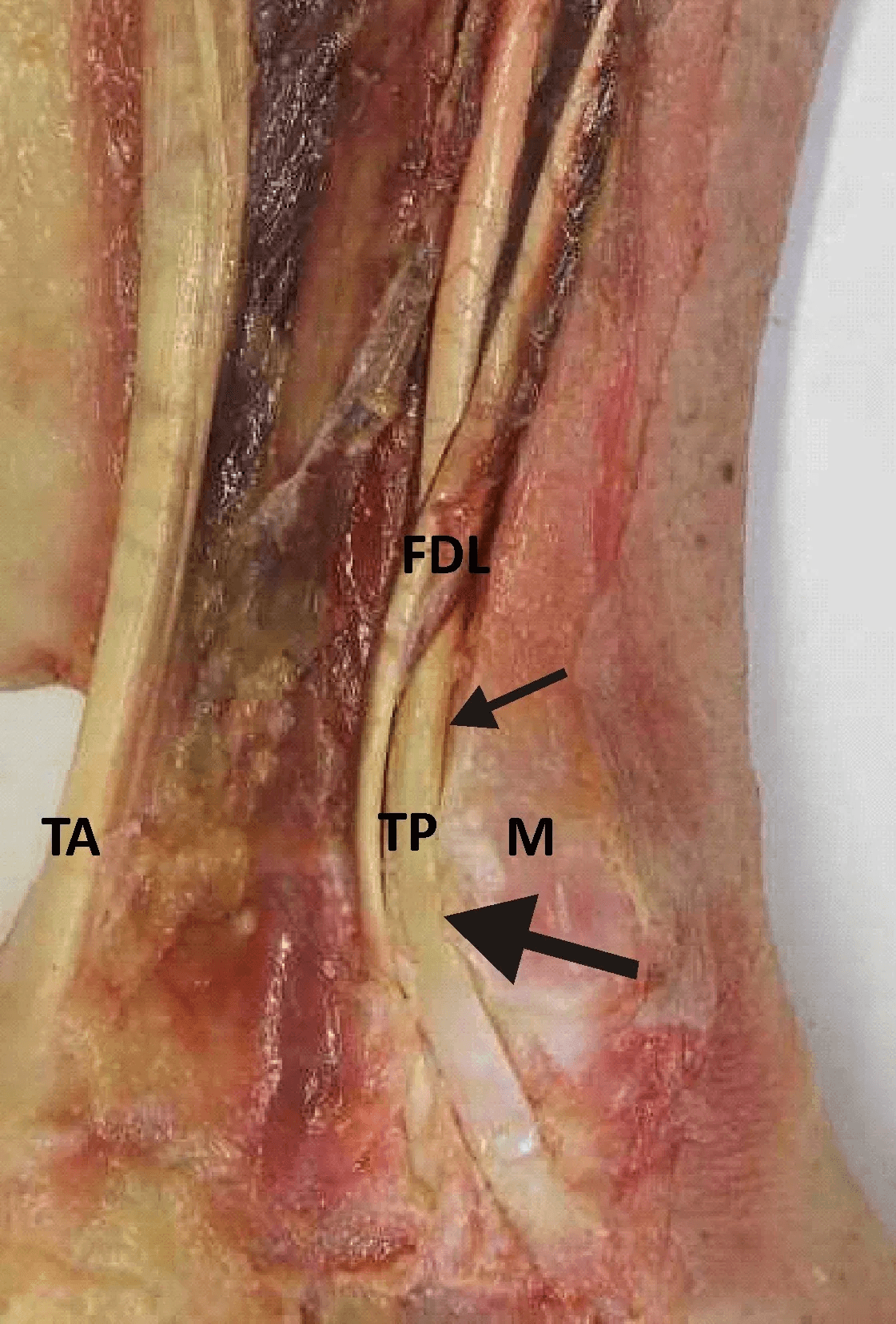
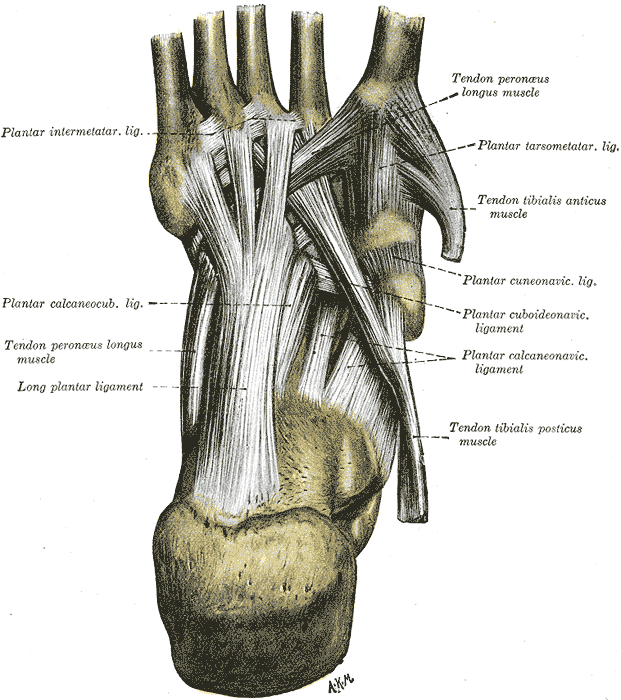
Where the ossicle sits. Medial and plantar to the navicular tuberosity. Tibialis posterior inserts partly onto it, and in Types II and III the tendon often inserts broad-based onto the accessory bone rather than onto the native navicular. The medial cutaneous nerve of the leg, or branches of the saphenous nerve, can be compressed over the lump.
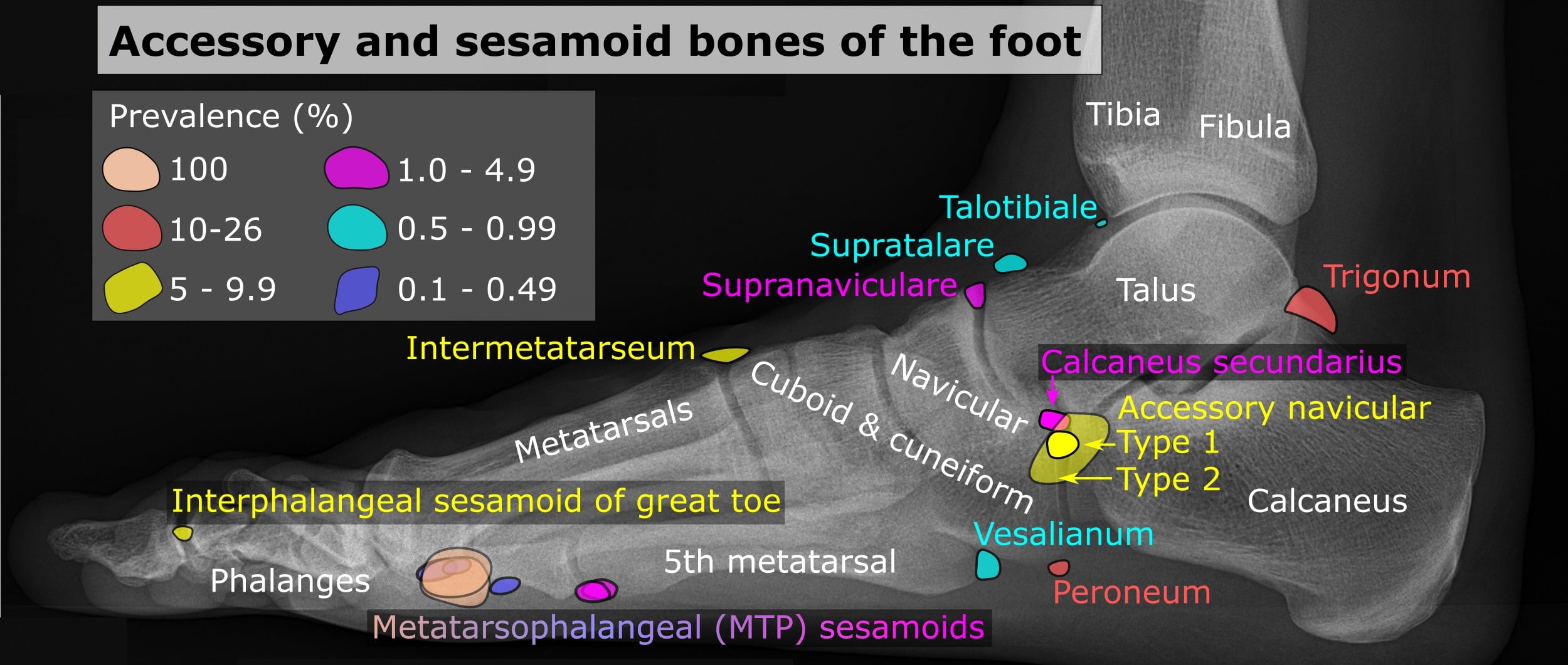
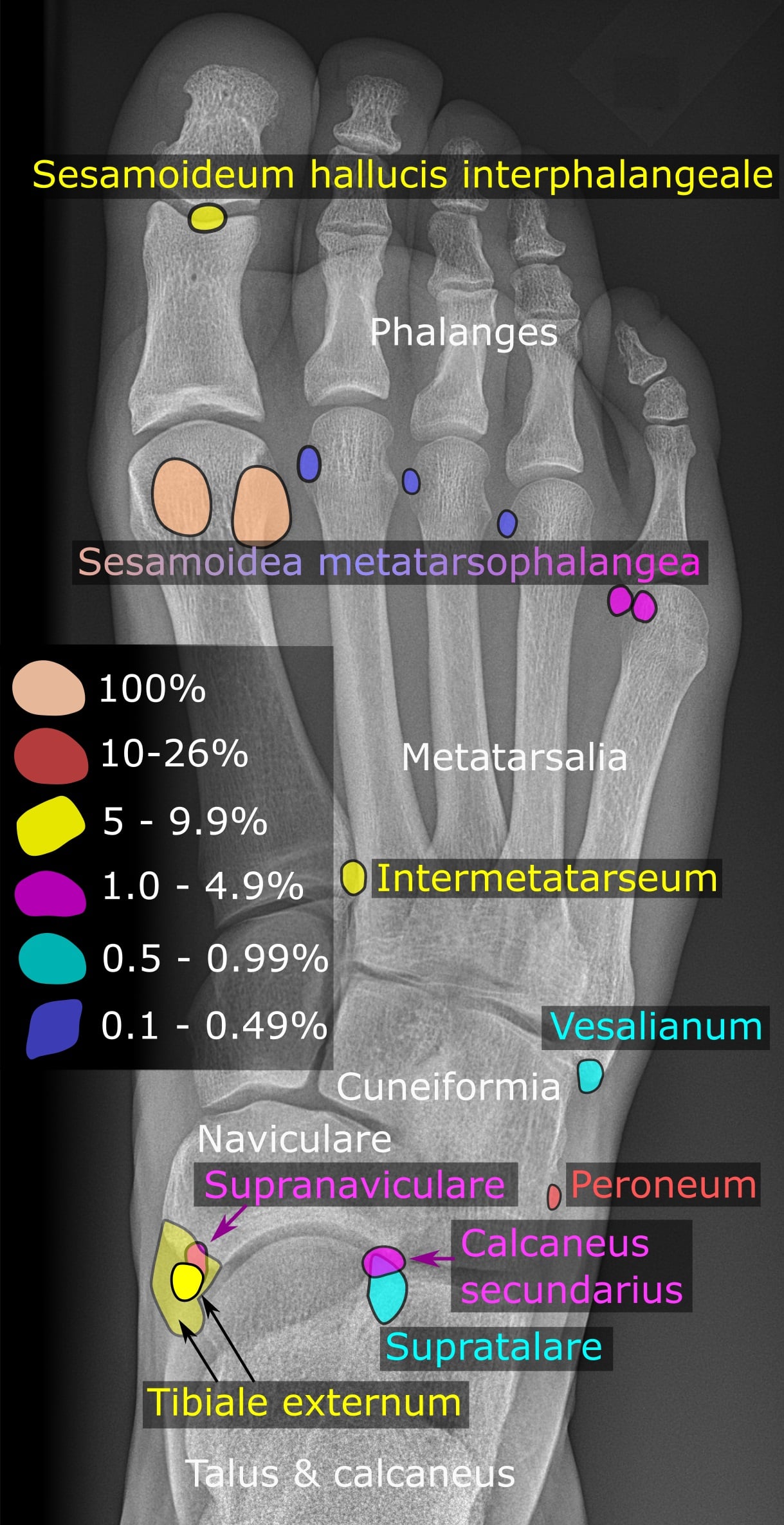
The pulley theory. Tibialis posterior uses the navicular tuberosity as a pulley, or fulcrum, to generate inversion torque. When it inserts on the accessory bone the insertion is more proximal, which shortens the lever arm and costs it leverage to plantarflex the navicular and support the arch, functionally like a lengthened tendon. The muscle has to work harder for the same inversion force, which can lead to earlier fatigue and may predispose to tendinopathy or to insufficiency and flatfoot.
Pain inhibition. If the insertion is painful, the patient avoids firing the muscle ("antalgic inhibition"). That leaves the foot flexible and flat during push-off, which adds further strain.
The flatfoot question. The association with pes planus is debated. Flatfoot is often found alongside a symptomatic ossicle, but large studies suggest its incidence is the same in those with and without the ossicle, and Kidner's claim that the ossicle disadvantages the tendon has not been substantiated (see the evidence below). Advancing the tendon distally onto the navicular body is meant to restore the mechanical advantage, yet simple excision performs comparably, so hold the pulley theory as the rationale for the operation rather than a proven mechanism.
Why It Hurts
Three triggers. Symptoms usually arise in adolescence, from one of three sources:
- Trauma - a twisted ankle or a direct blow disrupts the fragile Type II synchondrosis
- Overuse - repetitive traction from tibialis posterior on the ossicle it partly inserts into
- Pressure - direct conflict between footwear and the prominence
The weak link. In Type II the synchondrosis, a cartilage bridge, is a weak point, and motion occurs across it. Painful excised fragments often show chronic inflammation, micro-fractures and reparative change consistent with a prominent chronic non-union, which supports the theory that the pain is a "chronic stress fracture" of the synchondrosis. What the histology of an excised Type II typically shows:
- Irregular cartilage columns at the chondro-osseous junction
- Fibrosis, with scar tissue replacing hyaline cartilage
- Micro-fractures, evidence of acute-on-chronic trauma
- Sometimes zones of necrosis (avascular change)
Developmental Natural History and the Timing of Surgery
A secondary ossification centre. The navicular tuberosity has a separate ossification centre that normally appears around age 9-11 and fuses to the main navicular by about age 15. When it fails to fuse, the persistent centre becomes the accessory bone: a Type I sesamoid, a Type II synchondrosis, or a Type III cornuate navicular if it fuses late or incompletely as a beak.
Why adolescence. Symptoms cluster around skeletal maturity because that is when the synchondrosis is most stressed, with peak sport loading a tuberosity that is still cartilaginous. Hence the classic adolescent patient.
Why wait. A proportion of symptomatic Type II synchondroses spontaneously ossify and fuse into a painless Type III with skeletal maturity. That is a strong argument for a genuine trial of conservative care through adolescence, and for deferring excision until the skeleton is mature and non-operative treatment has clearly failed.
Both feet. Because the deformity is developmental it is bilateral in 50-90%, so the other foot often harbours the same ossicle. Examine and counsel about both sides even when only one is painful.
Classification Systems
Geist's classification (1914) describes how the ossicle is attached, and the attachment predicts the symptoms. The free sesamoid is usually silent, the synchondrosis moves and is the commonest cause of pain, and the fused beak hurts where the shoe rubs.
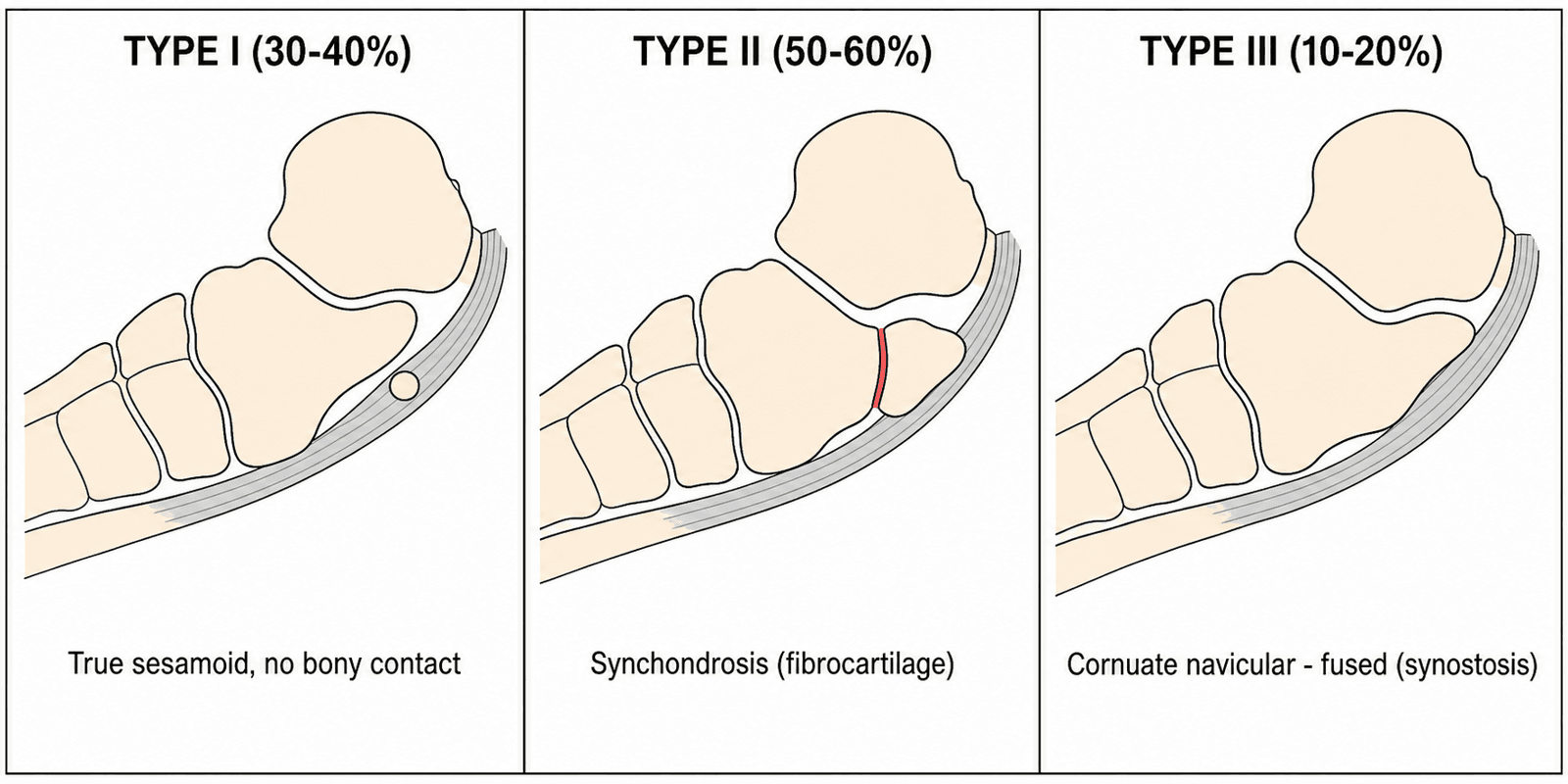
- Share
- 30-40%
- Anatomy
- True sesamoid within the tibialis posterior tendon; small (2-3mm) and round; separated from the navicular by more than 3mm
- Clinical
- Usually asymptomatic
- Share
- 50-60%
- Anatomy
- Bipartite navicular; triangular or heart-shaped ossicle joined by fibrocartilage or hyaline cartilage; gap less than 2mm
- Clinical
- Most common cause of pain
- Share
- 10-20%
- Anatomy
- Cornuate navicular: the ossicle is fused, leaving a prominent beak
- Clinical
- Pain from shoe rub (bunion-like)
SSSGeist Classification
Hook:The 3 S's of the Accessory Navicular.
The Sella modification. An MRI-based grading of the Type II ossicle. It is less commonly used, but useful for deciding whether the bump is actually the source of pain:
- Sella I - Type II on radiographs with high signal on MRI (active)
- Sella II - Type II on radiographs with low signal (inactive)
Clinical Assessment
History. Medial midfoot pain, either an intense "toothache"-like pain or a sharp pain with activity. The onset is often insidious, or follows a specific "sprain", an eversion injury that disrupts the synchondrosis. Narrow shoes, skates and ski boots provoke it.
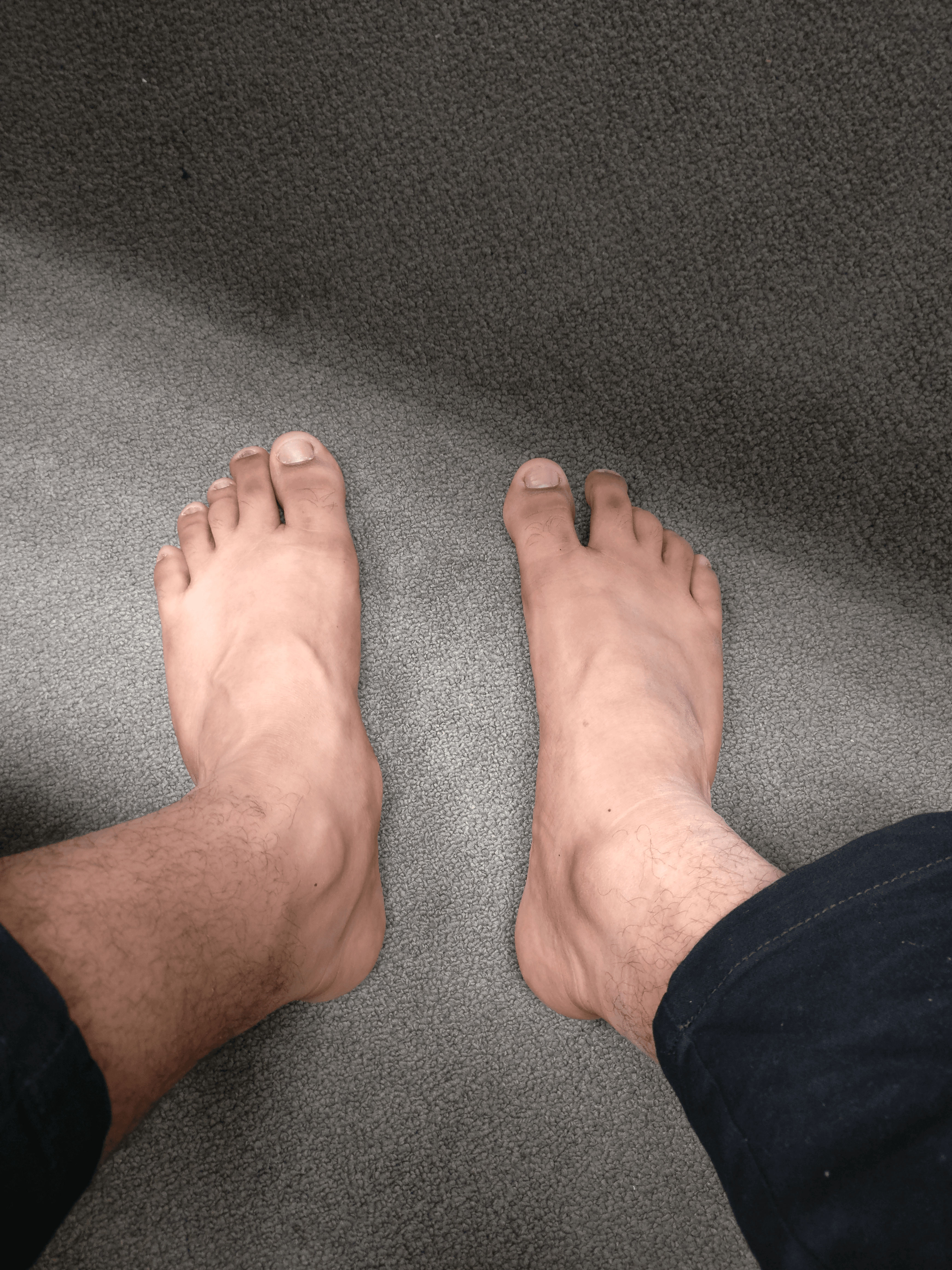
Examination. A visible bony prominence on the medial side of the foot, proximal to the first ray, gives the "double medial malleolus" appearance. The key sign is pinpoint tenderness directly over the prominence, not along the tendon itself. Then test the tendon and look at the arch:
- Resisted inversion reproduces the pain by engaging tibialis posterior, which pulls on the ossicle; resisted plantarflexion may also hurt
- The single-leg heel rise is usually normal because the tendon is intact, unlike adult acquired flatfoot
- A flexible flatfoot is often present and should be looked for
Investigations
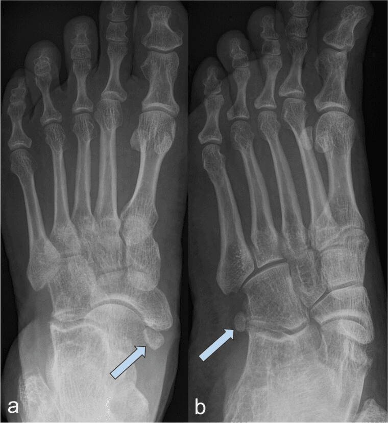
Radiographs. Weight-bearing views, of which the external oblique is the money view:
- AP foot - may miss the bone through overlap
- Lateral foot - shows the "double floor", an overlapping density
- External oblique (45°) - the key view, which clearly profiles the medial side of the navicular and the accessory bone
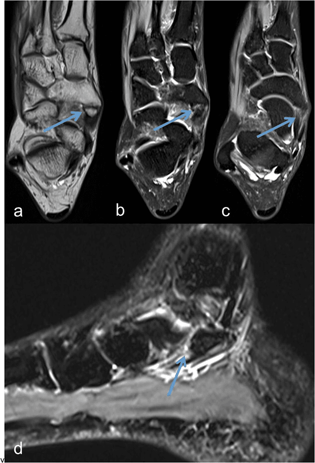
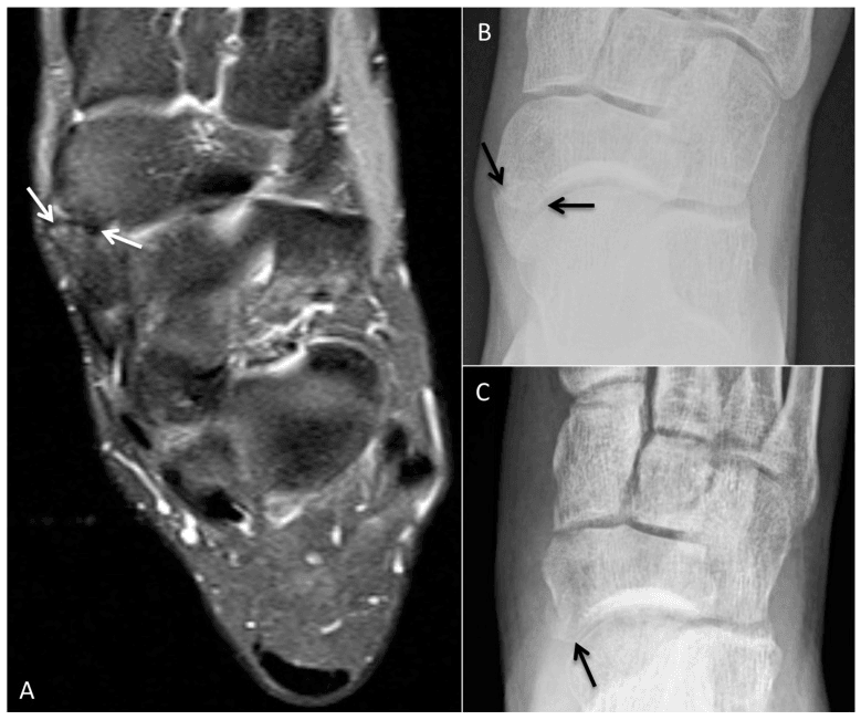
MRI. Indicated for a diagnostic dilemma, or for persistent pain with a normal radiograph (Type I). Bone marrow oedema in the accessory bone and the adjacent navicular body is diagnostic of a symptomatic synchondrosis; without oedema, look for another cause such as a stress fracture. In the symptomatic Type II, T1 shows the synchondrosis, and fluid-sensitive sequences show marrow oedema on both sides of the fibrocartilage and tibialis posterior insertional tendinosis.
CT. Confirms fusion (Type III) against a tight Type II.
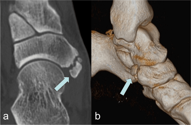
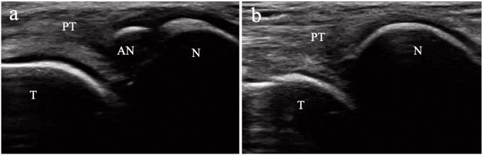
Bone scan and ultrasound. A bone scan (SPECT) shows a hot spot at the navicular but is historical, and MRI is preferred. Ultrasound shows the ossicle in relation to the tendon, and dynamic tenderness can be reproduced under the probe.
Confirming the Pain Generator: the Diagnostic Local Anaesthetic Injection
Why do it. An accessory navicular is common and often incidental. Before excising it you must be sure it is the pain generator, and not a coincidental finding beside a tarsal coalition, a navicular stress fracture or tibialis posterior tendinopathy.
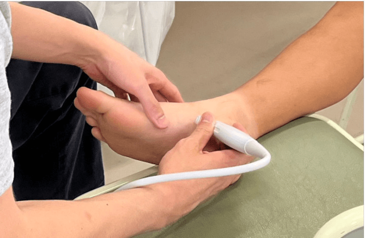
The test. An image-guided (ultrasound or fluoroscopic) injection of a small volume of local anaesthetic into or around the synchondrosis. Near-complete, temporary abolition of the patient's typical pain confirms the ossicle and synchondrosis as the source and predicts a good response to excision. It complements the MRI oedema pattern above. Under ultrasound, the probe lies longitudinally over the medial navicular tuberosity, following the posterior tibial tendon across the synchondrosis, and the same window guides the injection without entering the tendon.
No steroid here. A pure diagnostic block uses local anaesthetic alone. Adding corticosteroid near the tibialis posterior tendon risks tendon attrition or rupture and is generally avoided, because here the injection is a diagnostic, not a therapeutic, tool.
Differential Diagnosis
A navicular stress fracture is the killer differential, common in high-level sprinters and gymnasts. The accessory navicular hurts medially, over the prominence; the stress fracture hurts dorsally and deep, a vaguer midfoot pain. Radiographs can be subtle, and CT is often required.
Tibialis posterior tendinopathy. It differs from a painful Type II in age, site of pain, swelling and radiographs.
- Accessory Navicular (Type II)
- Adolescent (10-18y)
- Tib Post Tendinopathy
- Adult (greater than 40y)
- Accessory Navicular (Type II)
- Focal on bony prominence
- Tib Post Tendinopathy
- Along the tendon course
- Accessory Navicular (Type II)
- Bony lump (Hard)
- Tib Post Tendinopathy
- Soft tissue swelling (Boggy)
- Accessory Navicular (Type II)
- Associated (Secondary)
- Tib Post Tendinopathy
- Acquired (Progressive)
- Accessory Navicular (Type II)
- Ossicle visible
- Tib Post Tendinopathy
- Normal (or arthritis)
Tarsal coalition. A talonavicular coalition is rare, but a middle-facet calcaneonavicular (CN) coalition can present with vague medial or midfoot pain. The distinction matters because a Kidner on a coalition patient will fail: the pain comes from the rigid hindfoot, not the accessory bone. Always check subtalar motion before booking a Kidner.
- Motion - full subtalar motion with an accessory navicular (unless there is severe spasm); reduced or rigid with a coalition
- Age - a coalition often presents slightly older, at 12-16
- Radiographs - the "C-sign" and the "anteater nose", both for CN coalition
Management
The asymptomatic ossicle. An incidental accessory navicular needs observation only; no treatment is required.
Conservative care first. For a minimum of 3-6 months, and in the growing child through adolescence:
- Activity modification - temporarily avoid aggravating sports such as jumping and ballet
- Footwear - a wide toe box to reduce compression on the medial bump
- Orthoses - a medial arch support offloads tibialis posterior and reduces traction on the synchondrosis; a heel cup stabilises the hindfoot
- Immobilisation - a short leg walking cast or CAM boot for 4 weeks, for an acute flare-up or "fracture" of the synchondrosis
The athlete. Athletes settle less readily. In Jegal's series only 6.9% of athletes improved with conservative care against 34% of non-athletes, and earlier surgery can be considered in this group.
A dancer who settled. A 14-year-old ballet dancer had right medial foot pain, worse en pointe, of gradual onset over 3 months with no trauma. She had bilateral mild pes planus, a prominent and red navicular on the right, 10/10 pinpoint tenderness over the ossicle, and a heel rise that was painful but complete. The external oblique film showed a Type II ossicle with irregular sclerosis at the synchondrosis, a sign of stress, and MRI showed bone marrow oedema in the ossicle and navicular body.
Four weeks in a moon boot with no dancing, ice and NSAIDs resolved the pain. Progressive loading and a custom orthotic with a "navicular accommodation" (a sweet-spot cut-out) followed. She returned to dance at 3 months, with flare-ups managed by taping, and surgery was avoided.
Indications for surgery. Failure of conservative management with persistent pain localised to the accessory navicular. The features that point to operating:
- Pain - conservative care failed at 6 months
- Prominence - shoe-wear problems from the bump
- Palpable - tender on palpation
Which operation. Excision is the gold standard, either as a Kidner procedure (excision plus tendon advancement) or as simple excision. Wariach's systematic review found the two comparable, with recurrent pain after Kidner concentrated in feet with planovalgus or hindfoot valgus. Fusion is the rarely used alternative.
Surgical Technique: The Kidner Procedure
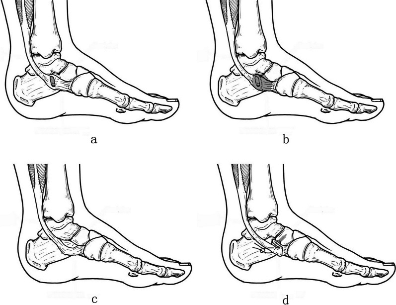
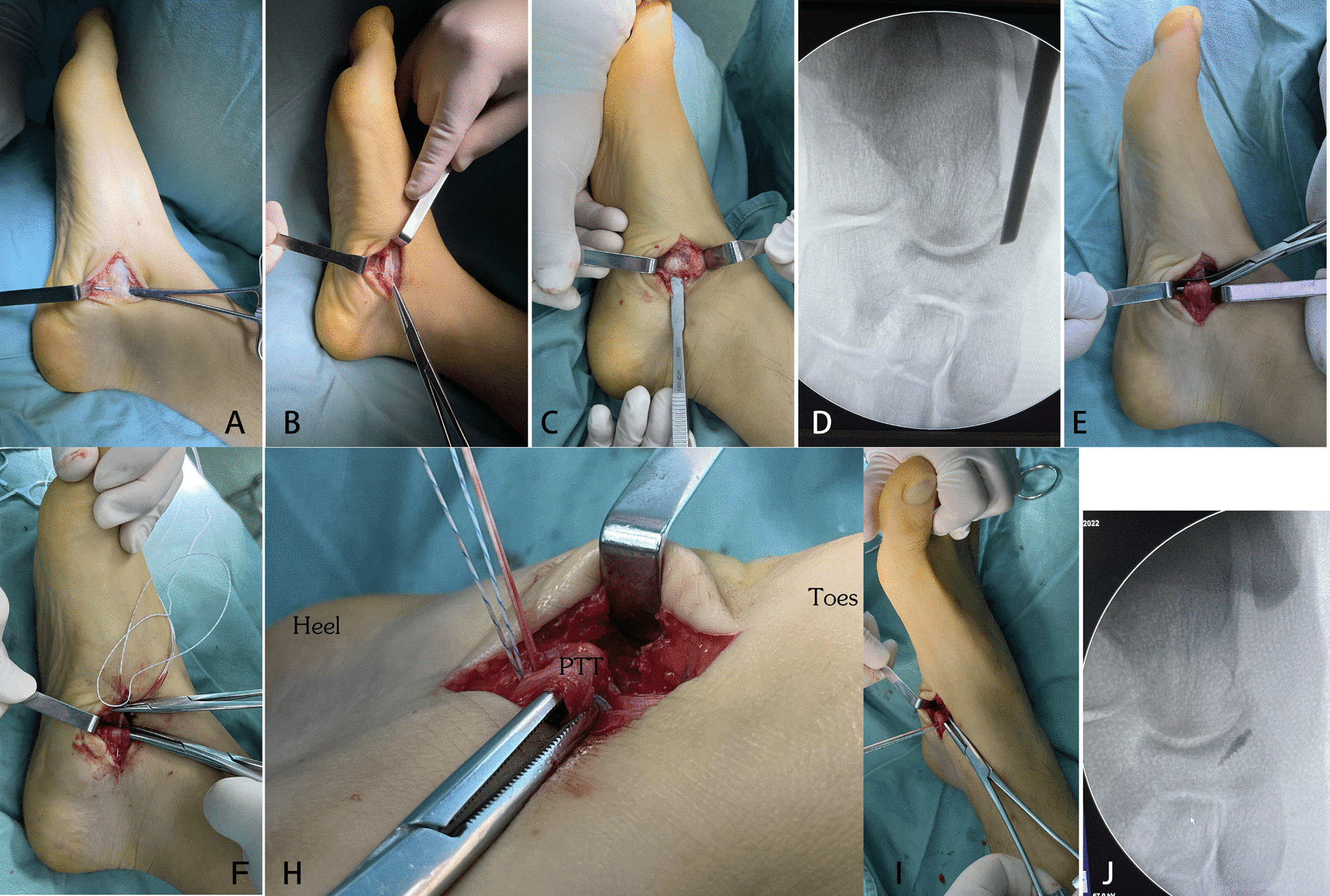
The Kidner procedure excises the ossicle and advances the tibialis posterior tendon. Kidner's original operation transposed the tendon to the plantar surface; the modern modification is simple advancement.
- Approach. A curvilinear 3-4cm incision at the medial prominence, placed slightly dorsal rather than directly over it, because the shoe line often hits the prominence and a scar there is miserable. Avoid the saphenous nerve and vein, which lie dorsal and anterior.
- Deep dissection. Incise the tibialis posterior tendon sheath; the tendon can be seen broadening to insert onto the accessory bone. Make a "T"- or "I"-shaped incision in the periosteum and tendon to preserve flaps for the repair.
- Excision. Shell out the ossicle (Type I or II) after identifying the synchondrosis line. The medial native navicular is often still beaked, so remove the prominence with an osteotome or saw flush with the cuneiform, judged by palpating the naviculocuneiform joint. Palpate for any remaining spur: it must be smooth, and a step left behind will rub. Confirm complete resection of the ossicle on fluoroscopy.
- Repair. The tendon insertion is now loose. Prepare the navicular bed, then place a suture anchor (SwiveLock or Corkscrew) in the plantar-medial aspect of the navicular body, then advance the tendon and suture it down under tension with the foot in inversion.
- Closure. Repair the tendon sheath, then close the skin.
Anchor or drill holes. Drill holes are the classic technique: two holes in the navicular with the suture passed through, inexpensive and strong but harder to do. Anchors are the modern technique, fast and reproducible with strong pull-out strength, but expensive, and most surgeons now use them for ease of use.
Simple excision. The bone is removed without completely detaching the tendon, for a Type I or small Type II where the tendon inserts primarily on the main body. Outcomes are similar to Kidner in some studies, but Kidner is preferred if the tendon insertion is compromised.
A tendon-preserving variant. Resect the accessory navicular and preserve the native tibialis posterior insertion when possible, then fold and re-tension the lax tendon onto an anchor in the navicular. The fold is adjusted to restore inversion without over-tensioning the medial arch.
Alternative: Fusion
The idea. Turn the painful Type II (synchondrosis) into a painless Type III (synostosis) with a percutaneous screw across the fragment. Historically some surgeons argued this was superior because it preserves the "full" insertion of the tendon, and it is minimally invasive.
The problem. The synchondrosis has poor blood supply. Non-union rates after percutaneous screw fixation are high, and the screw head can become symptomatic.
The verdict. Fusion is rarely performed. It is reserved for a large, congruent Type II with enough bone stock: very large ossicles (greater than 1.5cm) where excision would remove more than 30% of the articular surface (risk of instability). Even then many experts prefer excision with robust repair. Kidner is superior, being reliable and technically easier, with equivalent or better outcomes. Zeng's small comparison found Kidner and local arthrodesis comparable (80% against 83% good-to-excellent).
Complications
- Rate
- 5-10%
- Prevention/Management
- Usually due to inadequate resection (leaving a stump) or neuroma.
- Rate
- Common
- Prevention/Management
- Injury to medial cutaneous nerve. Bury nerve end if cut.
- Rate
- Rare
- Prevention/Management
- Failure to reattach tendon securely.
- Rate
- Common
- Prevention/Management
- Desensitisation massage.
- Rate
- Very Rare
- Prevention/Management
- Over-aggressive trimming of tendon.
Recurrence. True regrowth is rare if the excision is adequate. A small stump of bone left behind, however, can be bothersome, which is why the prominence is shaved flat.
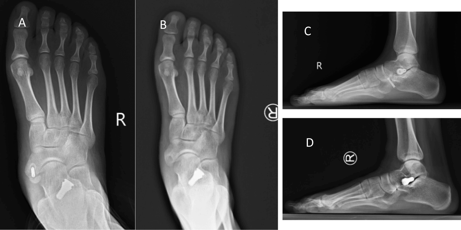
Fusion failure. The failure specific to fusion is persistent synchondrosis nonunion around the screw, at ten months in the case below. Revision removes the screw and ossicle, then restores the tibialis posterior insertion with a suture anchor.
Postoperative Care and Rehabilitation
The tendon repair is protected first, then loaded, then strengthened. Weight-bearing in weeks 2-6 varies between surgeons.
- Weeks 0-2, protection - backslab, cast or moon boot; strict non-weight-bearing; elevation above heart level, so the bone-tendon interface heals without shear stress. Keep the wound dry; sutures are removed at 2 weeks.
- Weeks 2-6, protected weight-bearing - transition to a CAM walker. After a Kidner, weight-bearing as tolerated if the anchor is strong; some surgeons prefer non-weight-bearing for 4 weeks, and a staged plan is touch-down on crutches for weeks 2-4 then partial to full in the boot for weeks 4-6. Active plantarflexion and dorsiflexion; active inversion is started to stimulate the tendon but kept limited. Toe yoga for the intrinsics, and static quadriceps and gluteal work.
- Weeks 6-12, weaning and strengthening - wean the boot into a supportive trainer with a medial arch support, which is essential to protect the repair. Full active inversion and eversion; Theraband inversion (yellow to red to green); double- then single-leg heel rises; eccentric loading with physiotherapy; proprioception on a balance board, since tibialis posterior is a key proprioceptor.
- Months 3-6, return to sport - progress walk to jog to run to cut to jump.
Return-to-sport criteria. A pain-free single-leg heel rise (3x10 reps), full range of motion, and running and hopping without pain. Return to sport is at 3-4 months, and contact sport is usually safe at 4 months. Dancers need specific en pointe training: basic dance usually returns at 3-4 months, but pointe work typically requires 6 months of strengthening, and full return often takes 6 months.
Follow-up imaging. After resection, imaging is reserved for persistent pain, not routine union assessment.
Outcomes and Prognosis
Success. The Kidner procedure has a high success rate, 90% good or excellent. Full resolution of swelling and scar tenderness can take 6-12 months.
The arch. The operation removes the pain; it does not typically restore the arch in a patient with flatfeet, a common misconception, and arch height usually stays the same, which is fine as long as it is painless. If the flatfoot is severe, a medialising calcaneal osteotomy may be needed at the same time. Kim's revision series is the warning: 20 of 21 patients revised for recurrent pain after Kidner had valgus heel alignment, so assess the hindfoot before the first operation.
Does excision cause flatfoot? The worry is that removing the bone detaches the arch support. The primary support of the arch is the spring ligament complex and the interosseous ligaments, not just the tibialis posterior insertion, and available medium-to-long-term follow-up studies show no significant radiographic progression of flatfoot deformity after the Kidner procedure compared with non-operated controls.
Arthritis. Long-term studies show minimal risk of naviculocuneiform osteoarthritis from the altered mechanics, and the operation is a safe, benign procedure.
Counselling the family. Tight shoes did not cause it: the extra bone is congenital, and shoes may have irritated it and brought it to attention without creating it. Removing it does not make the foot weak, because the accessory bone is "stealing" the power of tibialis posterior, and re-attaching the tendon to the main bone often improves the muscle's mechanical efficiency. A flat foot is a complex three-dimensional deformity, and the operation generally will not fix it.
Guidelines, Registries & Global Practice
Global epidemiology
- Accessory navicular is present in roughly 10-14% of the general population and is bilateral in 50-90% of cases, with a female predominance.
- The great majority are asymptomatic incidental findings; symptomatic accessory navicular syndrome clusters in adolescents and young adults, especially dancers, gymnasts and pivoting athletes.
Society / guideline positions (side by side)
No society publishes a formal guideline for accessory navicular syndrome. The rows below summarise typical practice patterns in each region's foot and ankle community, not published guideline recommendations.
- Typical practice pattern (no formal guideline)
- Conservative care first (rest, orthoses, immobilisation); surgery (excision or modified Kidner) for refractory pain. No dedicated standalone guideline.
- Typical practice pattern (no formal guideline)
- Same conservative-first pathway; foot and ankle surgeon referral after failed non-operative care; address coexisting planovalgus.
- Typical practice pattern (no formal guideline)
- Frames acute synchondrosis disruption as a stress-injury/non-union; immobilise acutely, excise if persistently symptomatic.
- Typical practice pattern (no formal guideline)
- Endorses excision or Kidner with equivalent results; recommends correcting hindfoot valgus to reduce recurrence.
Registry note
- No arthroplasty or implant registry tracks this condition (it is non-prosthetic). Evidence is limited to retrospective series and one systematic review; there are no randomised trials comparing excision with Kidner.
Resource-setting variation
- High-resource settings use weight-bearing radiographs plus MRI to confirm a symptomatic synchondrosis and suture anchors for tendon reattachment.
- Limited-resource settings rely on clinical examination and plain external-oblique radiographs, with transosseous suture (drill-hole) tendon repair as a low-cost, equally effective alternative to anchors.
- First-contact management is frequently led by physiotherapy/podiatry worldwide, with orthopaedic referral reserved for failed conservative care.
MCQ Practice Points
Q: Which type of accessory navicular is most commonly symptomatic? A: Type II. The movement at the synchondrosis causes pain. Type I is too small, Type III is fused.
Q: What is the primary purpose of the Kidner procedure? A: Pain relief. It is NOT primarily an arch-reconstruction procedure, although tendon advancement is performed.
Q: Which X-ray view best visualizes the accessory navicular? A: External Oblique (45 degree). It projects the ossicle away from the other tarsal bones.
Q: What sensory nerve is at risk during the medial approach to the navicular? A: Medial Cutaneous Nerve (branch of Saphenous).
Q: Which tendon inserts onto the accessory navicular? A: Tibialis Posterior. In Type II/III, the insertion is often broad-based and inserts onto the ossicle, losing its mechanical advantage.
Q: What is the approximate prevalence of an accessory navicular in the general population? A: 10-14%, bilateral in 50-90% of cases, but symptomatic in less than 1% - usually an incidental finding.
Q: A patient still has pain after a Kidner procedure - give three causes. A: Inadequate resection (residual stump or regrowth), neuroma of the medial cutaneous nerve, and a missed tarsal coalition.
Viva Scenarios
Practise clinical reasoning and management decisions out loud
“14-year-old female dancer. Medial foot pain. Visible lump. Failed physio.”
“Tennis player. Lateral lunge. Felt a 'pop' medial foot. Now painful lump.”
“30-year-old. Had surgery as a child. Now pain has returned in same spot.”
Evidence Base
Kidner (original description)
- Original description of excision of the accessory navicular with re-routing of the tibialis posterior tendon
- Proposed the (now disputed) concept that the ossicle disadvantages the tendon and contributes to flatfoot
- Modern practice has simplified this to excision plus tendon advancement/repair
Wariach et al — Excision vs Kidner (Systematic Review)
- Systematic review comparing simple excision with the Kidner procedure for symptomatic accessory navicular
- Both procedures were efficacious with low complication profiles and high patient satisfaction; minor complications were scar and wound irritation
- Recurrent pain after Kidner was concentrated in feet with planovalgus / hindfoot valgus, sometimes requiring revision
Jegal et al — Athletes vs General Population
- 79 patients with accessory navicular syndrome; only 6.9% of athletes improved with conservative care vs 34% of non-athletes (p less than 0.001)
- Bone marrow oedema in both navicular and accessory bone was seen in 100% of athletes vs 80% of non-athletes
- Trauma history and inter-ossicle movement were more frequent in athletes
Takahashi et al — MRI Bone Marrow Oedema
- Edema-like bone marrow pattern was present in all symptomatic navicular tuberosities and absent in asymptomatic ones
- Oedema intensity diminished as symptoms resolved, suggesting it tracks the active pain generator
- The pattern occurred even in patients without an accessory bone, implying an osteitis-like mechanism
Zeng et al — Kidner vs Local Arthrodesis
- 16 patients (20 feet) with symptomatic Type II accessory navicular; Kidner vs local arthrodesis
- Good-to-excellent outcomes were comparable: 80% (Kidner) vs 83% (arthrodesis), p=0.696
- Calcaneal pitch improved in both groups with no significant difference in radiographic indices
Kim et al — Revision After Failed Kidner
- 21 patients revised for recurrent pain after Kidner; 20/21 (95%) had valgus heel alignment
- Contralateral feet showed similar deformity, indicating a pre-existing alignment problem rather than surgical error
- Adding a medial displacement calcaneal osteotomy improved radiographic and clinical outcomes
Kakihana et al — Suture-Anchor Stabilisation
- 22 feet in 15 adolescents treated with suture-anchor osteosynthesis instead of excision
- Radiographic union in only 64% by 8 weeks, yet clinical scores rose from 54 to 96
- Bony union was modest but symptom relief was reliable in the short to mid term