End-Stage Ankle Arthritis | Position Critical | Open vs Arthroscopic | Tibiotalocalcaneal Option
Ankle Arthrodesis Types
Critical Must-Knows
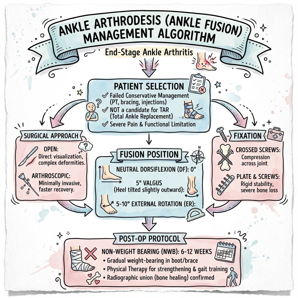
- Optimal position: Neutral dorsiflexion (5° plantar), 5° valgus, 5-10° ER (match contralateral)
- Arthroscopic: Faster recovery, similar union rates, best for non-deformed ankles
- Open: Better for deformity, bone loss, revision cases with bone grafting capability
- Union rate: 90-95% with optimal technique; malalignment increases nonunion 3-fold
- Compression critical: Minimum 250N compression across joint for reliable fusion
Clinical Pearls
- "Position errors cannot be revised - get it right first time (5-5-5 rule)
- "Arthroscopic vs open debate settled: equivalent union, arthroscopic faster recovery
- "Tibiotalocalcaneal nail salvages failed ankle replacement and severe deformity
- "TARVA RCT: TAR and fusion give comparable 1-year outcomes - choice is patient-specific
Clinical Imaging
Imaging Gallery


Critical Ankle Arthrodesis Exam Points
Position is Everything
Malalignment = guaranteed poor outcome. Sagittal position most critical: 5° neutral dorsiflexion (0-10° acceptable). Valgus 5° prevents varus collapse. External rotation must match contralateral side.
Indications Hierarchy
Post-traumatic arthritis number 1 (60% of cases). Also rheumatoid, avascular necrosis, failed ankle replacement, Charcot arthropathy. Age under 60 favors fusion over replacement.
Technique Selection
Open vs arthroscopic not superior/inferior - patient-specific. Arthroscopic: faster recovery, less wound issues. Open: bone grafting, deformity correction, revision.
Compression Fixation
Minimum 250N compression across joint. Crossed screws provide rotation control. Plate adds compression and stability in osteoporotic bone or revision.
Quick Decision Guide - Ankle Arthrodesis
| Patient Scenario | Technique | Fixation | Key Pearl |
|---|---|---|---|
| Young, post-traumatic, minimal deformity, good bone stock | Arthroscopic | 3 cannulated screws (2 lateral-medial, 1 anterior-posterior) | Fastest recovery, back to work 10-12 weeks |
| Moderate deformity (under 15°), some bone loss | Mini-open or open anterior | Plate plus screws for compression | Bone graft local or iliac crest if over 1cm defect |
| Failed ankle replacement, severe bone loss, Charcot | Tibiotalocalcaneal fusion | Intramedullary hindfoot nail | Bypass ankle, fuse through calcaneus, major salvage |
| Rheumatoid, osteoporotic bone, elderly | Open anterior approach | Plate with locking screws | Bone quality critical - consider bisphosphonate holiday |
FIVEOptimal Ankle Fusion Position (5-5-5 Rule)
| F | Five degrees neutral dorsiflexion 0-10° acceptable, sagittal position most critical for gait |
| I | Ipsilateral rotation match 5-10° external rotation matching contralateral ankle |
| V | Valgus five degrees Hindfoot alignment prevents varus collapse and load transfer |
| E | Examine with fluoroscopy Confirm position before final fixation in all planes |
| F | Five degrees neutral dorsiflexion 0-10° acceptable, sagittal position most critical for gait | V | Valgus five degrees Hindfoot alignment prevents varus collapse and load transfer |
| I | Ipsilateral rotation match 5-10° external rotation matching contralateral ankle | E | Examine with fluoroscopy Confirm position before final fixation in all planes |
Hook:Five-Five-Five: 5° neutral, 5° valgus, 5-10° ER - get position right because you cannot revise it!
PARTICAnkle Arthrodesis Indications
| P | Post-traumatic arthritis 60% of cases, young active patients, malunited fractures |
| A | AVN (avascular necrosis) Talar dome necrosis, sickle cell, steroid use |
| R | Rheumatoid arthritis Severe joint destruction, failed medical management |
| T | Total ankle replacement failure Aseptic loosening, infection, instability |
| I | Infection (septic arthritis) After eradication of infection, staged fusion |
| C | Charcot arthropathy Neuropathic destruction, diabetes, tibiotalocalcaneal fusion |
| P | Post-traumatic arthritis 60% of cases, young active patients, malunited fractures | R | Rheumatoid arthritis Severe joint destruction, failed medical management | I | Infection (septic arthritis) After eradication of infection, staged fusion |
| A | AVN (avascular necrosis) Talar dome necrosis, sickle cell, steroid use | T | Total ankle replacement failure Aseptic loosening, infection, instability | C | Charcot arthropathy Neuropathic destruction, diabetes, tibiotalocalcaneal fusion |
Hook:PARTIC = Participate in fusion when joint is destroyed - most common is Post-traumatic!
FASTERArthroscopic Ankle Fusion Advantages
| F | Faster recovery Return to work 10-12 weeks vs 16-20 weeks open |
| A | Avoid wound complications No anterior incision, lower infection rate 2% vs 5% |
| S | Same union rates 90-95% union equivalent to open technique |
| T | Time to union shorter 12 weeks vs 16 weeks for radiographic union |
| E | Easy debridement Direct visualization and removal of cartilage |
| R | Reduced hospital stay Day case or overnight vs 2-3 days open |
| F | Faster recovery Return to work 10-12 weeks vs 16-20 weeks open | S | Same union rates 90-95% union equivalent to open technique | E | Easy debridement Direct visualization and removal of cartilage |
| A | Avoid wound complications No anterior incision, lower infection rate 2% vs 5% | T | Time to union shorter 12 weeks vs 16 weeks for radiographic union | R | Reduced hospital stay Day case or overnight vs 2-3 days open |
Hook:FASTER recovery with arthroscopic fusion - but only for minimal deformity and good bone stock!
PAINSAnkle Arthrodesis Complications
| P | Position malunion Most common complication, cannot revise easily, equinus worst |
| A | Adjacent joint arthritis Subtalar 30% at 10 years, talonavicular 20%, midfoot stress |
| I | Infection Deep infection 2-5%, higher in diabetes and open technique |
| N | Nonunion 5-10% rate, risk factors: smoking, malalignment, poor compression |
| S | Stiffness and pain Residual pain 10-20%, hindfoot stiffness, difficulty on uneven ground |
| P | Position malunion Most common complication, cannot revise easily, equinus worst | N | Nonunion 5-10% rate, risk factors: smoking, malalignment, poor compression |
| A | Adjacent joint arthritis Subtalar 30% at 10 years, talonavicular 20%, midfoot stress | S | Stiffness and pain Residual pain 10-20%, hindfoot stiffness, difficulty on uneven ground |
| I | Infection Deep infection 2-5%, higher in diabetes and open technique |
Hook:Ankle fusion causes PAINS - position malunion and adjacent joint arthritis are the main long-term issues!
Overview and Epidemiology
Clinical Context
Ankle arthrodesis remains a gold-standard option for end-stage ankle arthritis, especially in young, active patients and those with significant deformity, and it avoids the risk of implant failure. Historically fusion showed better medium-term survivorship than early total ankle replacements, but the TARVA RCT found comparable 1-year patient-reported outcomes between fusion and modern TAR. The trade-off for fusion is permanent loss of ankle motion, increased stress on adjacent joints, and difficulty on uneven terrain.
Demographics
- Age: Most patients 40-60 years at time of fusion
- Gender: Male greater than female 2:1 (trauma predominance)
- Activity: Young active patients favor fusion over replacement
- Occupation: Heavy manual workers benefit from stability
Impact
- Function: 80% satisfied despite loss of motion
- Gait: Compensatory midfoot and hip motion
- Adjacent joints: 30% develop symptomatic arthritis by 10 years
- Revision rate: roughly 7-10% at 10 years (registry and RCT data)
Anatomy and Biomechanics
Ankle Joint Anatomy
Tibiotalar Joint
- Articular surface: 350-500mm² contact area
- Stability: Mortise and deep malleoli (intrinsic stability)
- Motion: 20° dorsiflexion, 30° plantarflexion (normal gait)
- Load: 5-6× body weight during walking, 13× running
Blood Supply
- Tibial side: Anterior tibial artery perforators
- Talar dome: Posterior tibial and dorsalis pedis branches
- Watershed area: Central talar dome (AVN risk)
- Fusion healing: Metaphyseal bone excellent vascularity
Biomechanics After Fusion
Gait Compensation Mechanisms
After ankle arthrodesis, patients compensate through:
- Subtalar motion: Increased stress (15° inversion/eversion becomes 25°)
- Midfoot motion: Talonavicular and midfoot joints increase 40% motion
- Hip and knee: External rotation of hip, increased knee flexion in swing phase
- Energy expenditure: 5-10% increase in metabolic cost of walking
These compensatory mechanisms lead to adjacent joint arthritis in 30% of patients by 10 years post-fusion.
| Joint | Normal Motion | After Fusion | Arthritis Risk |
|---|---|---|---|
| Tibiotalar (fused) | 50° total | 0° (fused) | N/A |
| Subtalar | 30° total | Increased to 40° | 30% at 10 years |
| Talonavicular | 10° total | Increased to 15° | 20% at 10 years |
| Midfoot (Lisfranc) | 5° total | Increased to 10° | 15% at 10 years |
Classification of Ankle Arthrodesis Types
Standard Ankle Arthrodesis
Indications:
- Post-traumatic arthritis isolated to tibiotalar joint
- Rheumatoid arthritis without subtalar involvement
- AVN of talus with preserved subtalar joint
- Failed ankle replacement with good talar bone stock
Contraindications:
- Active infection (relative, stage with antibiotics first)
- Severe peripheral vascular disease (relative)
- Charcot arthropathy (prefer tibiotalocalcaneal)
- Severe osteoporosis (relative, consider bisphosphonates)
Technical Point
Preserving the subtalar joint is essential - it provides 70% of hindfoot inversion/eversion. Never extend fusion to subtalar unless severely arthritic, as this doubles disability and adjacent joint stress.
Clinical Assessment
History
- Pain: Location (ankle vs subtalar vs midfoot)
- Function: Walking distance, stairs, uneven ground ability
- Prior surgery: Ankle fracture, ligament reconstruction, replacement
- Medical: Diabetes (Charcot risk), RA, smoking status
- Occupation: Manual labor vs sedentary (fusion vs replacement)
- Goals: Return to work, sports, daily activities
Examination
- Gait: Antalgic, hindfoot alignment, foot progression angle
- Alignment: Weightbearing varus/valgus, forefoot supination
- ROM: Ankle (tibiotalar) vs subtalar motion isolated
- Tenderness: Joint line, subtalar, talonavicular, midfoot
- Deformity: Fixed vs flexible with hindfoot block
- Neurovascular: Posterior tibial pulse, sensation
Special Tests
| Test | Technique | Interpretation |
|---|---|---|
| Intra-articular injection | Fluoroscopy-guided 5mL lignocaine | Pain relief greater than 80% confirms tibiotalar source |
| Hindfoot block test | Lateral hindfoot wedge under heel | Corrects supination = flexible deformity, good fusion candidate |
| Subtalar injection | Sinus tarsi approach with contrast | No relief = isolated tibiotalar arthritis (good for isolated fusion) |
Pitfall: Missing Subtalar Arthritis
Do not fuse ankle alone if subtalar arthritis present - this leads to persistent pain and early failure. Always:
- Examine subtalar motion separately (inversion/eversion with ankle blocked)
- Look for subtalar tenderness in sinus tarsi
- Consider CT if subtalar arthritis suspected on X-ray
- Inject subtalar separately if diagnosis unclear
If subtalar arthritis confirmed, offer tibiotalocalcaneal fusion from the start.
Differential Diagnosis of the Painful Arthritic Ankle
Distinguishing Causes of Hindfoot/Ankle Pain Before Committing to Fusion
| Diagnosis | Key features | Discriminator | Implication |
|---|---|---|---|
| Tibiotalar OA (fusion target) | Anterior/diffuse ankle pain, stiffness, joint-line tenderness | Pain abolished by intra-articular tibiotalar injection | Fusion or TAR indicated |
| Isolated subtalar arthritis | Sinus tarsi pain, pain on inversion/eversion with ankle held | Relief with subtalar (not tibiotalar) block | Subtalar fusion, not ankle fusion |
| Talar AVN / osteonecrosis | Deep pain, risk factors (steroid, alcohol, trauma) | MRI marrow oedema/collapse; talar body involvement | May need TTC fusion if body collapsed |
| Inflammatory arthritis (RA) | Polyarticular, morning stiffness, raised CRP/ESR/RF | Systemic features and serology | Optimise DMARDs; perioperative immunosuppression plan |
| Septic arthritis / infected nonunion | Rest pain, warmth, effusion, fevers, raised inflammatory markers | Aspiration culture; never fuse over active infection | Eradicate infection first, staged fusion |
| Charcot neuroarthropathy | Diabetic neuropathy, swelling, deformity, often painless | Neuropathy plus fragmentation/dislocation on imaging | TTC fusion/reconstruction, not standard fusion |
Investigations
Imaging Protocol for Ankle Arthrodesis Planning
Essential views:
- AP ankle, mortise, lateral ankle (weight-bearing mandatory)
- Hindfoot alignment view (Saltzman view for varus/valgus)
- Foot AP and lateral to assess midfoot arthritis
What to assess:
- Degree of joint space narrowing and osteophyte formation
- Subchondral cysts and sclerosis (severity markers)
- Deformity: varus/valgus angle, tibiotalar subluxation
- Bone stock: talar dome height, previous surgery defects
- Adjacent joints: subtalar, talonavicular, midfoot
Indications for CT:
- Deformity greater than 15° (need 3D planning)
- Prior surgery with hardware (assess bone stock)
- Suspected AVN or subchondral collapse
- Planning bone graft requirements
CT provides:
- Bone defect size and location (quantify graft need)
- Deformity in all planes (surgical correction plan)
- Hardware position if revision case
- Subtalar and talonavicular joint status
Indications:
- Suspected talar AVN (bone marrow edema, collapse)
- Rule out infection (enhancement, fluid collections)
- Soft tissue pathology (tendon tears, masses)
- Young patient considering ankle replacement vs fusion
MRI not routinely required for straightforward arthrodesis.
- FBC, CRP, ESR: Baseline inflammatory markers (infection screen)
- HbA1c: If diabetic (target under 7% for surgery)
- Bone density: If osteoporotic appearance on X-ray
- Group and save: Standard for major joint surgery
Management Algorithm
Conservative Management Role
Non-operative treatment is not curative for end-stage arthritis but delays surgery and helps patient selection. Trial for 3-6 months before offering fusion. If symptoms controlled, continue conservative; if refractory pain despite maximal non-operative treatment, proceed to surgery.
Non-Operative Treatment Options
Pharmacological
- Analgesia: Paracetamol, NSAIDs (intermittent use)
- Topical: NSAID gel, capsaicin cream
- Steroid injection: Intra-articular (3-6 months relief)
- Viscosupplementation: Hyaluronic acid (limited evidence)
Non-Pharmacological
- Bracing: AFO (ankle-foot orthosis) for support and pain relief
- Footwear: Rocker-bottom sole reduces ankle motion demand
- Physiotherapy: Strengthening, gait training, activity modification
- Weight loss: 1kg loss reduces ankle load by 3-4kg
Conservative Management Pathway
- Analgesia (paracetamol + NSAID)
- AFO brace trial
- Physiotherapy referral for strengthening
- Footwear modification (rocker sole, cushioned insoles)
- Intra-articular steroid injection (fluoroscopy-guided)
- Custom-molded AFO if off-the-shelf inadequate
- Weight loss program if BMI over 30
- Consider viscosupplementation (limited evidence)
Surgical fusion indicated if:
- Pain despite maximal conservative treatment
- Functional limitation affecting quality of life
- Patient willing to accept permanent stiffness
- Medically fit for surgery
This progressive approach ensures appropriate patient selection for surgery.
Surgical Technique
Pre-operative Planning
Consent Discussion Points
- Union rate: 90-95% (5-10% risk nonunion requiring revision)
- Adjacent joint arthritis: 30% symptomatic at 10 years
- Infection: 2% arthroscopic, 5% open technique
- Malunion: Position critical, cannot be revised easily
- Nerve injury: Superficial peroneal nerve 2-5% (dorsal portals)
- Pain: 10-20% residual pain despite solid fusion
- Revision rate: roughly 7-10% at 10 years (registry data)
Equipment Checklist
- Implants: 6.5mm or 7.0mm cannulated screws (3-4 screws) OR anterior plate with locking screws
- Arthroscopy: If arthroscopic technique, 30° scope, 4.0mm shaver, burr, curettes
- Power: Drill, reamer, sagittal saw (if open with bone cuts)
- Imaging: C-arm (AP, lateral, mortise views essential)
- Bone graft: Local bone mill OR iliac crest set if defect over 1cm
- Compression device: Interfragmentary compression clamp or use screws
Arthroscopic Ankle Arthrodesis
Indications for Arthroscopic Approach:
- Minimal deformity (under 10° in any plane)
- Good bone stock (no significant bone loss or AVN)
- Non-obese patient (BMI under 35 allows visualization)
- Isolated tibiotalar arthritis (no subtalar disease)
Contraindications:
- Deformity over 15° (cannot correct arthroscopically)
- Severe bone loss (need structural graft)
- Prior infection (open debridement safer)
- Revision surgery (open access required)
Arthroscopic Fusion Steps
- Position: Supine on radiolucent table
- Leg holder: Thigh post at mid-thigh (allows joint distraction)
- Tourniquet: High thigh tourniquet inflated to 300mmHg
- C-arm: Position for AP, lateral, mortise views before draping
- Distraction: 10-15 pounds traction with ankle distractor (opens joint 3-5mm)
Anteromedial portal:
- 1cm medial to tibialis anterior tendon at joint line
- Beware saphenous nerve and vein branches
Anterolateral portal:
- Lateral to peroneus tertius, at joint line
- Risk: superficial peroneal nerve (identify with plantar flexion/inversion)
Posterolateral portal (if needed):
- Lateral to Achilles, lateral to peroneal tendons
- Used for posterior debridement and screw insertion
Confirm portal position with C-arm before enlarging.
- Debride tibial plafond: Remove all cartilage to subchondral bone with shaver and burr
- Debride talar dome: Aggressive burring to bleeding bone (critical for fusion)
- Create flat surfaces: Fish-scale appearance (bleeding cancellous bone)
- Debride medial and lateral gutters: Remove all synovium and osteophytes
- Bone graft: Mill excised bone and pack into defects for graft
Goal: Flat, congruent, bleeding bony surfaces for fusion.
Position (5-5-5 rule):
- Neutral dorsiflexion (0-5° plantar flexion on lateral X-ray)
- 5° valgus hindfoot alignment (compare contralateral)
- 5-10° external rotation (match contralateral foot progression angle)
Fixation technique:
- Remove distraction, compress joint manually
- Insert 2× screws lateral malleolus to medial tibia (crossed configuration)
- Insert 1× screw anterior tibia to posterior talus (3rd screw for rotation control)
- Alternative: Add 4th screw if large patient or poor bone quality
- Confirm compression with C-arm (no joint line visible)
Screw placement:
- Lateral malleolus to medial: Start 4cm proximal to joint, aim for medial cortex
- Anterior to posterior: Start 2cm above joint, perpendicular to fusion surface
- Use 6.5mm or 7.0mm partially threaded cannulated screws
- Compress with washer on lateral screws
- Release tourniquet, achieve hemostasis
- Close portals with single nylon suture
- Below-knee backslab (plaster or fiberglass)
- Position: Neutral ankle, slight internal rotation (matches target position)
Arthroscopic Pitfalls
Common errors:
- Inadequate debridement - most common cause of nonunion (must see bleeding bone)
- Equinus position - foot drops into plantar flexion during fixation (hold neutral!)
- Varus malposition - easiest to correct before final screw tightening
- Superficial peroneal nerve - identify and protect during lateral portal placement
Arthroscopic Advantage
Recovery timeline arthroscopic vs open:
- Time to union: 12 weeks vs 16 weeks
- Return to work: 10-12 weeks vs 16-20 weeks
- Hospital stay: Day case vs 2-3 days
- Wound complications: 2% vs 8%
- Union rate: 93% vs 92% (equivalent)
Main advantage is speed of recovery, not union rate.
Complications
| Complication | Incidence | Risk Factors | Management |
|---|---|---|---|
| Nonunion | 5-10% | Smoking, malalignment, poor compression, infection | Revision with bone graft, plate fixation, consider TTC nail |
| Malunion | 5-8% | Inadequate position verification, loss of position during healing | Corrective osteotomy if symptomatic (major surgery), usually observe |
| Deep infection | 2-5% | Open surgery, diabetes, steroid use, prior surgery | Debridement, antibiotics 6 weeks, may need staged revision |
| Wound dehiscence | 3-5% | Anterior incision, poor soft tissue, smoking | Local wound care, VAC therapy, may need skin graft or flap |
| Nerve injury | 2-5% | Superficial peroneal nerve (dorsal portals) | Usually neuropraxia, resolves 3-6 months, neuroma excision if persistent |
| Adjacent joint arthritis | 30% at 10 years | Increased motion demand, malalignment | Conservative initially (braces, injections), fusion extension if severe |
| Persistent pain | 10-20% | Subtalar arthritis, midfoot arthritis, nerve injury | Investigate source: injections, CT/MRI, treat underlying cause |
| Hardware prominence | 5-10% | Screw heads, plate edges | Removal after union (12 months minimum) |
Nonunion Management Algorithm
Investigation:
- Weight-bearing X-rays at 12 weeks (if no union, repeat at 16 and 20 weeks)
- CT scan if X-ray unclear (assess union in all planes)
- Blood tests: CRP, ESR (rule out infection)
- Consider aspiration if elevated inflammatory markers
Management based on scenario:
- Aseptic nonunion, good position: Revision screws + iliac crest bone graft
- Aseptic nonunion, malposition: Corrective osteotomy + rigid plate fixation
- Infected nonunion: Staged - debridement + antibiotic spacer, then TTC nail after infection cleared
- Atrophic nonunion, bone loss: TTC nail with structural bone graft
Success rate revision arthrodesis: 70-80% (lower than primary).
Adjacent Joint Arthritis
Long-term Consequence
Adjacent joint arthritis is the price of fusion:
- Subtalar arthritis: 30% symptomatic at 10 years
- Talonavicular: 20% at 10 years
- Midfoot stress: 15% at 10 years
Mechanism: Increased motion demand on adjacent joints (subtalar increases motion 40%, talonavicular 50%). Worse with malalignment - varus accelerates lateral column stress.
Management: Usually conservative (bracing, injections). If severe and disabling, consider fusion extension (pantalar fusion = complete hindfoot stiffness).
Postoperative Care and Rehabilitation
Arthroscopic Fusion Rehabilitation
- Below-knee backslab in neutral position
- Elevate leg (foot higher than heart) for 48 hours
- Ice packs 20 minutes every 2 hours while awake
- Analgesia: Paracetamol + oxycodone, transition to oral day 1
- DVT prophylaxis: LMWH (enoxaparin 40mg daily) for 2 weeks
- Mobilize non-weight-bearing with crutches before discharge
- Discharge day 0-1 for arthroscopic, day 2-3 for open
- Strict non-weight-bearing (crutches, knee scooter)
- Convert backslab to fiberglass cast at 2 weeks (if wounds healed)
- Remove sutures 14 days (open), 10 days (arthroscopic)
- Continue DVT prophylaxis until mobile
- Physiotherapy: Quadriceps strengthening, core stability
- X-ray at 6 weeks (AP, lateral, mortise)
- If early callus forming, transition to weight-bearing boot
- If no callus, continue cast and non-weight-bearing
- Physiotherapy: Hip and knee ROM, trunk strengthening
- X-ray at 12 weeks (assess union)
- If bridging bone 3/4 cortices, allow full weight-bearing
- Transition from boot to supportive shoe with rocker sole
- Physiotherapy: Gait retraining, balance, proprioception
- Return to sedentary work 10-12 weeks (arthroscopic), 16 weeks (open)
- X-ray at 6 months (confirm solid union)
- Remove hardware if prominent and causing pain (after 12 months minimum)
- Return to full activities including manual labor
- Ongoing: Subtalar and midfoot stretching to preserve motion
Weight-bearing Progression
Union assessment timeline:
- 6 weeks: Early callus (transition to boot if present)
- 12 weeks: Bridging bone 3/4 cortices (full weight-bearing)
- 6 months: Complete remodeling (return to full activity)
Do NOT advance weight-bearing without radiographic evidence of healing - premature loading causes nonunion.
Physiotherapy Goals
Early phase (0-6 weeks):
- Maintain quadriceps strength (straight leg raises)
- Core stability and trunk control
- Contralateral leg strengthening
Middle phase (6-12 weeks):
- Progressive weight-bearing gait training
- Hip and knee ROM maintenance
- Subtalar and midfoot mobility
Late phase (3-6 months):
- Gait optimization with rocker-sole shoes
- Balance and proprioception
- Return to activity-specific training
Red Flags During Recovery
- Increasing pain at 6-12 weeks: Suggests nonunion (X-ray + CT)
- Wound drainage: Infection until proven otherwise (urgent review)
- Loss of position: Cast loosening, repeat X-ray
- Calf pain/swelling: DVT (urgent Doppler ultrasound)
- Numbness/tingling: Nerve compression or neuropathy
- New midfoot pain: Stress reaction (reduce loading)
Outcomes and Prognosis
Functional Outcomes
| Outcome Measure | Pre-operative | 2 Years Post-op | 10 Years Post-op |
|---|---|---|---|
| Pain (VAS 0-10) | 8/10 | 2/10 | 3/10 (adjacent joint arthritis) |
| AOFAS Ankle Score | 35/100 | 75/100 | 70/100 |
| SF-36 Physical Function | 40/100 | 70/100 | 65/100 |
| Return to work | Unable | 85% return | 80% still working |
Patient Satisfaction Factors
Predictors of satisfaction:
- Union achieved: 95% satisfied vs 40% if nonunion
- Position correct: Neutral sagittal critical (equinus = dissatisfied)
- Pain relief: 80% have significant pain reduction
- Realistic expectations: Understanding stiffness and adjacent joint risk
Predictors of dissatisfaction:
- Malunion (especially equinus)
- Nonunion requiring revision
- Adjacent joint arthritis (30% at 10 years)
- Persistent pain despite solid fusion
Comparison: Fusion vs Total Ankle Replacement
| Factor | Ankle Arthrodesis | Total Ankle Replacement |
|---|---|---|
| 1-year patient-reported outcome | Equivalent (TARVA RCT) | Equivalent (TARVA RCT) |
| Specific risks | Nonunion (around 7% symptomatic), VTE | Wound healing, nerve injury, late loosening/wear |
| Patient age | Younger / high-demand favoured | Older / lower-demand favoured |
| Activity level | Heavy labour possible | Light-to-moderate activity |
| Adjacent arthritis | Near-universal ipsilateral hindfoot/midfoot long term | Likely less (motion preserved) - long-term data limited |
| Gait | Stiff ankle, compensatory motion | Near-normal ankle motion |
TARVA Take-Home
The landmark TARVA RCT (Goldberg 2022, Ann Intern Med) randomised 303 patients and found no clinically significant difference in the MOXFQ walking/standing score at 1 year. The historical claim of clear fusion superiority no longer holds for modern fixed-bearing implants - decide by patient age, demand, deformity, bone quality and complication tolerance.
Long-term Considerations
10-year outcomes:
- Revision roughly 7-10%: Mainly nonunion and symptomatic malunion (registry/RCT data)
- Adjacent joint arthritis: 30% symptomatic subtalar arthritis requiring intervention
- Patient satisfaction: 80% still satisfied despite stiffness (pain relief dominant factor)
- Function: Most patients return to work and activities, but struggle with uneven ground
Counsel patients: Ankle fusion is durable and reliable, but not normal - permanent stiffness and increased adjacent joint stress are the trade-offs for pain relief and stability.
Controversies and Areas of Uncertainty
Fusion vs Total Ankle Replacement
The central debate. The TARVA RCT (Goldberg 2022) found no clinically significant difference in patient-reported outcome at 1 year, challenging the long-held assumption of fusion superiority. Open questions: long-term implant survivorship, who benefits most from each, and whether motion preservation truly reduces adjacent-joint arthritis in the long run.
Arthroscopic vs Open
Comparative series favour arthroscopic for faster recovery and fewer wound problems, but there is no large RCT and case selection (minimal deformity) biases results. The limit of correctable deformity arthroscopically is debated; experienced surgeons push beyond the classic 10-15° threshold.
Optimal Fixation Construct
Crossed screws, anterior plating, posterior blade plate and external fixation all achieve high union. No construct is proven superior in well-aligned ankles; plates/external fixation are favoured for poor bone, deformity and revision. The minimum compression needed is an estimate, not a hard rule.
Biologics and Adjuncts
Routine use of BMP, bone-stimulators (PEMF/ultrasound), PRP and graft substitutes for primary fusion is not supported by high-level evidence. Reserve adjuncts for high-risk (smokers, diabetics, revision) cases, where benefit is plausible but unproven.
How to Handle Controversy in the Viva
State the position honestly: "The evidence is evolving." Cite TARVA as the landmark RCT showing equivalence at 1 year, acknowledge the absence of RCT data for arthroscopic vs open, and frame your answer around patient-specific decision-making (age, demand, deformity, bone quality, infection history) rather than a single "right" answer. Examiners reward balanced, evidence-aware reasoning over dogma.
Evidence Base and Key Trials
Arthroscopic vs Open Ankle Arthrodesis (Multicentre Comparative Series)
- Two-centre comparative case series: 30 arthroscopic vs 30 open fusions, 2-year follow-up
- Both groups significantly improved Ankle Osteoarthritis Scale and SF-36 physical scores
- Arthroscopic group had significantly greater AOS improvement at 1 and 2 years
- Shorter hospital stay in the arthroscopic group
- Complications, surgical time and radiographic alignment similar between groups
Optimum Position of Ankle Arthrodesis (Landmark Gait Study)
- Biomechanical gait analysis of 19 patients followed mean 10.4 years after ankle fusion
- Plantar-flexed fusion was associated with genu recurvatum (knee hyperextension)
- Medial collateral ligament laxity noted in 12 of 19 patients (63%)
- Valgus position gave more normal gait, particularly on uneven ground
- Defined the optimum position: neutral flexion, 0-5° valgus, 5-10° external rotation
Long-Term Adjacent-Joint Arthritis After Ankle Fusion
- Longest follow-up series to date: 23 isolated fusions for post-traumatic arthritis, mean 22 years
- Ipsilateral subtalar, talonavicular, calcaneocuboid, naviculocuneiform, TMT and first MTP arthritis all significantly worse than the contralateral side (each p less than 0.01)
- No excess osteoarthritis in the ipsilateral knee versus contralateral
- Involved foot consistently more symptomatic for activity limitation, pain and disability (p less than 0.0001)
- Foot pain, not knee pain, was the dominant long-term functional limitation
TARVA Trial - Total Ankle Replacement vs Arthrodesis (RCT)
- First multicentre RCT (17 UK centres): 303 patients aged 50-85 with end-stage ankle OA randomised to TAR or fusion
- Both groups improved on the MOXFQ walking/standing score at 52 weeks
- No clinically or statistically significant difference between TAR and fusion (adjusted difference -5.6, 95% CI -12.5 to 1.4)
- Similar total adverse events (109 vs 104) but different profiles: more wound and nerve problems with TAR, more thromboembolism and nonunion with fusion
- Symptomatic nonunion rate after fusion was 7%; post-hoc analysis favoured fixed-bearing TAR
Tibiotalocalcaneal Fusion with Retrograde Intramedullary Nail
- Single-surgeon series of 33 consecutive TTC fusions stabilised with a dynamically locked retrograde IM nail
- Union in 29 of 33 feet (88%) at a mean of 3.7 months
- Indications were complex hindfoot disease: Charcot, talar osteonecrosis, combined ankle/subtalar arthritis and failed total ankle arthroplasty
- Cortical hypertrophy at the nail tip or proximal interlock in roughly half (13 of 27)
- A higher nail-tibial angle trended toward higher nonunion
Perioperative Complications - Fusion vs Replacement (NSQIP)
- ACS-NSQIP database study, 2012-2017: 1214 patients (1027 TAR, 187 ankle fusion)
- Fusion patients were younger, heavier and more often had insulin-dependent diabetes
- Multivariate analysis: ankle fusion independently associated with adverse perioperative outcomes (p = 0.049)
- Fusion carried higher rates of deep surgical site infection and reoperation
- No difference between groups in superficial infection, wound dehiscence or 30-day readmission
Exam Viva Scenarios
Use these scenarios to practise clinical reasoning and management decisions
Scenario 1: Indications and Technique Selection (2-3 min)
"A 45-year-old male tradie presents with severe ankle pain limiting his ability to work. He has post-traumatic arthritis following a pilon fracture 5 years ago. X-rays show complete loss of joint space, subchondral cysts, and minimal deformity (5° valgus). His subtalar joint appears normal on X-ray. He asks about ankle replacement vs fusion. What is your assessment and management?"
Scenario 2: Surgical Technique and Position (3-4 min)
"You are performing an arthroscopic ankle arthrodesis. Walk me through your technique, with particular emphasis on achieving optimal position and fixation."
Scenario 3: Nonunion Management (2-3 min)
"A 50-year-old woman underwent ankle arthrodesis 6 months ago. She continues to have pain with weight-bearing. X-rays show no bridging bone and lucency around the screws. She is a smoker. How do you manage this?"
MCQ Practice Points
Indication Question
Q: What is the most common indication for ankle arthrodesis worldwide? A: Post-traumatic arthritis (around 60% of cases), typically following pilon fractures, malunited ankle fractures, or severe ligamentous injuries. Other indications include rheumatoid/inflammatory arthritis, AVN of the talus, failed total ankle replacement, and Charcot arthropathy. The trauma predominance distinguishes the ankle from the hip and knee, where primary OA dominates.
Position Question
Q: What is the optimal sagittal plane position for ankle arthrodesis and why? A: Neutral to 5° plantar flexion (acceptable range 0-10° plantar flexion). This allows normal heel strike in gait. Equinus over 10° causes inability to heel strike, leading to knee hyperextension, back pain, and patient dissatisfaction. Sagittal position is the most critical plane - errors cannot be revised and guarantee poor outcomes.
Technique Question
Q: What are the advantages of arthroscopic ankle arthrodesis compared to open technique? A: Faster recovery and fewer wound complications, with equivalent union rates. Arthroscopic: 93% union, 12 weeks to union, 10-12 weeks return to work, 2% infection. Open: 92% union, 16 weeks to union, 16-20 weeks return to work, 5% infection. However, arthroscopic requires minimal deformity (under 10°) and good bone stock - open allows bone grafting and deformity correction.
Complication Question
Q: What is the incidence and timeline of adjacent joint arthritis after ankle arthrodesis? A: 30% symptomatic at 10 years (subtalar most common, followed by talonavicular). Radiographic changes occur in nearly 100% by 20 years, but only 30-35% become symptomatic requiring intervention. Mechanism is increased motion demand on adjacent joints - subtalar motion increases 40%, talonavicular 50%. Malalignment accelerates arthritis development.
Revision Question
Q: What are the main reasons for revision after ankle arthrodesis and how does it compare with replacement? A: Registry series report roughly 7-10% revision at 10 years, historically lower than total ankle replacement. Main reasons: nonunion (smoking, inadequate compression, infection) and symptomatic malunion (position error, especially equinus). The landmark TARVA RCT found comparable 1-year patient-reported outcomes between fusion and TAR, with a 7% symptomatic nonunion rate after fusion, so the historical survivorship gap is narrowing with modern implants.
Evidence Question
Q: What did the key comparative study of arthroscopic vs open ankle fusion show? A: Townshend et al 2013 (JBJS Am), a two-centre comparative series of 30 arthroscopic vs 30 open fusions, found both groups improved significantly, with greater Ankle Osteoarthritis Scale improvement at 1 and 2 years and a shorter hospital stay in the arthroscopic group; complications, surgical time and radiographic alignment were similar. Conclusion: arthroscopic fusion gives at least equivalent outcome with faster recovery in suitable (non-deformed) ankles.
Guidelines, Registries & Global Practice
Global Epidemiology
- Leading cause: Post-traumatic arthritis (around 60-80%) - unlike hip/knee, primary OA is uncommon
- Age: Typically 40-60 years, a decade younger than arthroplasty cohorts
- Sex: Male predominance (trauma-driven), roughly 2:1
- Burden: End-stage ankle OA carries disability comparable to end-stage hip OA
Registry Signals
- Joint registries (e.g. NJR UK, AOANJRR Australia, SHAR Sweden) primarily track total ankle replacement, against which fusion is the historical comparator
- Fusion has historically shown lower medium-term revision than first/second-generation TAR
- Modern third-generation implants are narrowing this gap (see TARVA RCT)
- Reliable population-level fusion-specific revision data remain limited - most evidence is cohort/RCT
Society Guidance, Side by Side
How Major Bodies Frame Fusion vs Replacement
| Body | Position on fusion vs TAR | Practical emphasis |
|---|---|---|
| AOFAS / American consensus | Both accepted; TAR increasingly favoured in older, lower-demand, well-aligned ankles | Patient selection and deformity correction |
| BOA / BOFAS (UK) | Fusion remains workhorse; TAR in selected patients at specialist centres | Centralisation of TAR, MDT decision-making |
| NICE (UK) | Supports informed shared decision-making, citing TARVA equivalence at 1 year | Realistic counselling on trade-offs |
| AO Foundation | Technique-focused: rigid internal fixation and optimal position are paramount | Compression, alignment, stable constructs |
Global Practice Variation
High-resource settings: Ready access to arthroscopic equipment, total ankle replacement, hindfoot nails, CT planning and bone-graft substitutes; greater use of arthroscopic fusion and TAR.
Limited-resource settings: Open fusion with crossed screws or compression external fixation predominates; arthroscopy and TAR are often unavailable. Fusion remains the dependable, low-cost, durable solution requiring no implant supply chain - a key reason it stays the global default for young, high-demand and neglected/post-traumatic ankles.
Medicolegal and Consent Essentials
Consent and Documentation
1. Position malunion (the commonest source of dissatisfaction and litigation): document intra-operative position verification in all planes (fluoroscopy) and counsel pre-operatively that position is permanent and cannot easily be revised.
2. Nonunion: document smoking status and cessation advice, quote a roughly 5-10% nonunion risk (around 7% symptomatic in the TARVA RCT), and record the compression technique used.
3. Adjacent-joint arthritis: counsel explicitly that ipsilateral hindfoot/midfoot degeneration is near-universal long term (Coester) and may require further surgery.
4. Infection and VTE: document antibiotic prophylaxis, wound-care and follow-up plans, and thromboprophylaxis (fusion carried higher thromboembolism than TAR in TARVA).
Ankle Arthrodesis
Clinical summary
Key Indications
- •Post-traumatic arthritis = 60% (most common, young active patients)
- •Rheumatoid arthritis = 15% (severe joint destruction)
- •Failed total ankle replacement = 8% (salvage procedure)
- •Charcot arthropathy = tibiotalocalcaneal fusion (hindfoot nail)
Position (5-5-5 Rule)
- •Neutral dorsiflexion = 0-5° plantar flexion (sagittal most critical)
- •5° valgus hindfoot alignment (prevents varus collapse)
- •5-10° external rotation = match contralateral side
- •Equinus over 10° = worst malposition (cannot heel strike, knee pain)
Technique Selection
- •Arthroscopic = minimal deformity under 10°, good bone stock, faster recovery
- •Open = deformity over 10°, bone loss, revision, need bone grafting
- •Tibiotalocalcaneal = failed TAR, Charcot, severe bone loss (hindfoot nail)
- •Union rate equivalent: 93% arthroscopic vs 92% open
Surgical Pearls
- •Debridement critical = bleeding cancellous bone fish-scale appearance
- •Compression minimum 250N = crossed screws with washers or plate
- •Verify position before final fixation = cannot revise after fusion heals
- •Protect superficial peroneal nerve = identify before lateral portal (arthroscopic)
Complications
- •Nonunion 5-10% = smoking cessation mandatory, rigid fixation, bone graft
- •Malunion 5-8% = position verification critical, equinus worst error
- •Adjacent joint arthritis 30% at 10 years = counsel pre-operatively
- •Infection 2-5% = arthroscopic lower risk than open (2% vs 5%)