Rarest Benign Cartilaginous Tumor | Eccentric Metaphyseal Lesion | Lobular Myxoid Pattern
- THE RAREST BENIGN CARTILAGINOUS TUMOUR - less than 1% of primary bone tumours, and often misdiagnosed
- ECCENTRIC METAPHYSEAL LOCATION - the classic pattern, with endosteal scalloping and cortical expansion
- LOBULAR HISTOLOGY - characteristic lobules with a myxoid matrix, a hypercellular periphery and sparse central cells
- HIGH RECURRENCE RATE - 25% after simple curettage, so consider adjuvants (phenol, PMMA, cryotherapy)
- IT CAN MIMIC CHONDROSARCOMA both clinically and histologically, so expert pathology review is required
- “Know the classic imaging triad: eccentric metaphyseal location, a scalloped margin and a sclerotic rim
- “Histology pearl: a hypercellular periphery with spindle cells against a hypocellular myxoid centre - the opposite pattern to chondrosarcoma
- “Be prepared to discuss the differential: chondroblastoma, aneurysmal bone cyst, chondrosarcoma and fibrous dysplasia
- “Understand the treatment controversy: curettage alone (high recurrence) versus curettage with adjuvants versus en bloc resection
Overview and Epidemiology
Chondromyxoid fibroma (CMF) is the rarest benign cartilaginous tumour: under 1% of all primary bone tumours and about 2% of benign bone tumours. Jaffe and Lichtenstein described it in 1948, and it remains one of the most diagnostically challenging bone lesions, partly because it is so rare and partly because it can mimic chondrosarcoma both radiographically and histologically. Most orthopaedic surgeons will meet only a handful of cases in a career, and the lesion is often misdiagnosed at first, so a high index of suspicion and expert pathology review are the foundation of getting it right.
Who. The peak age is 10-30 years, the second to third decade, with a male predominance of 2:1.
Where. The metaphysis of a long bone is the usual site (70%), particularly around the knee. The proximal tibia is the single most common location:
- Proximal tibia - 25-30%
- Distal femur - 15%
- Pelvis - 15%, especially the iliac wing; pelvic involvement is more common than with other benign cartilaginous tumours
- Rare sites - skull, ribs, small bones of the hands and feet
Rare axial lesions (ribs, skull base) bring their own problems: anatomical constraints, and difficulty distinguishing them from chordoma or chondrosarcoma.
Natural history. CMF grows slowly and is locally aggressive. It is often asymptomatic and found incidentally; where there are symptoms they have usually been present for months to years. Local recurrence after curettage is the practical problem, and malignant transformation is exceedingly rare.
Pathophysiology and Histopathology
Origin. The cell of origin is still debated. The theories:
- Cartilage rest - aberrant cartilage differentiation from primitive mesenchyme
- Metaplastic - fibrous tissue undergoing cartilaginous metaplasia
- Neoplastic chondroblast - a benign proliferation of immature cartilage-forming cells
Behaviour. CMF is benign but locally aggressive, growing slowly by expansion and endosteal erosion. Unlike the true cartilaginous neoplasms (enchondroma, chondrosarcoma) it lacks classic hyaline cartilage and instead contains abundant myxochondroid matrix, both myxoid and chondroid. That composition places it between fibrous and cartilaginous lesions, which is what the name "chondro-myxo-fibroma" records.
Molecular hallmark. CMF is now defined molecularly by recurrent rearrangement of the GRM1 gene (encoding metabotropic glutamate receptor 1), in which GRM1 is placed under a constitutively active promoter and over-expressed (Torrence D et al., Histopathology 2024 — DOI). Break-apart FISH detects the rearrangement in roughly 75% of cases, while GRM1 immunohistochemistry is the most sensitive surrogate and is positive in around 97% of CMF yet negative in chondrosarcoma, enchondroma, chondroblastoma, giant cell tumour and other mimics (Toland AMS et al., Am J Surg Pathol 2022 — DOI). This makes GRM1 testing the single most useful ancillary tool when CMF must be separated from low-grade chondrosarcoma on a difficult biopsy.
Gross appearance. The tumour is grey-white to tan-yellow, firm and lobulated, rubbery to gritty, and well circumscribed, sometimes with a thin shell; there may be occasional haemorrhage and focal cystic degeneration. At surgery the cortex is thinned and expanded from within with the characteristic endosteal scalloping, there is usually no soft-tissue extension, and a pseudocapsule is often present. Curettage yields gelatinous to gritty tissue: the gelatinous consistency is the myxochondroid matrix, so the surgeon feels what the pathologist will see.
The lobule. Lobular architecture is the most distinctive histological feature. Lobules are separated by fibrous septa, and each shows the same zonal variation in cellularity:
- Central zone - sparse stellate and spindle cells in abundant myxoid or chondroid matrix
- Intermediate zone - increased cellularity with chondroblast-like cells
- Peripheral zone - a hypercellular rim of spindle cells, stellate cells, osteoclast-like multinucleated giant cells and reactive bone
The pattern repeats throughout the lesion. Zonation is an architectural feature and is best appreciated on low power; a pathologist who starts on high power sees only the alarming cellular rim.
Why it is misread. The rim can look pleomorphic, but there is no true anaplasia: nuclei are bland and there is no permeative growth. That zone, sampled in isolation on a small biopsy, is the reason CMF is mistaken for chondrosarcoma.
The hypercellular peripheral zone of CMF can closely mimic chondrosarcoma on small biopsy samples. The key distinguishing features are: (1) CMF has lobular architecture with hypocellular myxoid centres, (2) CMF lacks the permeative growth pattern of chondrosarcoma, (3) CMF shows bland cytology despite cellularity, and (4) clinical and radiographic correlation shows benign features (sclerotic rim, eccentric location). Expert pathology review is essential - misdiagnosis as chondrosarcoma can lead to unnecessary amputation.
Calcification. Uncommon, unlike other cartilage tumours. When present it is focal and stippled within the myxoid matrix, not the ring-and-arc pattern of enchondroma, so a heavily calcified cartilage lesion should make you reconsider the diagnosis.
Immunohistochemistry. Staining is variable:
- S100 protein - positive in chondroid areas, confirming cartilaginous differentiation
- Vimentin - diffusely positive
- Keratin - usually negative
- Sox9 - positive in chondrogenic cells
- Ki-67 - low proliferation index (under 5%), confirming the benign nature
- GRM1 - overexpressed; a sensitive, relatively specific marker for CMF
Classification
WHO. Chondromyxoid fibroma is classified as a benign cartilaginous tumour with low risk of local recurrence and no metastatic potential.
Enneking. Benign tumours are staged by behaviour, and the stage points at the treatment. S2 (active) lesions require treatment with extended curettage and an adjuvant; S1 (latent) lesions may be observed if incidental and asymptomatic; S3 (aggressive) features should raise suspicion of chondrosarcoma and call for expert review.
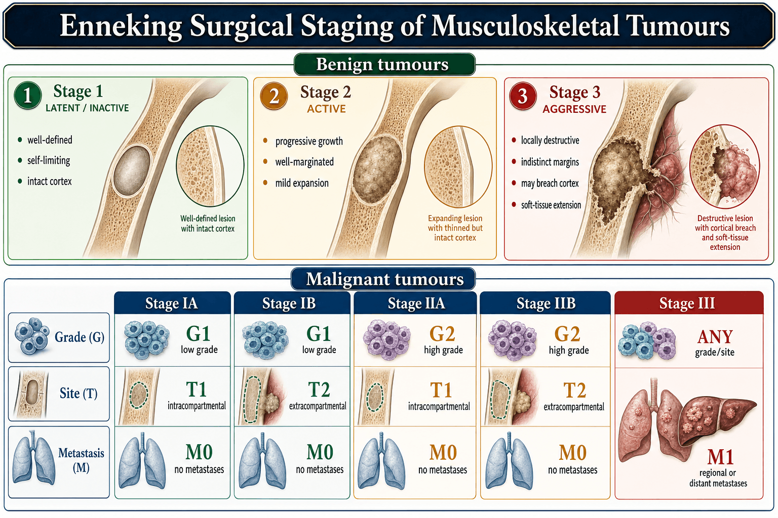
- Behavior
- Inactive
- Features
- Well-defined, sclerotic rim, stable
- CMF Applicability
- Incidental CMF, minimal symptoms
- Behavior
- Growing slowly
- Features
- Defined margins, thin rim, may progress
- CMF Applicability
- Typical symptomatic CMF
- Behavior
- Locally aggressive
- Features
- Poorly defined, cortical destruction
- CMF Applicability
- Rare - consider chondrosarcoma
By site within the bone. Metaphyseal lesions are the most common (90%) and have the typical appearance. Diaphyseal lesions are less common and may be larger at presentation; epiphyseal lesions are rare and more difficult to reach surgically.
By behaviour. A primary lesion at first presentation is the usual case. A recurrent lesion after prior treatment carries a higher suspicion of grade if it behaves aggressively.
Clinical Presentation
Symptoms. Pain is the most common symptom: dull, aching and intermittent, of gradual onset over months to years. A palpable swelling appears in superficial locations, and motion may be limited when the lesion is near a joint. Night pain is uncommon, unlike osteoid osteoma. 20-30% are discovered incidentally on imaging done for another reason.
Examination. There may be mild swelling over the metaphysis, and the lesion is firm, fixed to bone and non-tender or mildly tender. The joint usually has a normal range of motion, the limb is typically neurovascularly intact, the skin shows no warmth, erythema or other change, and there is no regional lymphadenopathy.
- Typical Symptoms
- Knee pain, limp, activity-related pain
- Physical Findings
- Tenderness over proximal tibia, palpable mass
- Special Considerations
- DDx includes osteosarcoma, GCT, chondroblastoma
- Typical Symptoms
- Deep pelvic pain, gluteal region discomfort
- Physical Findings
- Mass difficult to palpate, check hip ROM
- Special Considerations
- MRI essential for defining extent, can mimic chondrosarcoma
- Typical Symptoms
- Localized pain, swelling, difficulty with footwear
- Physical Findings
- Visible swelling, point tenderness
- Special Considerations
- High recurrence risk in confined space
Pathological fracture. Unlike the more aggressive lesions (ABC, GCT), pathological fracture through a CMF is rare, because growth is slow and the cortical shell is preserved. When a fracture does occur it suggests one of three things:
- A large lesion with significant cortical thinning
- Trauma to weakened bone
- More aggressive behaviour warranting wide excision
A fracture does not change the benign nature of the lesion, but it may necessitate staged treatment: healing first, then definitive surgery.
Unusual presentations. Rarely CMF presents through its location:
- Spine - neurological symptoms from cord or nerve root compression
- Sacrum - bowel or bladder symptoms mimicking chordoma
- Rib - chest wall mass, respiratory symptoms
- Skull base - cranial nerve palsies, headaches
Imaging and Diagnosis
Radiographs. The classic triad in a patient aged 10-30 should raise suspicion of CMF:
- Eccentric metaphyseal location
- Endosteal scalloping with cortical expansion
- Sclerotic rim
Imaging alone cannot distinguish CMF from low-grade chondrosarcoma, so biopsy is mandatory before definitive treatment. Each element of the triad carries its own reasoning.
Eccentric location. CMF sits eccentrically in the metaphysis, expanding outward and thinning the overlying cortex, whereas enchondroma sits centrally in the medullary canal. Location alone therefore does much of the diagnostic work before you look at the matrix.
Endosteal scalloping. The cortex is thinned from within, giving a scalloped or festooned inner margin. This is the signature of a lesion growing slowly enough for bone to remodel around it rather than destroy it; frank cortical breakthrough is a different message and should raise concern.
Sclerotic rim. Reactive sclerosis at the margin means the host bone has had time to wall the lesion off: a narrow zone of transition, the radiographic definition of indolent behaviour (Lodwick IA). Loss of that rim, with a widening zone of transition, is the red flag for chondrosarcoma.
Matrix. The lesion is usually purely lytic. When calcification appears it is faint, amorphous and irregular, not the ring-and-arc of enchondroma nor the fluffy clouds of chondrosarcoma. The reason is that CMF's matrix is myxoid rather than mature hyaline cartilage, so it does not mineralise in the lobular pattern: mineralisation was identified radiographically in only about 13% of the Mayo series. The absence of organised chondroid calcification in an eccentric metaphyseal cartilage lesion is itself a clue toward CMF over enchondroma.
Rings and arcs. "Rings and arcs" (also described as punctate, stippled, comma-shaped, flocculent or "popcorn") is the radiographic fingerprint of a chondroid matrix; its presence on radiograph or CT essentially confirms that a lesion is a cartilage tumour. Cartilage lobules are avascular, so mineralisation (enchondral ossification) occurs at the periphery and septa of the lobules, ringing each one. Dense, organised rings-and-arcs favour a benign or low-grade cartilage tumour, whereas destruction of previously visible calcification, a new lytic area, deep endosteal scalloping (more than two thirds of the cortex), or a soft-tissue mass signal chondrosarcomatous behaviour.
Lodwick-Madewell margins. The margin grades a lytic lesion by how fast it is growing:
- Geographic (grade I) - a single, well-defined lytic area implying slow growth: IA with a sclerotic rim (the most indolent, and the classic CMF pattern), IB well-defined without a sclerotic rim, IC a partly ill-defined margin
- Moth-eaten (grade II) - multiple confluent lucencies
- Permeative (grade III) - ill-defined tiny lucencies infiltrating between trabeculae
Grades II and III both imply a rapid, aggressive or malignant process. The unifying concept is the zone of transition, the width of the border between lesion and normal bone: a narrow zone (a sharp, often sclerotic edge) means slow, benign growth, and a wide zone (no clear line where the lesion ends) means rapid, aggressive growth. It is the single most useful plain-film discriminator of benign from aggressive bone lesions.
CT. CT adds precision to what the radiograph suggests:
- Cortical integrity - thinning versus breakthrough
- Matrix calcification - subtle calcification the radiograph misses
- Sclerotic margins - quantification of reactive sclerosis
- Bone destruction - geographic versus permeative pattern
- Surgical planning - 3D reconstruction for complex anatomy (pelvis, spine)
On CT a CMF is an eccentric, lobulated, expansile mass with endosteal scalloping, cortical thinning and a sclerotic rim, with no or minimal internal calcification, no periosteal reaction unless fractured, and no soft-tissue mass unless aggressive. CT and MRI complement each other: CT for calcification and cortical bone detail, MRI for soft-tissue extent, cartilage cap assessment and intramedullary involvement.
MRI. MRI is superior for intramedullary extent (marrow involvement), soft-tissue extension beyond the cortex, proximity to nerves and vessels, and the heterogeneous signal that reflects the lobular myxoid composition.
- Signal Intensity
- Low to intermediate signal
- Interpretation
- Reflects myxoid and cartilaginous matrix
- Signal Intensity
- Very high signal (bright)
- Interpretation
- Myxoid matrix has high water content, bright on T2
- Signal Intensity
- Heterogeneous enhancement
- Interpretation
- Peripheral lobular enhancement, septal enhancement
MRI does not resolve the central question either. CMF and low-grade chondrosarcoma share high T2 signal, lobulated margins and heterogeneous enhancement, so clinical correlation (age, symptoms) and histology are essential and biopsy is always required.
Nuclear medicine. A Tc-99m MDP bone scan shows moderate to marked uptake in CMF; it is of limited diagnostic value but useful for detecting multifocal disease or as a metastatic survey. PET-CT is not routinely used, and CMF can show FDG uptake, which complicates interpretation.
Biopsy. Image-guided core needle biopsy is preferred over open biopsy for initial diagnosis:
- Multiple cores (3-5) to sample the lobular architecture
- Avoid contaminating neurovascular structures with the biopsy tract
- CT or fluoroscopic guidance for deep lesions (pelvis, spine)
- Send fresh tissue for cytogenetics if available
Pathology review. Expert musculoskeletal pathology review is mandatory: CMF is rare and easily misdiagnosed, a high-volume centre reduces misdiagnosis as chondrosarcoma, correlation with radiology is essential, and any atypical feature deserves a second opinion. Small cores may sample only the hypercellular rim of the lobules, so the pathologist must recognise the low-power lobular architecture and read it against the imaging (sclerotic rim, eccentric location) and the clinical context (young age). If doubt remains, an open biopsy with a larger sample may be warranted before proceeding to ablative surgery.
Differential Diagnosis
Cartilaginous Lesion Differentials
- Age
- 10-30 years
- Location
- Eccentric metaphysis
- Imaging
- Scalloped, sclerotic rim
- Histology
- Lobules, myxoid, hypercellular periphery
- Age
- 10-20 years (younger)
- Location
- Epiphysis/apophysis
- Imaging
- Lytic, rim sclerosis, ABC component
- Histology
- Chondroblasts, chicken-wire calcification, giant cells
- Age
- 20-40 years
- Location
- Central medullary (hands common)
- Imaging
- Ring-and-arc calcification
- Histology
- Hyaline cartilage lobules, no atypia
- Age
- Over 40 years (older)
- Location
- Medullary or surface
- Imaging
- Permeative, cortical thickening
- Histology
- Permeation, nuclear atypia, myxoid change
CMF versus chondroblastoma. Both occur in young patients and both can show giant cells on histology, so the separation rests on where and when. Chondroblastoma is epiphyseal or apophyseal and peaks at 10-20 years, before physeal closure; CMF is metaphyseal and peaks slightly older, after physeal closure. On imaging chondroblastoma is more central, whereas CMF is eccentric and cortically based; on histology chondroblastoma has uniform chondroblasts with chicken-wire calcification, and CMF has the lobular myxoid pattern.
Distinguishing CMF from low-grade chondrosarcoma is the most important clinical challenge and has enormous treatment implications (curettage versus amputation or wide resection). Favouring CMF: age under 30, eccentric cortical location, sclerotic rim, no soft tissue mass, lobular histology with bland cytology. Favouring chondrosarcoma: age over 40, central medullary or surface location, permeative pattern, cortical thickening, true nuclear atypia with permeative growth. When in doubt, seek expert pathology review and consider multidisciplinary tumour board discussion.
Management Algorithm
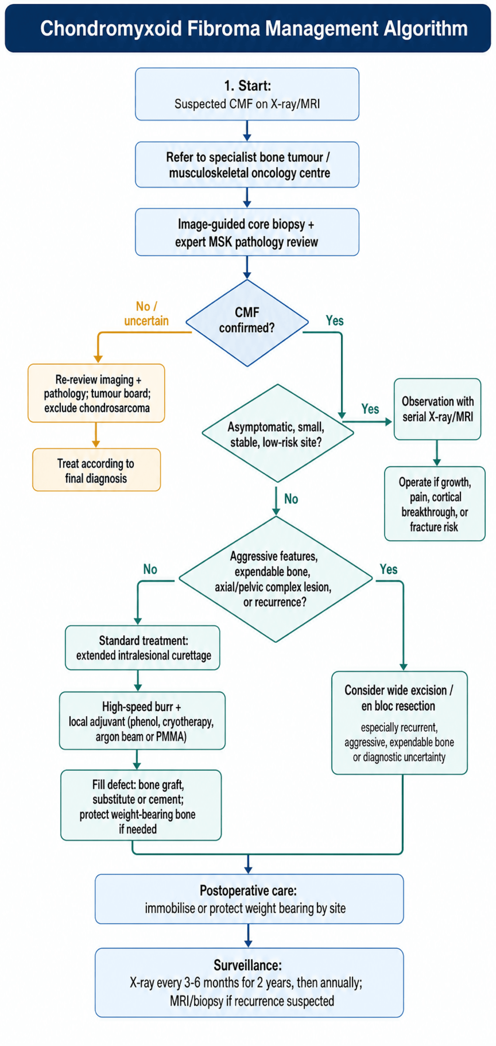
Diagnosis first. Tissue comes before treatment: an image-guided core needle biopsy with expert musculoskeletal pathology review and radiological correlation, confirming the lobular myxoid pattern before any definitive surgery.
Then staging and selection. Complete the imaging with plain radiographs and MRI (or CT if MRI is contraindicated) to assess cortical integrity, intramedullary extent, soft-tissue extension and proximity to neurovascular structures; on the Enneking system CMF is benign, stage 3 if it shows aggressive features. Treatment is then chosen on lesion size, location, symptoms, cortical involvement, patient age and functional demands, from observation, extended curettage with adjuvants, wide excision or en bloc resection, and every patient enters surveillance afterwards.
- Imaging Pattern
- Eccentric, sclerotic rim, no soft tissue
- Management
- Observation with serial radiographs
- Key Pearl
- Many are asymptomatic incidental findings
- Imaging Pattern
- Metaphyseal, scalloped, well-defined
- Management
- Extended curettage with adjuvant
- Key Pearl
- Use phenol or cryotherapy to reduce recurrence
- Imaging Pattern
- Cortical breakthrough, soft tissue mass
- Management
- Wide excision with reconstruction
- Key Pearl
- Consider en bloc for expendable bones (fibula, rib)
Observation. Most cases warrant surgery, because of the risk of continued growth and pathological fracture and because of diagnostic uncertainty; given how rarely CMF is seen and how easily it is misdiagnosed, most surgeons advocate curettage both as treatment and to obtain adequate tissue for a definitive histological diagnosis. Observation is suitable only for truly asymptomatic, stable, small lesions in low-risk locations where biopsy has definitively confirmed the diagnosis. The indications:
- Small, asymptomatic, incidental finding
- Lesion in a stable phase (no growth on serial imaging)
- Patient unwilling to undergo surgery
- High surgical risk (medical comorbidities)
- Elderly patient with limited life expectancy
An observed lesion is examined every 3-6 months for 2 years, radiographed every 6 months for 2 years and then annually, and imaged with MRI if symptoms change or the radiograph shows growth. Surgery follows if the lesion enlarges, symptoms worsen or cortical breakthrough develops.
Extended curettage with adjuvants. This is the gold-standard treatment for most CMF. The goals are to remove all tumour tissue, destroy residual microscopic disease with an adjuvant, fill the defect to restore structural integrity, and keep morbidity below that of wide excision. The operation, a cortical window, aggressive curettage of all lobular tissue, high-speed burring of the cavity walls and a local adjuvant (phenol, liquid nitrogen, argon beam or PMMA), is set out step by step in the Surgical Technique section.
Wide excision or en bloc resection. Reserved for specific scenarios:
- Recurrent disease - after failed curettage, especially multiple recurrences
- Aggressive features - soft-tissue extension, cortical destruction, pathological fracture
- Expendable bone - fibula, rib, distal ulna, where excision has minimal morbidity
- Diagnostic uncertainty - concern for low-grade chondrosarcoma on imaging or biopsy
- Failed adjuvant therapy - multiple recurrences despite adjuvants
Wide excision carries significantly higher morbidity (loss of bone, need for reconstruction, functional deficit) but the lowest recurrence risk, under 5%.
- Surgical Approach
- Excise fibula segment with margins
- Reconstruction
- None (non-essential bone)
- Functional Outcome
- Excellent, protect peroneal nerve
- Surgical Approach
- En bloc pelvic resection
- Reconstruction
- Depends on extent (may not reconstruct)
- Functional Outcome
- Good for Type I resection, limp possible
- Surgical Approach
- Segmental tibial resection
- Reconstruction
- Intercalary allograft or vascularized fibula
- Functional Outcome
- Fair to good, prolonged protected weight-bearing
Recurrence. A recurrence is picked up on surveillance imaging (the signs are described under Postoperative Care); biopsy if the imaging is unclear. A first recurrence is treated by repeat extended curettage with a more aggressive adjuvant, cryotherapy if phenol was used initially, with fresh tissue sent to confirm the diagnosis and rule out malignant transformation. After a second recurrence consider en bloc resection, especially if the bone is expendable or there is diagnostic uncertainty, since multiple recurrences may indicate sampling error with an underlying low-grade chondrosarcoma. If biopsy of a recurrence shows chondrosarcoma, proceed to wide resection with margins.
Surgical Technique
Planning. Review all imaging (X-ray, CT, MRI) to plan the cortical window, identify the neurovascular structures at risk, decide on reconstruction (bone graft, PMMA or allograft) and consent the patient for a possible pathological fracture during curettage. Position according to the lesion, with a tourniquet for extremity lesions.
1. Exposure and window. The incision lies directly over the lesion, avoiding a weight-bearing surface, with subperiosteal dissection that preserves the periosteum for closure. The window is cut with osteotomes or a saw directly over the lesion through the thinnest cortex, sized to expose the entire lesion so that every margin can be seen, and kept for use as autograft. In the skeletally immature, avoid the physis.
2. Intralesional curettage. Remove all visible tumour piecemeal with curettes of varying sizes, large first then small, confirming the lobular, gelatinous appearance consistent with CMF. Send multiple samples for histology, with a frozen section to confirm the diagnosis if this is the first surgery. Curettage must reach healthy bone circumferentially, extending 1-2mm beyond the visible tumour margin.
3. Extended curettage with the high-speed burr. A high-speed pneumatic burr removes a further 1-2mm of the cavity wall over the whole 360 degrees (floor, walls and ceiling), leaving smooth walls and removing microscopic residual tumour, followed by copious irrigation to clear bone debris. The burr is the standard adjuvant with a low complication rate, and extended curettage with it reduces recurrence from 25% to approximately 10%; adding phenol or cryotherapy decreases recurrence further but increases complication risk (pathological fracture, wound-healing problems). A combination may be used for high-risk lesions.
4. Local adjuvant. Apply the adjuvant to the cavity walls; the options, how to choose between them and the technique for each are set out below.
5. Filling and reconstruction. Fill the cavity completely, to prevent haematoma and provide structural support, with a filler chosen by cavity size, location and load:
- Small cavities (under 3cm) - cancellous autograft or allograft chips
- Large cavities - PMMA cement (immediate weight-bearing) or structural allograft
- Metaphyseal cavities near a joint - consider cancellous graft to preserve future arthroplasty options
Large cavities near joints may require structural support, a cortical strut or plate fixation, and prophylactic plate fixation should be considered where the cortex is extensively thinned.
6. Closure. Replace the cortical window as autograft if preserved, close the periosteum over it where possible to enhance healing, and close the subcutaneous tissue and skin in layers. A drain is optional; some surgeons use one for large cavities.
Local Adjuvant Selection
- Mechanism
- Chemical cytotoxicity
- Advantages
- Low cost, widely available, easy application
- Disadvantages
- Soft tissue toxicity if spills, neurotoxic
- Mechanism
- Freezing cellular necrosis
- Advantages
- Highly effective, minimal recurrence
- Disadvantages
- Fracture risk, equipment needed, technique-sensitive
- Mechanism
- Exothermic heat (60-80°C)
- Advantages
- Structural support, detects recurrence (radiopaque)
- Disadvantages
- Precludes future arthroplasty, risk of thermal necrosis
- Mechanism
- Thermal ablation
- Advantages
- Precise application, hemostasis
- Disadvantages
- Equipment cost, learning curve
The evidence. Studies comparing curettage alone with curettage plus an adjuvant show:
- Curettage alone - 25-30% recurrence
- Curettage with phenol - 10-15% recurrence
- Curettage with cryotherapy - 5-10% recurrence
- Curettage with PMMA - 5-10% recurrence
Choosing. Phenol is the most commonly used, because it is available and easy to apply, and suits most cases. Cryotherapy offers the lowest recurrence but a higher fracture risk, so it is best for expendable bones (fibula, rib) or where structural reconstruction is planned. PMMA gives both an adjuvant effect and structural support, ideal for weight-bearing metaphyseal lesions that need immediate stability, and argon beam is useful in the spine or pelvis where precise application near neurovascular structures is needed.
Phenol. Use 5% liquefied phenol, not crystalline, applied to the cavity walls on gauze pledgets held with forceps for a contact time of 2 minutes minimum, then neutralised with 95% alcohol and copious saline irrigation. Protect the soft tissues with moist laparotomy pads.
Cryotherapy. A double freeze-thaw cycle (freeze 3 minutes, thaw 5 minutes, freeze 3 minutes) using cryoprobes or direct application of liquid nitrogen, monitored with thermocouples where available to a target under -20°C. Allow complete thawing before bone grafting.
PMMA. Mix with vancomycin powder for antibiotic elution and apply during the dough phase, mouldable rather than runny. The polymerisation is exothermic, and the temperature can exceed 70°C centrally, so leave the cortical window open until polymerisation is complete to vent the heat.
Argon beam. Coagulate the cavity surface at a high setting.
Complications
- Incidence
- 25% curettage alone, 10% with adjuvant
- Prevention
- Extended curettage, adjuvant use, adequate margins
- Management
- Repeat curettage or wide excision
- Incidence
- 5-10% (higher with cryotherapy)
- Prevention
- Assess cortical integrity pre-op, prophylactic fixation
- Management
- ORIF with plate/screws, bone grafting
- Incidence
- 2-5%
- Prevention
- Perioperative antibiotics, sterile technique
- Management
- Antibiotics, debridement if deep
- Incidence
- Under 2%
- Prevention
- Careful dissection, identify structures, avoid phenol spill
- Management
- Immediate exploration and repair if recognized
- Incidence
- 10-20% (iliac crest harvest)
- Prevention
- Limit harvest size, preserve outer table, good closure
- Management
- Pain management, physical therapy
- Incidence
- Under 5%
- Prevention
- Adequate graft fill, protected weight-bearing, avoid NSAIDs
- Management
- Revision grafting, consider BMP
Phenol. A spill causes a chemical burn with soft-tissue necrosis, contact with a major nerve causes neurotoxic damage, and systemic toxicity (metabolic acidosis) is rare with local use. Prevention is protecting the soft tissues with moist pads, keeping phenol off nerves and limiting the volume.
Cryotherapy. Bone necrosis raises the pathological fracture rate to up to 10-15%, adjacent nerves suffer cold injury (the peroneal nerve at the proximal tibia), and the skin necroses if the cryoprobe is too close to it. Prevention is prophylactic fixation in weight-bearing bones, identifying the nerves and monitoring the freeze.
PMMA. The exothermic heat (60-80°C) can damage soft tissues and nerves, cement can extravasate into the joint or soft tissues, and the cement is difficult to remove if a joint replacement is needed later. Vent the heat, protect the soft tissues, and avoid PMMA in young patients near joints.
Bone graft. Graft resorbs, especially allograft; fracture through the graft follows inadequate incorporation or premature loading; and infection is higher with allograft (0.5-1%). Prevention is adequate fill, protected weight-bearing and antibiotics for allograft.
Long-term. Some patients are left with chronic pain or reduced function, especially in weight-bearing locations:
- Stiffness - prolonged immobilisation or adhesions
- Weakness - muscle atrophy during non-weight-bearing
- Pain - altered biomechanics, hardware irritation
- Limb length discrepancy - premature physeal closure in the skeletally immature
These are managed with aggressive physiotherapy, pain management, hardware removal if symptomatic and osteotomy for limb length inequality.
Malignant transformation. Exceedingly rare, with fewer than 10 reported cases. When it occurs it is unclear whether it represents:
- True malignant transformation of a benign CMF
- Initial misdiagnosis with an underlying low-grade chondrosarcoma
- Radiation-induced sarcoma, if prior radiation was given
Most experts believe cases of "malignant CMF" are sampling errors at the initial diagnosis, which is another argument for adequate tissue sampling and expert pathology review.
Postoperative Care and Rehabilitation
The wound. A sterile dressing stays on for 48-72 hours, a drain, if placed, comes out when output is under 30mL in 24 hours, and sutures or staples are removed at 10-14 days. Watch for erythema, drainage or fever.
Analgesia. Multimodal, with paracetamol and opioids as required; ice and elevation control swelling, and a regional nerve block suits upper-limb surgery. Avoid NSAIDs for the first 6 weeks, as they impair bone healing.
- Graft Type
- Autograft or allograft
- Initial Status
- Sling for comfort, early ROM
- Full Weight-Bearing
- Immediate for ADLs, avoid heavy lifting 6 weeks
- Graft Type
- Any
- Initial Status
- Weight-bearing as tolerated
- Full Weight-Bearing
- Immediate
- Graft Type
- Cancellous autograft
- Initial Status
- Touch-down weight-bearing (10-20 lbs)
- Full Weight-Bearing
- 6-12 weeks (when X-ray shows incorporation)
- Graft Type
- PMMA cement
- Initial Status
- Weight-bearing as tolerated immediately
- Full Weight-Bearing
- Immediate (cement provides structural support)
- Graft Type
- Intercalary allograft
- Initial Status
- Non-weight bearing 6 weeks, then progressive
- Full Weight-Bearing
- 12-24 weeks (slower incorporation)
Rehabilitation Phases
The aims are wound healing, minimal swelling and maintained motion in the adjacent joints, with no weight-bearing (unless PMMA was used) and no resistance training.
- Upper extremity - pendulum exercises for the shoulder, elbow and wrist motion, grip strengthening
- Lower extremity - ankle pumps, quad sets, hip and knee motion without weight-bearing
Weight-bearing advances from touch-down to partial (50%) to full once the radiograph shows trabecular bridging and protected weight-bearing is painless. Progressive resistance, proprioception and gait training restore motion and begin strengthening.
Functional training, sport-specific drills and endurance building aim at full motion, normal strength and return to activities. Progression requires pain-free full motion and strength at 80% of the contralateral side; return to sport is unrestricted at 6 months if those criteria are met.
A home exercise programme with activity modification as needed, alongside surveillance imaging for recurrence.
Surveillance. Most recurrences occur within 24 months. Lifelong surveillance is recommended, with the most intensive monitoring in the first 5 years.
- Clinical Exam
- Every 3 months
- Radiographs
- Every 3-6 months
- MRI Indications
- Any pain, swelling, or X-ray concern
- Clinical Exam
- Every 6 months
- Radiographs
- Every 6-12 months
- MRI Indications
- Clinical symptoms or radiographic changes
- Clinical Exam
- Annually
- Radiographs
- Annually
- MRI Indications
- New symptoms only
Signs of recurrence. On the plain radiograph:
- New lysis - lucency within the previously grafted area
- Loss of trabeculation - disappearance of the bone graft trabecular pattern
- Cortical erosion - scalloping or thinning of the cortex
- Soft-tissue mass - visible soft-tissue swelling
On MRI, if obtained:
- High T2 signal - the very bright myxoid recurrent tumour
- Lobulated mass - the characteristic lobular pattern
- Cortical breach - extension beyond the original cavity
- Soft-tissue extension - an extra-osseous component
Recurrence or healing? Early post-operative radiographs may show apparent lysis as graft resorbs during creeping substitution, which is normal healing. Four things separate the two:
- Timing - normal resorption occurs at 6-12 weeks; recurrence typically after 6 months
- Pattern - normal resorption is diffuse throughout the graft; recurrence is a focal lucency
- Stability - normal resorption stabilises and then remodels; recurrence progressively enlarges
- Symptoms - normal healing is painless; recurrence may cause pain
When in doubt, MRI or biopsy settles it.
Prognosis and Outcomes
Overall. CMF is a benign tumour with an excellent prognosis. There are no cases of metastasis, and the primary concern is local recurrence, which can be managed with repeat curettage or wide excision without any impact on survival.
- Low Risk (under 10%)
- Extended curettage with adjuvant
- High Risk (over 25%)
- Simple curettage alone
- Low Risk (under 10%)
- Cryotherapy or PMMA
- High Risk (over 25%)
- No adjuvant
- Low Risk (under 10%)
- Expendable bones (fibula, rib)
- High Risk (over 25%)
- Complex anatomy (pelvis, spine)
- Low Risk (under 10%)
- Under 3cm diameter
- High Risk (over 25%)
- Over 5cm diameter
- Low Risk (under 10%)
- Intact cortex with window
- High Risk (over 25%)
- Pathological fracture, extensive destruction
Function. Outcomes are excellent for upper-limb lesions (near-normal function after healing), for the fibula (no deficit if the peroneal nerve is preserved), for small lesions (full return to the pre-morbid activity level) and in young patients, whose bone remodels and adapts well. The limitations come with large metaphyseal lesions (residual stiffness or weakness), pathological fracture (prolonged recovery, possible chronic pain), multiple recurrences (cumulative surgical morbidity) and wide excision (loss of bone and reconstruction-related problems).
The long-term data. Long-term studies are limited by the rarity of CMF, but the available evidence shows:
- 10-year tumour-free survival - 90-95% with curettage and adjuvant
- Functional scores - MSTS (Musculoskeletal Tumor Society) scores average 25-28/30, excellent
- Return to sport - most patients return to their pre-injury activity level by 6-12 months
- Quality of life - comparable to the general population after successful treatment
Largest Clinicopathologic Series (Mayo Clinic, 278 cases)
- Largest single-institution series; slight male predominance, peak in the second decade
- Almost half involved long bones; ilium and small bones also common sites
- Eccentric metaphyseal lucent lesion with thinned, expanded cortex; soft-tissue extension uncommon
- Approximately one quarter of patients developed local recurrence after conservative surgical removal
Treatment Experience at the Istituto Ortopedico Rizzoli
- Surgical series correlating treatment modality (curettage vs resection) with local recurrence
- Recurrence concentrated in lesions treated by intralesional curettage alone
- Marginal/wide excision associated with lower recurrence than simple curettage
- Supports extended curettage with adjuvants or marginal excision over intralesional curettage alone
Guidelines, Registries & Global Practice
Global Epidemiology
Chondromyxoid fibroma is genuinely rare worldwide. In the Mayo Clinic series of 278 cases (Wu CT et al., Hum Pathol 1998 — DOI) CMF accounted for well under 1% of all primary bone tumours, with a slight male predominance and a peak in the second decade. Almost half of lesions arose in the long bones (classically around the knee), with the ilium and the small bones of the hands and feet as other characteristic sites. These distribution and demographic patterns are consistent across North American, European (Rizzoli — Gherlinzoni F et al., J Bone Joint Surg Am 1983 — DOI) and other international series, so the same diagnostic and surgical principles apply globally rather than being region-specific.
Because any individual surgeon sees very few cases, the single most important global practice point is referral to a specialist bone-and-soft-tissue sarcoma/musculoskeletal oncology centre before biopsy or definitive surgery. This mirrors the principle behind national sarcoma networks (e.g. UK sarcoma "diagnostic and treatment" centres under NICE NG10/IOG guidance, US NCCN-affiliated sarcoma centres, and ISG/EORTC referral pathways in Europe).
Guidelines and Standards — Side-by-Side
CMF is too rare to have a dedicated single-disease guideline; it is managed within broader bone-tumour/sarcoma frameworks. The table summarises how the major bodies converge on the same core message — refer, biopsy correctly, avoid radiotherapy.
- Relevant Guidance
- NG10 / Improving Outcomes for Sarcoma (IOG)
- Position Relevant to CMF
- Suspected bone tumours referred to a designated bone sarcoma centre; biopsy only at the treating centre
- Evidence Basis
- Guideline / expert consensus
- Relevant Guidance
- BOAST: Suspected bone tumour & metastatic bone disease
- Position Relevant to CMF
- Do not perform definitive surgery or biopsy a likely primary bone tumour outside a specialist unit
- Evidence Basis
- Standard of care / consensus
- Relevant Guidance
- Bone sarcoma clinical practice guidelines
- Position Relevant to CMF
- Multidisciplinary diagnosis; image-guided core biopsy planned with the surgeon; expert bone pathology review
- Evidence Basis
- Level III-IV, expert consensus
- Relevant Guidance
- Bone Cancer guidelines (benign/borderline pathways)
- Position Relevant to CMF
- Curettage with adjuvants for benign aggressive lesions; wide excision reserved for diagnostic doubt
- Evidence Basis
- Category 2A consensus
- Relevant Guidance
- Classification of Tumours: Soft Tissue and Bone, 5th ed (2020)
- Position Relevant to CMF
- CMF defined as a benign cartilaginous tumour; GRM1 rearrangement now recognised as the molecular hallmark
- Evidence Basis
- Reference standard
Across all frameworks, radiotherapy is not recommended for CMF. The Zillmer & Dorfman series (Hum Pathol 1989 — DOI) documented a radiation-associated sarcoma arising after irradiation of a benign CMF. Radiotherapy is reserved only for genuinely unresectable lesions in critical locations after multidisciplinary discussion.
Registry and Practice Variation
- No dedicated CMF registry exists in any country; arthroplasty registries (NJR, AJRR, AOANJRR, SHAR, NZJR) do not capture primary benign bone tumours
- Epidemiology is therefore derived from single-institution pathology archives (Mayo Clinic, Rizzoli, Memorial Sloan Kettering) and national bone-tumour databases
- Recommendation: enter cases into local/national bone-tumour or rare-cancer registries to improve population-level data
- Adjuvant choice varies by availability: phenol and high-speed burr near-universal; cryotherapy and argon-beam more common in well-resourced units
- Observation of small asymptomatic lesions is emerging but not yet standard (Butler Z et al., Cureus 2026 — DOI)
- GRM1 IHC/FISH access varies; where available it materially reduces CMF-versus-chondrosarcoma misdiagnosis
Perioperative Medication Principles (Generic, Globally Applicable)
- Multimodal analgesia: paracetamol plus short-course opioid as required
- Avoid NSAIDs during the bone-healing phase (first ~6 weeks) where graft incorporation is required
- Use locally available agents per national formularies
- Single-dose first-generation cephalosporin (e.g. cefazolin) at induction is the international standard for clean orthopaedic surgery
- Follow local antimicrobial stewardship guidelines for choice and duration
- Consider antibiotic-loaded cement/graft for larger reconstructions per unit protocol
Return to Function (Surgery-Driven, Not Jurisdiction-Driven)
Return-to-activity timelines depend on reconstruction type and weight-bearing status rather than any specific health or compensation system:
- Sedentary work / desk-based activity: approximately 2-4 weeks (upper limb), 6-8 weeks (lower limb)
- Light manual work: approximately 6-12 weeks, guided by radiographic graft incorporation
- Heavy manual / impact loading: approximately 3-6 months, after radiographic union and adequate graft incorporation
MCQ Practice Points
High-Yield MCQ Topics
- Rarest benign cartilage tumor (under 1% of bone tumors)
- Peak age 10-30 years (slightly older than chondroblastoma)
- Male predominance 2:1
- Most common site: proximal tibia (25-30%)
- Classic triad: eccentric metaphyseal, scalloped margin, sclerotic rim
- Very high T2 signal on MRI (myxoid matrix)
- Minimal or absent calcification (unlike enchondroma)
- Cannot distinguish from chondrosarcoma on imaging alone
- Lobular architecture with fibrous septa (pathognomonic)
- Hypocellular myxoid center, hypercellular periphery
- S100 positive, low Ki-67 (under 5%)
- Reverse pattern compared to chondrosarcoma
- Standard: curettage with adjuvant (phenol, cryo, or PMMA)
- Recurrence: 25% without adjuvant, 10-15% with adjuvant
- Wide excision for recurrent or expendable bones
- Prognosis: excellent, no metastases
Classic Exam Vignettes
"A 22-year-old male presents with knee pain. X-ray shows an eccentric, lytic lesion in the proximal tibia with endosteal scalloping and sclerotic rim. Biopsy shows lobules with myxoid matrix and hypercellular periphery. What is the diagnosis?"Answer: Chondromyxoid fibroma (lobular architecture with myxoid matrix is pathognomonic)
"Which feature BEST distinguishes chondromyxoid fibroma from chondroblastoma?"Answer: Chondroblastoma is epiphyseal/apophyseal, CMF is metaphyseal (or: chondroblastoma has chicken-wire calcification, CMF has myxoid matrix)
"What is the recurrence rate after simple curettage alone for CMF?"Answer: 25-30% (versus 10-15% with adjuvant)
"Core needle biopsy of a proximal tibial lesion shows hypercellular cartilaginous tissue. The pathologist is concerned about chondrosarcoma. What feature would favor CMF?"Answer: Lobular architecture with hypocellular myxoid centers (CMF has reverse pattern - hypocellular center, hypercellular periphery; chondrosarcoma has permeative pattern with nuclear atypia throughout)
"A 25-year-old has an eccentric metaphyseal lesion with very high T2 signal on MRI. Differential diagnosis includes CMF and low-grade chondrosarcoma. Which clinical feature most favors CMF?"Answer: Age under 30 years (chondrosarcoma typically over 40 years)
Common Examiner Questions
-
Why is CMF easily misdiagnosed as chondrosarcoma?
- Hypercellular peripheral zones can mimic malignancy on small biopsies
- Both show high T2 signal on MRI
- Expert pathology review essential
-
What is the significance of the lobular architecture?
- Pathognomonic feature of CMF
- Lobules separated by fibrous septa
- Central myxoid zone, peripheral hypercellular zone
-
How do you reduce recurrence risk?
- Extended curettage (not simple curettage)
- High-speed burr to remove 1-2mm of cavity walls
- Local adjuvant (phenol, cryotherapy, or PMMA)
- Complete filling of defect with bone graft
-
When would you recommend wide excision over curettage?
- Recurrent disease (especially second recurrence)
- Expendable bone (fibula, rib)
- Diagnostic uncertainty with concern for chondrosarcoma
- Patient preference for lower recurrence risk
-
What is the prognosis?
- Excellent (benign tumor)
- 100% 5-year survival
- No metastatic potential
- Recurrence does not affect survival
Q: What is the defining molecular feature of chondromyxoid fibroma, and which ancillary test exploits it? A: A recurrent GRM1 gene rearrangement (promoter swapping) drives CMF; GRM1 immunohistochemistry is positive in ~97% of CMF and negative in chondrosarcoma, enchondroma, chondroblastoma, GCT and fibrous dysplasia - resolving the CMF-versus-chondrosarcoma biopsy dilemma.
Q: Describe the classic radiographic appearance of CMF and the one thing imaging cannot do. A: An eccentric metaphyseal lytic lesion with a scalloped sclerotic rim, lobulated margins and septation, typically in the second decade and around the knee. Imaging is suggestive but cannot exclude chondrosarcoma - biopsy is mandatory.
Q: Which treatment variable most drives local recurrence in CMF? A: Intralesional curettage alone - recurrence concentrates in curettage-only cases (about one quarter in the Mayo series), so extended curettage with an adjuvant (phenol/cryotherapy) plus grafting is standard; wide excision is reserved for recurrence, expendable bones or diagnostic uncertainty.
Exam Viva Scenarios
Practise clinical reasoning and management decisions out loud
“A 22-year-old male presents with a 6-month history of mild knee pain. X-ray shows an eccentric, lytic lesion in the proximal tibial metaphysis with endosteal scalloping and a sclerotic rim. MRI demonstrates a lobulated mass with very high T2 signal and cortical thinning without breakthrough. Describe your approach.”
“A 28-year-old female presents with deep pelvic pain. CT shows a 6cm expansile, lytic lesion in the iliac wing with cortical thinning and minimal internal calcification. Core needle biopsy shows hypercellular cartilaginous tissue with focal myxoid areas. The reporting pathologist suggests 'low-grade chondrosarcoma cannot be excluded.' How do you proceed?”
“A 25-year-old returns 18 months after extended curettage (with phenol and bone grafting) for proximal tibial CMF. He reports new onset of pain and swelling. X-ray shows a lucent area in the previously grafted site with loss of trabecular pattern. MRI demonstrates a 3cm lobulated mass with high T2 signal in the proximal tibia metaphysis. How do you manage this recurrence?”
Key Epidemiology
- The rarest benign cartilage tumour - under 1% of all bone tumours
- Peak age 10-30 years, with a male to female ratio of 2:1
- Proximal tibia 25-30%, distal femur 15%, pelvis 15%
- Metaphyseal location (eccentric, cortical-based)
Classic Imaging Triad
- Eccentric metaphyseal location
- Endosteal scalloping with cortical expansion
- A sclerotic rim at the margins - a benign feature
- MRI: very high T2 signal (myxoid matrix), lobulated
Pathognomonic Histology
- Lobular architecture, with fibrous septa separating the lobules
- A myxoid matrix centrally within the lobules, which are hypocellular
- A hypercellular periphery containing spindle cells and giant cells
- S100 positive, Ki-67 under 5%, and no nuclear atypia
Key Differentials
- Chondroblastoma: epiphyseal, younger age (10-20 years), chicken-wire calcification
- Low-grade chondrosarcoma: older age (over 40), a permeative pattern, and true atypia
- Enchondroma: central medullary location with ring-and-arc calcification
- Aneurysmal bone cyst: fluid-fluid levels, blood-filled spaces, and no lobules
Standard Treatment
- Extended curettage through a cortical window
- High-speed burr to remove 1-2 mm of the cavity walls
- Local adjuvant: phenol (most commonly used), cryotherapy (lowest recurrence), or PMMA
- Bone grafting with cancellous autograft or allograft chips
Recurrence and Outcomes
- 25-30% recurrence after curettage alone
- 10-15% recurrence with curettage plus an adjuvant
- Under 5% recurrence after wide excision
- Excellent prognosis: no metastases and 100% survival
Management of Recurrence
- Always biopsy to confirm the diagnosis and rule out malignancy
- First recurrence: repeat curettage using a different adjuvant, such as cryotherapy
- Second recurrence: give strong consideration to wide excision
- Prophylactic fixation if cryotherapy is used in a weight-bearing bone
Exam Pearls
- The most important distinction is CMF versus low-grade chondrosarcoma, which requires expert pathology
- The lobular pattern is pathognomonic - fibrous septa with myxoid centres
- Reverse cellular pattern: hypocellular centre with hypercellular periphery, the opposite of chondrosarcoma
- It cannot be diagnosed on imaging alone - biopsy is mandatory despite a typical appearance
Evidence Base and Guidelines
Characteristic Radiographic Appearance (38 cases + literature review)
- CMF has a characteristic but not specific radiographic appearance and often mimics commoner tumours
- Can occur anywhere in the skeleton, but almost half of cases occur around the knee
- Consider CMF for a focal lesion with geographic bone destruction, sclerotic rim, lobulated margins and septation
- Diagnosis most likely when the patient is in the second decade of life
Clinicopathologic Correlation Including Aggressive Variants (36 cases)
- Long-bone lesions appeared benign radiographically; vertebral lesions showed aggressive bone destruction
- Pseudolobulated tumour with myxoid and chondroid regions; cells occasionally bizarre/binucleate but rarely mitotic
- Majority of recurrences occurred in patients treated by curettage alone
- No malignant transformation; one radiation-associated sarcoma, cautioning against radiotherapy
GRM1 Immunohistochemistry Distinguishes CMF From Mimics
- GRM1 immunohistochemistry positive in 97% of CMF specimens (34/35), usually diffuse
- GRM1 negative in chondrosarcoma, enchondroma, chondroblastoma, GCT, fibrous dysplasia and other mimics
- Acid decalcification can reduce staining intensity - interpret with care
- Serves as a sensitive surrogate marker for the defining GRM1 gene rearrangement
Molecular Basis: GRM1 Gene Rearrangement
- GRM1 (metabotropic glutamate receptor 1) is recurrently rearranged and up-regulated in CMF via promoter swapping
- GRM1 break-apart FISH detected rearrangement in ~75% of testable cases (9/12)
- GRM1 protein overexpression by immunohistochemistry detected in 13/13 cases - the most sensitive method
- Targeted RNA sequencing did not reliably detect the fusion, so it is not yet a standalone diagnostic test
Contemporary Treatment Outcomes and Role of Observation
- Eight patients managed by en bloc resection (n=2), intralesional curettage with adjuvants (n=4) or serial imaging (n=2)
- All surgically treated patients remained free of local recurrence
- Two en bloc resections (25%) had complications requiring revision arthroplasty
- First report to document successful non-operative management with serial imaging in selected asymptomatic patients
Expert Recommendations and Guidelines
The WHO Soft Tissue and Bone Tumour Classification and Musculoskeletal Tumor Society guidelines recommend:
- Diagnosis: Image-guided biopsy with expert musculoskeletal pathology review
- Standard treatment: Extended intralesional curettage with local adjuvant and bone grafting
- Adjuvant selection: Phenol or cryotherapy based on surgeon preference and location
- Wide excision: Reserved for recurrent disease, expendable bones, or diagnostic uncertainty
- Surveillance: Serial imaging every 3-6 months for 2 years, then annually for 5 years
Controversial Areas and Research Needs
- Optimal adjuvant (phenol vs cryotherapy vs PMMA)
- Role of denosumab or bisphosphonates as adjuvants
- Genetic markers to predict recurrence risk
- Long-term outcomes (over 20 years) data lacking
- Molecular characterization (identify driver mutations)
- Less invasive techniques (radiofrequency ablation, MRI-guided focused ultrasound)
- Targeted therapies if molecular targets identified
- International registry for rare tumor outcomes tracking
References
-
Jaffe HL, Lichtenstein L. Chondromyxoid fibroma of bone: a distinctive benign tumor likely to be mistaken especially for chondrosarcoma. Arch Pathol. 1948;45:541-551.
-
Wu CT, Inwards CY, O'Laughlin S, et al. Chondromyxoid fibroma of bone: a clinicopathologic review of 278 cases. Hum Pathol. 1998;29(5):438-446. PMID:9596266. doi:10.1016/s0046-8177(98)90058-2
-
Rahimi A, Beabout JW, Ivins JC, Dahlin DC. Chondromyxoid fibroma: a clinicopathologic study of 76 cases. Cancer. 1972;30(3):726-736.
-
Gherlinzoni F, Rock M, Picci P. Chondromyxoid fibroma. The experience at the Istituto Ortopedico Rizzoli. J Bone Joint Surg Am. 1983;65(2):198-204. PMID:6337162. doi:10.2106/00004623-198365020-00008
-
Wilson AJ, Kyriakos M, Ackerman LV. Chondromyxoid fibroma: radiographic appearance in 38 cases and in a review of the literature. Radiology. 1991;179(2):513-518. PMID:2014302. doi:10.1148/radiology.179.2.2014302
-
Schajowicz F, Gallardo H. Chondromyxoid fibroma (fibromyxoid chondroma) of bone. A clinico-pathological study of thirty-two cases. J Bone Joint Surg Br. 1971;53(2):198-216.
-
Zillmer DA, Dorfman HD. Chondromyxoid fibroma of bone: thirty-six cases with clinicopathologic correlation. Hum Pathol. 1989;20(10):952-964. PMID:2793160. doi:10.1016/0046-8177(89)90267-0
-
WHO Classification of Tumours Editorial Board. Soft Tissue and Bone Tumours. 5th ed. Lyon: International Agency for Research on Cancer; 2020.
-
Xu H, Nugent D, Monforte HL, et al. Chondromyxoid fibroma of bone: a retrospective study of 107 cases. Clin Orthop Relat Res. 2015;473(3):1097-1104. doi:10.1007/s11999-014-4066-7
-
Marcove RC, Kambolis C, Bullough PG, Jaffe HL. Fibromyxoma of bone. A report of 23 cases. Clin Orthop Relat Res. 1976;(118):156-162.
-
Bloem JL, Mulder JD. Chondromyxoid fibroma: a clinical and radiological study of 76 cases. Skeletal Radiol. 1985;14(1):7-13. doi:10.1007/bf00361194
-
Springfield DS, Rosenberg AE, Mankin HJ, Mindell ER. Relationship between osteofibrous dysplasia and adamantinoma. Clin Orthop Relat Res. 1994;(309):234-244.
-
Huvos AG. Bone Tumors: Diagnosis, Treatment, and Prognosis. 2nd ed. Philadelphia: WB Saunders; 1991:283-294.
-
Unni KK, Inwards CY. Dahlin's Bone Tumors: General Aspects and Data on 10,165 Cases. 6th ed. Philadelphia: Lippincott Williams & Wilkins; 2010:91-98.
-
Murphey MD, Walker EA, Wilson AJ, Kransdorf MJ, Temple HT, Gannon FH. From the archives of the AFIP: imaging of primary chondrosarcoma: radiologic-pathologic correlation. Radiographics. 2003;23(5):1245-1278. doi:10.1148/rg.235035134
-
Kilpatrick SE, Wenger DE, Gilchrist GS, et al. Langerhans' cell histiocytosis (histiocytosis X) of bone. A clinicopathologic analysis of 263 pediatric and adult cases. Cancer. 1995;76(12):2471-2484.
-
Bertoni F, Unni KK, Beabout JW, Sim FH. Chondrosarcomas of the synovium. Cancer. 1991;67(1):155-162.
-
Fletcher CDM, Bridge JA, Hogendoorn PCW, Mertens F, eds. WHO Classification of Tumours of Soft Tissue and Bone. 4th ed. Lyon: IARC Press; 2013.
-
Douis H, Saifuddin A. The imaging of cartilaginous bone tumours. I. Benign lesions. Skeletal Radiol. 2012;41(10):1195-1212. doi:10.1007/s00256-012-1427-0
-
Giudici MA, Moser RP Jr, Kransdorf MJ. Cartilaginous bone tumors. Radiol Clin North Am. 1993;31(2):237-259.