Notochord Remnant | Sacrum and Skull Base
- Notochordal origin - remnants of embryonic notochord
- Sacrum (50%), skull base (35%), mobile spine (15%)
- Brachyury = pathognomonic nuclear marker (T-box transcription factor)
- Wide resection is treatment of choice (intralesional = local recurrence)
- Slow-growing but locally aggressive with high recurrence
- “Physaliferous cells (bubbly cytoplasm) on histology
- “S100 and cytokeratin positive
- “Proton beam radiotherapy for unresectable
- “10-year survival approximately 40-60%
Overview and Epidemiology
Chordoma is a rare, slow-growing but locally aggressive malignant bone tumour arising from remnants of the embryonic notochord. It accounts for approximately 1-4% of primary malignant bone tumours and approximately 20% of primary spine tumours. Its histology is classed as low grade, yet it behaves aggressively, with high rates of local recurrence and a potential for distant metastasis.
Who. The annual incidence is approximately 0.08 per 100,000, about 300 new cases a year in the United States. It typically presents in adults, peaking in the fifth to seventh decades (50-70 years), with a male-to-female ratio of approximately 2:1. Children make up approximately 5% of cases, and in them the skull base is the more common site.
Where. Chordoma is confined to the axial skeleton, the embryological territory of the notochord:
- Sacrum - 50% (sacrum and coccyx), the most common site
- Skull base (clivus) - 35%
- Mobile spine - 15%, cervical more often than thoracic, and thoracic more often than lumbar
These are the classic surgical-series figures; population data give a more even split between the three sites, as the Guidelines section below sets out.
Pathogenesis and Molecular Biology
The notochord. A transient embryonic structure that appears in the third week of gestation and serves as the primitive axial skeleton. It induces formation of the vertebral column and skull base, then normally regresses completely by 8 weeks of gestation. Its only normal remnant is the nucleus pulposus of the intervertebral discs.
The tumour. Chordoma arises from ectopic notochordal cell rests that persist after embryogenesis. These rests lie in the axial skeleton, which is why chordoma occurs exclusively in midline structures: the sacrum, the clivus and the vertebrae.
Brachyury. The defining molecular feature is expression of brachyury, a T-box transcription factor critical for notochord development, encoded by the TBXT gene. It marks the tumour's notochordal differentiation, is considered pathognomonic, and is not expressed in morphological mimics such as chondrosarcoma.
Genetics. Most chordomas are sporadic, with no identified hereditary pattern. Familial chordoma is rare but described, and is associated with duplications of TBXT on chromosome 6q27. Compared with other sarcomas the karyotype is relatively simple; the common alterations are:
- Loss of chromosome 1p, associated with a worse prognosis
- Loss of 9p (the CDKN2A/p16 locus)
- Amplification or mutation of the EGFR and PDGFR pathways
- TP53 mutations in some cases
Behaviour. The mitotic rate is low and the cytology bland, yet the tumour grows slowly and persistently, invades local bone and soft tissue and entraps neurovascular structures. Local recurrence runs at 30-50% even after adequate resection. Distant metastasis is a late event in 10-40%, to the lungs most often, then bone and liver, with a median time to metastasis of 5-7 years after diagnosis.
Histopathology and Diagnosis
Gross. A lobulated, gelatinous, grey-white to tan mass with a mucoid, glistening cut surface. Haemorrhage and necrosis are uncommon except in dedifferentiated tumours, and the soft-tissue component is often extensive.
Architecture. Lobules separated by fibrous septa, containing cords, nests and sheets of tumour cells in an abundant extracellular myxoid matrix. The matrix is rich in proteoglycans and stains with Alcian blue (blue) and mucicarmine (pink).
The cells. The physaliphorous cell (also written physaliferous; the word means bubble-bearing) is the pathognomonic feature. It is large, with abundant vacuolated, bubbly cytoplasm, the vacuoles containing mucin and glycogen; in some cells they push the nucleus to the periphery and give a signet-ring appearance. The nuclei are small, round to oval, central or eccentric, and in the conventional type pleomorphism is minimal and mitoses are rare, fewer than 1 per 10 high-power fields.
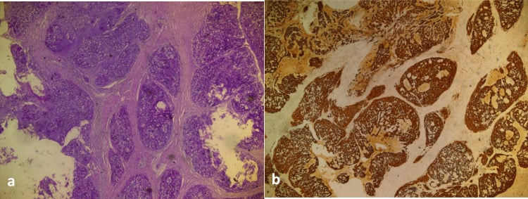
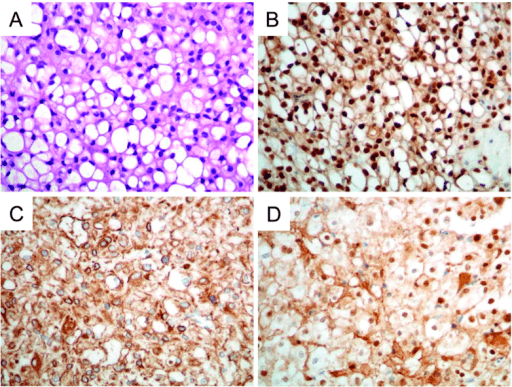
Immunohistochemistry. Brachyury is the diagnostic marker and the single most important immunostain: nuclear expression in over 95% of chordomas, and highly specific, positive only in chordoma and normal notochord. The rest of the panel:
- S100: positive in 80-100%, strong and diffuse, nuclear and cytoplasmic; also positive in chondrosarcoma, so not specific
- Cytokeratin: positive in 80-100%, the low-molecular-weight keratins (CAM 5.2, AE1/AE3), with a characteristic dot-like paranuclear pattern
- EMA: positive in most cases (70-90%)
- Vimentin: variably positive
- Negative: desmin, smooth muscle actin, chromogranin and synaptophysin, which helps exclude other tumour types
PBSChordoma Histology Features
Hook:PBS staining = Physaliferous, Brachyury, S100 for chordoma diagnosis!
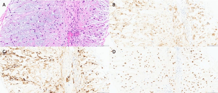
Subtypes. The conventional (classic) chordoma described above makes up 80% of cases. Three others matter:
- Chondroid - 5-15%. Areas of hyaline cartilage alongside the typical chordoma features, more common in the skull base. It was historically thought to carry a better prognosis, but several modern series suggest outcomes equal to conventional chordoma once margin and dose are controlled for, so the variant is now seen more as a histological than a prognostic distinction. Brachyury separates it from chondrosarcoma.
- Dedifferentiated - under 5%. Areas of high-grade sarcomatous transformation, with increased cellularity, atypia and mitotic activity. It arises de novo or in recurrent tumours, behaves more aggressively with a higher metastatic potential, and carries the worst prognosis: median survival approximately 1 year.
- Poorly differentiated. A rare variant, described in detail below.
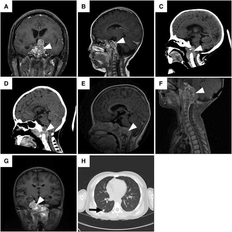
Poorly differentiated chordoma. A distinct, aggressive entity now defined by loss of nuclear SMARCB1 (INI1) expression on immunohistochemistry, from deletion or inactivation of the SMARCB1 gene on 22q11; the same tumour suppressor is lost in atypical teratoid/rhabdoid tumour and epithelioid sarcoma. It remains brachyury-positive, confirming its notochordal origin, but grows as solid epithelioid or rhabdoid sheets without the classic physaliphorous cells or myxoid matrix.
Who it affects. It occurs disproportionately in children and young adults and favours the skull base and cervical spine, unlike the sacral predominance of conventional chordoma. It is substantially more aggressive, with earlier metastasis and shorter survival, so recognising INI1 loss changes prognosis and surveillance and flags a paediatric skull-base tumour that behaves like a high-grade malignancy.
A therapeutic target. Its SMARCB1 biology has made it a candidate for EZH2 inhibition, as trialled in other SMARCB1-deficient tumours, one of the few rational targeted avenues in chordoma.
The pitfall. Without physaliphorous cells or myxoid matrix it is easily mistaken for another poorly differentiated round-cell or rhabdoid tumour. Brachyury positivity plus INI1 loss is the combination that secures the diagnosis.
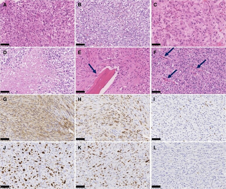
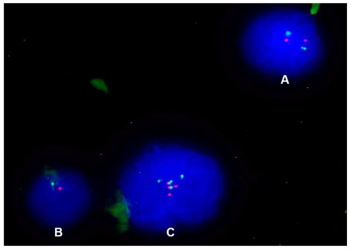
Differential diagnosis. Chondrosarcoma is the differential that matters most, especially for chondroid chordoma: both can contain cartilaginous matrix and both are S100-positive, so brachyury decides, and cytokeratin is also typically negative in chondrosarcoma. Axial location and cytokeratin positivity can mimic metastatic adenocarcinoma, where clinical history and additional immunostains help. Myxopapillary ependymoma can have a myxoid matrix but lacks physaliphorous cells, and GFAP, positive in ependymoma, is negative in chordoma. Ecchordosis physaliphora, a benign notochordal remnant typically found incidentally at the clivus, has identical histology and is told apart by clinical and imaging features.
- Key feature
- Physaliferous cells, lobular myxoid matrix, midline
- Brachyury
- Positive (nuclear)
- Cytokeratin
- Positive (dot-like)
- Other markers
- S100+, EMA+
- Key feature
- Malignant chondrocytes in hyaline cartilage
- Brachyury
- Negative
- Cytokeratin
- Negative
- Other markers
- S100+ (not discriminatory)
- Key feature
- Glandular/epithelial pattern, known primary
- Brachyury
- Negative
- Cytokeratin
- Positive
- Other markers
- Site-specific markers (TTF1, CDX2 etc)
- Key feature
- Sacrococcygeal, perivascular myxoid pseudorosettes
- Brachyury
- Negative
- Cytokeratin
- Negative
- Other markers
- GFAP+
- Key feature
- Benign clival notochordal rest, under 2 cm, no bone destruction
- Brachyury
- Positive
- Cytokeratin
- Positive
- Other markers
- Identical histology - distinguished by size/imaging
Clinical Presentation
Sacral chordoma. The symptoms are nonspecific, so diagnosis is delayed, with an average symptom duration of 6-12 months before diagnosis.
Pain is the most common presenting symptom, in 70-80%: mild, intermittent sacral or coccygeal pain at first, progressively worsening until constant, and it may radiate to the buttocks, hips or lower limbs.
Neurological symptoms are present in approximately 50% at presentation:
- Radicular pain, most commonly in the L5 and S1 dermatomes
- Motor weakness, including foot drop from L5 involvement
- Sensory change in a saddle distribution
- Bowel dysfunction, constipation most commonly
- Bladder dysfunction, retention or incontinence
- Sexual dysfunction
Anterior extension. In a sacral tumour it can cause constipation or a change in bowel habit, urinary symptoms from bladder compression and dyspareunia in women; rectal bleeding is rare and suggests mucosal invasion.
Skull base chordoma. The location is critical: even modest growth compromises the cranial nerves, the carotids and the brainstem, so these tumours present earlier. Cranial nerve palsy is the most common presentation (60-70%), and the abducens nerve (CN VI) is the nerve most commonly affected, causing diplopia; other cranial nerves are involved depending on the direction of extension.
Headache is present in 40-50% of patients, typically retro-orbital or at the vertex. Visual symptoms include diplopia, visual field defects and reduced visual acuity.
A large tumour can affect pituitary function and cause hypopituitarism, and anterior extension brings nasal obstruction, epistaxis or dysphagia.
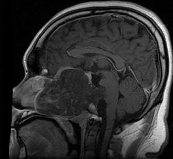
Mobile spine chordoma. Neck or back pain localised to the tumour, mechanical at first and progressively worse. Nerve root compression causes radiculopathy, with dermatomal pain and neurological deficit. Spinal cord compression causes myelopathy:
- Upper motor neuron signs below the level of the lesion
- Gait disturbance
- Bladder and bowel dysfunction
- A sensory level
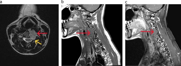
Examination. A visible or palpable mass may be present in advanced cases. Rectal examination is essential for a sacral lesion; a large tumour may be palpable as a presacral mass, typically firm and fixed, smooth or lobulated.
The neurological examination includes a comprehensive motor examination, looking for lower motor neuron signs from nerve root involvement, and sensory testing of the dermatomes and for saddle anaesthesia; reflexes may be diminished with nerve root compression. Rectal tone and perianal sensation complete it.
Investigations
Blood tests. Routine results are generally normal, and there is no specific serum tumour marker; alkaline phosphatase may be elevated but is nonspecific.
A full blood count, a basic metabolic panel and renal and hepatic function establish a baseline for treatment planning.
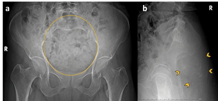
Radiographs. Of limited use, and they often underestimate the extent of the tumour, so a suspected chordoma should always proceed to cross-sectional imaging. When films do show something, it is sacral destruction with a soft-tissue shadow, loss of the normal sacral anatomy, calcification within the mass, or, uncommonly, a pathological fracture.
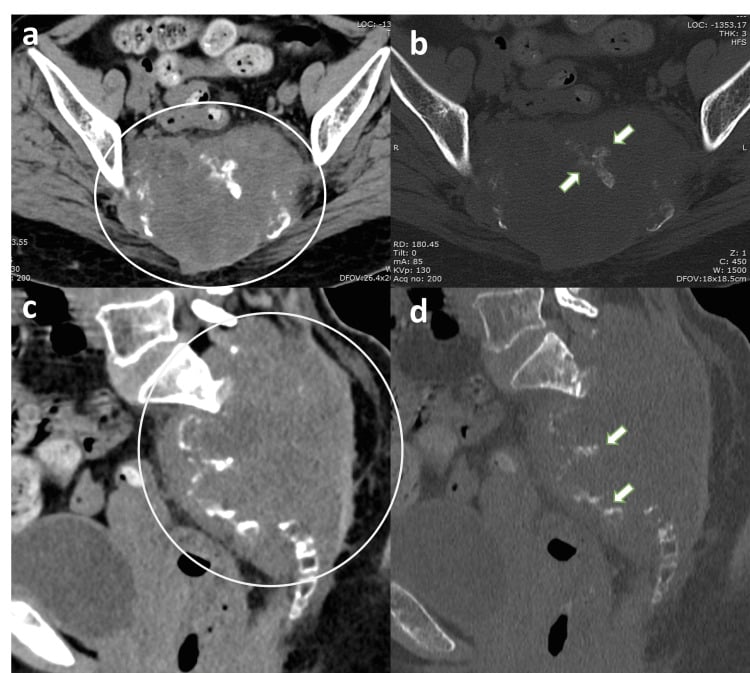
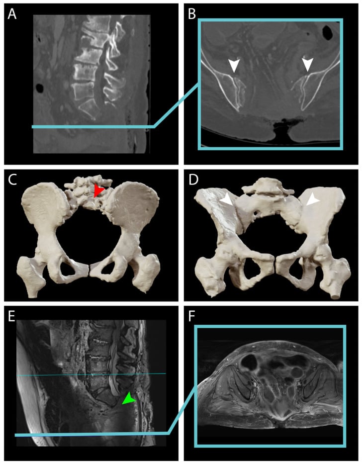
CT. Superior for the pattern of bone destruction and for matrix mineralisation, it is complementary to MRI and essential for bone involvement and staging. It defines the bony margins for resection planning and shows whether the sacral neural foramina are involved. The features:
- An expansile, midline lytic lesion centred in bone, with cortical disruption and trabecular destruction
- A soft-tissue mass containing bone fragments, often larger than the bone destruction suggests
- Calcification in 30-70%, flocculent or irregular
- Anterior soft-tissue extension, common into the presacral space with sacral tumours, and posterior extension through the sacral foramina
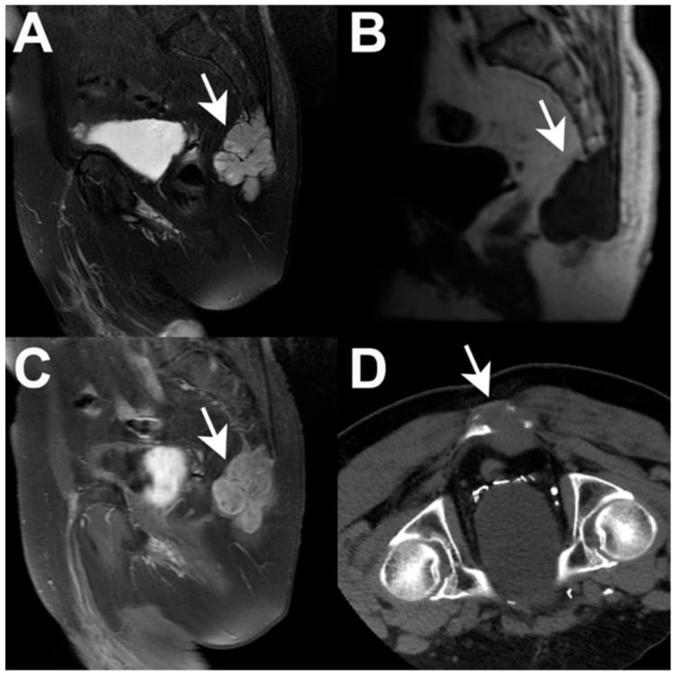
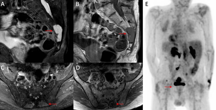
MRI. The gold standard, for its soft-tissue detail, its view of the neural structures and the extent of marrow involvement. It defines the extent of the tumour and its involvement of the dura, nerve roots and vessels, which is the basis of surgical planning, and it is the tool of post-treatment surveillance. The typical appearance:
- T1: hypointense to isointense relative to muscle, from the high water content of the myxoid matrix
- T2: very hyperintense, from the abundant mucoid and myxoid matrix
- Post-contrast T1: heterogeneous, irregular enhancement
- A midline or paracentral destructive lesion with a lobulated contour and internal septations
- A soft-tissue component that is usually large and often exceeds the bony component
The protocol:
- Sagittal T1 and T2 of the entire spine, for skip lesions
- Axial T1 and T2 through the lesion
- Coronal STIR or fat-suppressed T2
- T1 post-gadolinium with fat saturation
PET and other imaging. FDG-PET/CT has a limited role in primary diagnosis because chordomas typically have low metabolic activity. It may help identify dedifferentiated areas, which have a higher SUV, and in follow-up it can detect distant metastases and is useful for telling recurrence from post-treatment change. Whole-body MRI has an emerging role for skip lesions and distant bone metastases. Angiography is rarely needed; it may be considered for preoperative embolisation of a hypervascular tumour or to map the vascular anatomy for a complex resection.
Staging. Chest CT is mandatory to exclude pulmonary metastases; 30% of patients who develop metastases have pulmonary involvement.
The whole-spine MRI looks for skip metastases, which are rare but described; skip lesions and metastases change the resection field and the intent of treatment. The whole local compartment is imaged before biopsy, to map neurovascular and visceral extension. A skeletal survey or bone scan is considered if symptoms suggest other skeletal involvement.
Biopsy. Tissue diagnosis is mandatory before definitive treatment, because clinical and imaging features alone cannot definitively diagnose chordoma. Image-guided core needle biopsy, under CT or fluoroscopy, is preferred:
- Plan the tract so that it can be excised en bloc with the specimen at definitive surgery
- Avoid contaminating uninvolved tissue planes
- Take enough tissue for histology and immunohistochemistry
- A posterior approach is typical for sacral lesions
- Send fresh tissue if molecular studies are planned
Open biopsy is reserved for a nondiagnostic or inadequate core, and risks tumour contamination of the surgical field.
Management Principles
The margin decides. Wide surgical resection is the only curative treatment, and the margin achieved is the single most important prognostic factor for local control and survival. After marginal or intralesional surgery (intralesional meaning the tumour is violated during resection), 5-year local recurrence is 50-90%, against 10-30% with wide margins. En bloc resection with negative margins achieves the best outcomes but remains technically challenging because of the anatomical location.
The goals of surgery:
- En bloc resection with wide margins, negative on all surfaces
- Preservation of neurological function when possible
- Structural stability
- Minimal morbidity
The other modalities. Radiotherapy is used when wide resection is not feasible or the margins are positive, with proton beam or carbon ion therapy preferred over conventional photons. Conventional chemotherapy has no proven role, because chordomas are not chemosensitive; targeted therapies are under investigation for recurrent disease.
Surgery or radiotherapy. For a high sacral or skull-base tumour, where wide resection would cost bladder, bowel and ambulatory function, the threshold for offering definitive high-dose particle therapy in place of en bloc resection remains a genuine multidisciplinary judgement rather than a settled algorithm.
- 1Clinical suspicion (midline axial tumour)
MRI plus CT imaging, chest CT for staging
Confirm location and extent
- 2Image-guided biopsy
Core biopsy with immunohistochemistry
Brachyury confirmation for diagnosis
- 3Multidisciplinary tumour board
Oncology, neurosurgery/spine surgery, radiation oncology discussion
Treatment plan consensus
- 4Assess resectability
Wide margins feasible? Consider functional outcomes
Determine surgical candidacy
- 5Resectable with acceptable morbidity
En bloc wide resection with reconstruction
Curative intent surgery
- 6Unresectable or high morbidity
Definitive radiation (proton beam preferred) or palliative intent
Non-surgical management
- 7Post-operative assessment
Margin status evaluation
If positive margins, consider adjuvant radiation
- 8Surveillance
MRI of primary site every 3-4 months for 2 years, every 6 months to 5 years, then annually; chest CT annually
Monitor for recurrence and metastases
Radiation Therapy
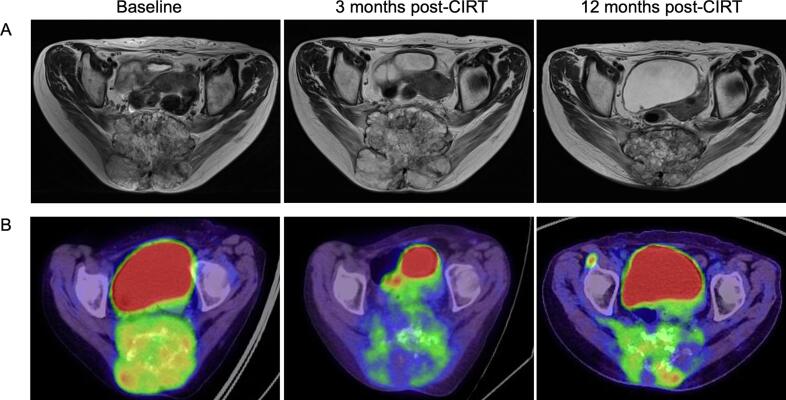
Photons. Chordomas are relatively radioresistant. The doses needed for control, greater than 70 Gy, exceed the tolerance of the surrounding normal tissues (brainstem, spinal cord, bowel, bladder), so conventional radiation has limited efficacy as primary treatment.
It may be used for palliation, for pain and symptom control in metastatic disease or in patients who are not candidates for surgery or proton therapy. After intralesional surgery, adjuvant conventional radiation may reduce the risk of recurrence but is inferior to proton therapy.
Protons. Protons deposit their maximum energy at a specific depth, the Bragg peak, with minimal exit dose, which allows a high dose to the tumour while sparing adjacent normal tissue.
Typical doses are 70-80 Gy (RBE) in 35-40 fractions, and the therapy is limited to specialised centres with proton beam facilities. The indications:
- Unresectable chordoma, at the skull base or in the sacrum with major vascular involvement
- Positive margins after surgery (adjuvant)
- Recurrent tumour not amenable to further surgery
- Patient choice when surgery is declined
Five-year local control is 60-80% for skull base and 50-70% for sacral chordomas, with the best results when proton therapy is combined with maximal safe surgery.
Carbon ions. The same concept, with theoretical radiobiological advantages and a higher relative biological effectiveness, which may suit radioresistant tumours.
Emerging evidence suggests local control similar to or slightly better than protons, but no randomised trial compares them, and carbon ion therapy is available at even fewer centres. The choice between the two is largely driven by access rather than proven superiority.
Systemic Therapy
Chemotherapy. Conventional cytotoxic chemotherapy has no established role, because chordomas are not chemosensitive. Multiple agents have been tested in clinical trials, among them ifosfamide, doxorubicin, cisplatin and temozolomide, with minimal response rates.
Targeted agents. Molecular profiling showing activation of receptor tyrosine kinase pathways led to the investigation of several targeted agents.
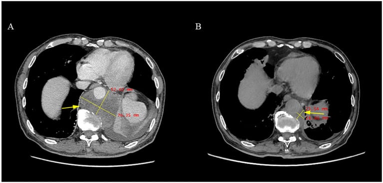
Imatinib, a PDGFR inhibitor, gave disease stabilisation in some patients in small series, without objective responses. The phase II trial found a clinical benefit of about 64% but an objective response rate of only 2%, and it may have a role in metastatic or unresectable disease.
EGFR inhibitors (erlotinib, cetuximab) have produced occasional responses in case reports, with no large trials. mTOR inhibitors are under investigation, based on pathway activation in some chordomas.
Checkpoint inhibitors are being explored, though response rates appear low given the low mutational burden of chordomas, and a brachyury-targeted vaccine is in clinical trials.
In practice. No standard systemic therapy exists: EGFR inhibitors, brachyury-targeted vaccines and immune checkpoint blockade remain investigational, and none is standard of care. For metastatic or unresectable progressive disease, consider:
- Clinical trial enrolment (preferred)
- Targeted therapy, imatinib or an EGFR inhibitor, with an understanding of the limited evidence
- A palliative care focus
Surgical Management
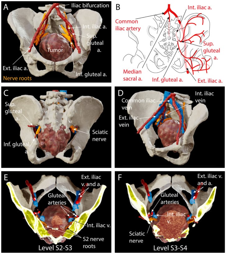
Planning. The MRI defines the extent of the tumour, and its relationship to the S2-S3 junction is critical. The vascular anatomy, the iliac vessels and the presacral plexus, is assessed, and preoperative embolisation is considered for a large hypervascular tumour.
The patient is counselled on the expected bowel, bladder and sexual function, and their medical status is optimised before surgery.
Walking depends on S1. The S1 roots carry the main motor supply to gastrocnemius-soleus and the gluteals, and bilateral preservation is essential for walking. Unilateral S1 sacrifice causes foot drop; bilateral S1 sacrifice causes major weakness, with foot drop and loss of push-off and hip extension, so preserving at least the S1 roots is the threshold for useful walking.
Continence depends on S2-S4: the Gunterberg rule. Bladder, bowel and sexual function depend on the S2-S3-S4 parasympathetic outflow. The classic teaching, level by level:
- Bilateral preservation of S1-S3: near-normal function
- Both S3 roots preserved (resection below S3): continence usually preserved
- Even one S3 root retained: often acceptable bladder and bowel control
- Sacrifice of S3 and below: varying degrees of dysfunction, generally better tolerated than loss of S2
- Both S2 roots preserved: maintains bladder and bowel function
- Bilateral sacrifice of S2 and above: reliably a neurogenic bladder needing permanent catheterisation, and bowel dysfunction, often requiring a colostomy
Bilateral loss is what counts. The roots are partly redundant across the midline, so unilateral sacrifice, a hemisacrectomy sparing the contralateral roots, generally preserves bladder, bowel and sexual function. It is bilateral sacrifice at a given level that is functionally decisive.
Margin against function. This is why the S2-S3 junction is the pivotal planning line on MRI: a resection that must cross above both S3 roots commits the patient to bladder, bowel and sexual dysfunction. Weighing that against margin adequacy is the central preoperative conversation, and margins should not be compromised for function when they are needed for cure.
High and low sacrectomy. A high sacrectomy, above S2-S3, sacrifices the S2-S3 roots with a significant functional cost, is reserved for high tumours where it is necessary for margins, and needs lumbopelvic (spinopelvic) reconstruction. A low sacrectomy, at S3 and below, preserves the S1-S2 roots; bowel, bladder and sexual dysfunction may still follow, but less severely.
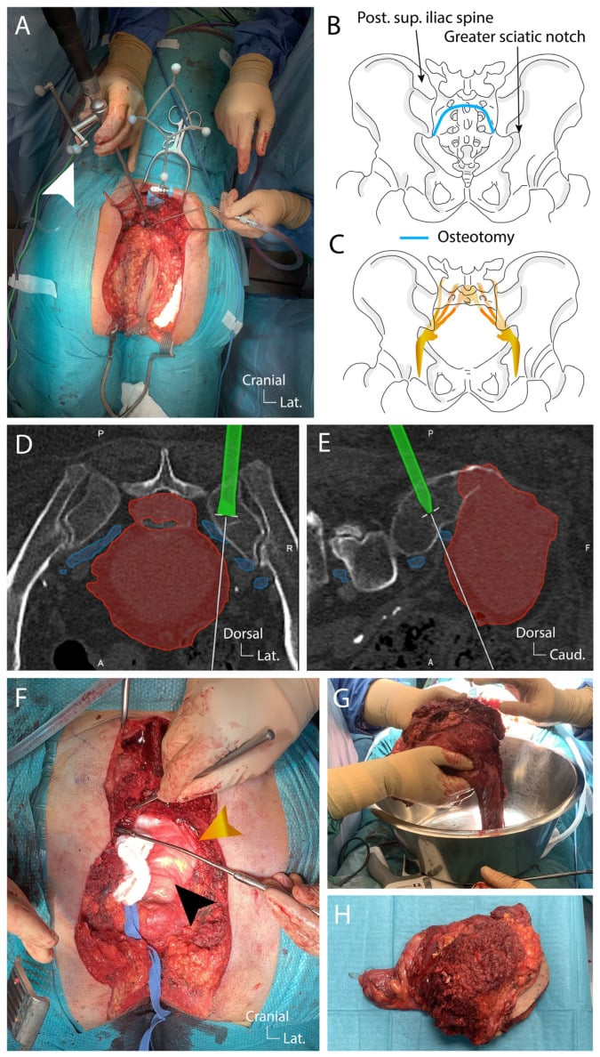
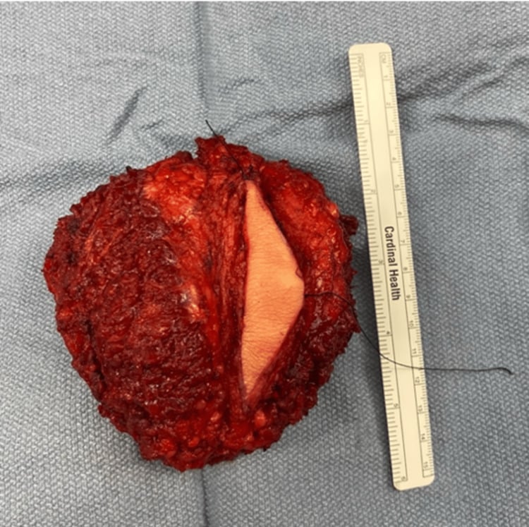
The approach. A combined anterior-posterior approach is the most common for a large sacral chordoma. The anterior phase, abdominal or pelvic, mobilises the rectum, ligates the vessels and dissects the presacral component; the patient is then turned prone and the posterior phase completes the osteotomy and removes the specimen en bloc. Both staged operations, anterior first and posterior days later, and single-stage procedures are described.
A posterior-only approach is feasible for smaller tumours without a large anterior extension.
Technique. Frozen sections confirm negative margins, and as many nerve roots are preserved as is feasible while achieving them. Soft-tissue coverage is critical and may need flap reconstruction.
Outcomes. Five-year local recurrence-free survival is 60-80% with wide margins and 20-40% with intralesional margins. Overall survival is 60-80% at 5 years and 40-60% at 10 years.
Complications
During surgery. Sacral resection can involve significant blood loss from the presacral venous plexus and iliac vessels, and transfusion is commonly required. The iliac vessels are at risk in sacral surgery, so vascular surgical availability is required. Dural tear is a high risk in spinal cases, with a risk of CSF leak. Nerve injury may be the intended sacrifice of a root or inadvertent injury to a root that was meant to be preserved.
The wound. Wound complications (infection, dehiscence, seroma) occur at a high rate, 20-40%, given the large dead space and possible contamination. Deep infection of the hardware or reconstruction may require removal and prolonged antibiotics.
Other early problems. Neurological deficit, lower limb weakness or bladder and bowel dysfunction, follows if roots were sacrificed or injured. Venous thromboembolism is a high risk after pelvic surgery and with immobility.
Late function. Bladder dysfunction after bilateral S2 sacrifice needs intermittent catheterisation or an indwelling catheter. Bowel dysfunction, constipation most commonly, may need a bowel regimen or, if severe, a colostomy, and sexual dysfunction means erectile dysfunction in men and dyspareunia in women.
Late pain and structure. Chronic neuropathic pain is common, especially after nerve root sacrifice. Spinopelvic fixation fails in 5-10% of cases, and stress fractures of the remaining sacrum reflect sacral insufficiency.
After radiotherapy. Acute effects are skin erythema, mucositis with skull base treatment and diarrhoea with sacral treatment.
Proton and carbon ion therapy reduce the risk of late complications compared with conventional radiation, because they spare normal tissue. The late effects:
- Radiation necrosis: brain or brainstem after skull base treatment, bowel injury after sacral treatment
- A long-term risk of radiation-induced sarcoma
- Hypopituitarism after skull base radiation
- Cranial neuropathies and a risk of myelopathy
From the disease. Local progression brings pain, neurological deterioration and mass effect on the pelvic organs.
Prognosis and Follow-Up
Prognostic factors. The margin, discussed under Management Principles, dominates.
Location and size. Sacral tumours generally have a better prognosis, being more amenable to wide resection; at the skull base margins are harder to achieve and adjuvant radiation is often required, and the mobile spine is intermediate. Larger tumours do worse, being harder to excise with margins and more likely to involve critical structures.
Biology and history. Chondroid chordoma behaves like conventional chordoma when adequately treated, and the dedifferentiated subtype is the worst. Younger age is generally associated with better survival, and a recurrent tumour has a worse prognosis than a primary one.
Survival. Five-year overall survival is 65-80% and 10-year 40-60%; 5-year progression-free survival with optimal treatment is 50-70%.
Cause of death. Most patients who die of chordoma do so from local progression rather than distant metastasis, and uncontrolled local disease in the sacrum or skull base causes significant morbidity.
Surveillance. Recurrence can come 10-20 years after initial treatment, so lifelong follow-up is recommended.
Because late recurrences and metastases occur well beyond 10 years, the ideal length and intensity of imaging follow-up is not evidence-defined and varies between centres. A typical schedule:
- Years 1-2: MRI of the primary site every 3-4 months
- Years 3-5: MRI every 6 months
- After 5 years: annual MRI
- Chest CT annually, for pulmonary metastases
- Whole-body imaging if symptoms suggest a new site of disease
Each visit assesses symptoms of local recurrence, neurological function, pain control and function, including bowel and bladder.
Guidelines, Registries & Global Practice
Global Epidemiology
Chordoma is uniformly rare worldwide, with a population-based incidence of approximately 0.08 per 100,000 (SEER, McMaster 2001). There is a consistent male predominance (~1.7:1) and a peak in the fifth to seventh decades, with cranial disease over-represented in younger patients and children. Population (SEER) data show a roughly even axial distribution (cranial ~32%, mobile spine ~33%, sacrum ~29%), whereas surgical series quote the classic sacrum 50% / skull base 35% / mobile spine 15% because surgically resected sacral tumours are over-represented.
Side-by-Side Guideline Comparison
- Surgery
- En bloc resection with wide margins at a reference centre
- Radiation
- High-dose particle therapy (proton/carbon ion) if margins inadequate or unresectable
- Systemic / Other
- No standard chemo; favour clinical trials and molecular profiling
- Surgery
- Wide en bloc resection is the goal; avoid intralesional violation
- Radiation
- Definitive high-dose RT when surgery would be mutilating or incomplete
- Systemic / Other
- Imatinib/EGFR agents only within trial or compassionate use
- Surgery
- Wide excision; spondylectomy/sacrectomy by expert teams
- Radiation
- RT (proton preferred) for residual, recurrent or unresectable disease
- Systemic / Other
- Targeted therapy or trial for progressive systemic disease
- Surgery
- Centralised management in designated sarcoma/spinal tumour MDTs
- Radiation
- Access to proton therapy via national high-energy proton service
- Systemic / Other
- Trial enrolment; no routine cytotoxic chemotherapy
The unifying message across all bodies is identical: wide en bloc resection at a high-volume reference centre, with high-dose particle radiotherapy reserved for inadequate margins or unresectable disease, and no role for routine cytotoxic chemotherapy.
Registry and Reference-Centre Notes
- Chordoma is too rare for arthroplasty-style implant registries; the most robust outcome data come from national sarcoma databases and reference-centre series (e.g. Rizzoli, MD Anderson, Massachusetts General, Swedish Musculoskeletal Tumour Centre).
- The Chordoma Foundation maintains an international patient registry and biobank that increasingly drives multi-centre collaborative evidence.
High- vs Limited-Resource Practice Variation
- Particle therapy (proton/carbon ion) is concentrated in a small number of centres in North America, Europe and East Asia; many regions require interstate or international referral, creating access inequity and treatment delay.
- In limited-resource settings, conventional photon radiotherapy and the quality of the index surgical resection carry disproportionate weight, since a contaminated first operation cannot be undone and dominates lifelong local-control prospects.
- Early referral before biopsy to a specialist tumour centre is the single most transferable, resource-independent quality measure, ensuring a correctly planned, excisable biopsy tract.
MCQ Practice Points
Q: What is the embryologic origin of chordoma?
A: Notochordal remnants. The notochord is an embryonic structure that forms the primitive axial skeleton and normally regresses completely by 8 weeks of gestation, leaving only the nucleus pulposus. Chordoma arises from ectopic notochordal cell rests that persist after embryogenesis. This explains the exclusive location of chordoma in midline axial skeleton structures.
Q: What is the pathognomonic immunohistochemical marker for chordoma?
A: Brachyury - a nuclear T-box transcription factor encoded by the TBXT gene. Brachyury is positive in over 95% of chordomas and is the single most specific marker. It is negative in chondrosarcoma and other differential diagnoses. Also positive: S100 (80-100%), cytokeratin (80-100%). The combination of brachyury, S100, and cytokeratin positivity confirms chordoma.
Q: What is the characteristic cell type in chordoma histology?
A: Physaliferous cells - large cells with abundant vacuolated, bubbly cytoplasm containing glycogen and mucin. The cytoplasmic vacuoles push the nucleus to the periphery, sometimes creating a signet-ring appearance. These cells are pathognomonic for chordoma when present. The tumor grows in lobules within abundant myxoid matrix.
Q: What is the most important prognostic factor for chordoma?
A: Surgical margin status. Wide negative margins are associated with significantly better local control and overall survival. Intralesional surgery (tumor violation) results in 50-90% local recurrence at 5 years, compared to 10-30% recurrence with wide en bloc resection. En bloc resection with negative margins is the only curative treatment.
Q: Why is proton beam therapy preferred over conventional photon radiation for chordoma?
A: Chordomas are relatively radioresistant and require high radiation doses (greater than 70 Gy) for local control. These doses exceed the tolerance of surrounding normal tissues (brainstem, spinal cord, bowel) with conventional photon radiation. Proton beam therapy allows delivery of high doses to the tumor with minimal exit dose due to the Bragg peak phenomenon, thereby sparing adjacent critical structures. This achieves better tumor control with fewer complications than conventional radiation.
Exam Viva Scenarios
Practise clinical reasoning and management decisions out loud
“A 55-year-old man presents with 12 months of progressive sacral pain and recent onset constipation. MRI shows a large midline sacral mass destroying S3-S5 with extensive anterior soft tissue extension. Biopsy shows physaliferous cells and is brachyury positive. How do you manage him?”
“A 48-year-old woman had sacral chordoma treated 3 years ago with intralesional surgery and conventional radiation. She now presents with recurrent pain. MRI shows tumor recurrence involving S2-S4. What is your approach?”
“A 60-year-old presents with a destructive midline sacral lesion on MRI that is very bright on T2 sequences. The differential diagnosis includes chordoma versus chondrosarcoma. How do you differentiate these pathologically?”
Key Facts (Must Know)
- Notochord remnant origin - an embryonic structure that normally regresses
- Location: sacrum 50%, skull base 35%, mobile spine 15%
- Incidence: 1-4% of bone tumors, 0.08 per 100,000 population
- Peak age: 50-70 years, male-to-female ratio 2:1
- Slow-growing but locally aggressive malignancy
Histology (PBS Mnemonic)
- P - Physaliferous cells with bubbly vacuolated cytoplasm (pathognomonic)
- B - Brachyury positive, a nuclear T-box transcription factor (diagnostic)
- S - S100 and cytokeratin positive, and also EMA positive
- Lobulated architecture in abundant myxoid matrix
- Brachyury distinguishes from chondrosarcoma (negative in CS)
Imaging Features
- MRI: T1 hypointense, T2 very hyperintense (myxoid matrix)
- CT: Midline lytic destruction with soft tissue mass
- Calcification in 30-70% (flocculent pattern)
- Heterogeneous enhancement post-contrast
- Chest CT mandatory for staging (lungs common metastatic site)
Treatment
- Wide margins; en bloc resection is the only approach offering cure, though 10-30% still recur by 5 years
- Intralesional excision means recurrence, with a 50-90% 5-year recurrence rate
- Sacral and skull-base surgery is complex
- Proton beam for unresectable disease or positive margins
- No role for conventional chemotherapy (not chemosensitive)
Sacral Surgery Pearls
- S1 roots - bilateral preservation is essential for walking
- S2 roots - bilateral preservation is needed for bladder and bowel function
- S3 sacrifice: Some dysfunction but better tolerated than S2
- Combined anterior-posterior approach for large tumors
- Margin status most important prognostic factor
Prognosis
- 5-year survival: 65-80%, 10-year survival: 40-60%
- Wide margins: 10-30% recurrence at 5 years
- Intralesional: 50-90% recurrence at 5 years
- Metastases occur in 10-40% (lungs, bone, liver)
- Dedifferentiated variant: Median survival 1 year
Differential Diagnosis
- Chondrosarcoma - brachyury negative, cytokeratin negative
- Metastatic adenocarcinoma - clinical history, brachyury negative
- Myxopapillary ependymoma - GFAP positive, no physaliferous cells
- Ecchordosis physaliphora - benign notochord remnant, small, no destruction
High-Yield Exam Points
- Brachyury is THE diagnostic marker (know this)
- En bloc wide resection is only cure (emphasize margins)
- Proton beam preferred over conventional radiation (radioresistance)
- S2 root preservation critical for bladder/bowel function
- No role for chemotherapy (but mention targeted trials)
Evidence Base
Stacchiotti & Sommer (ESMO/Chordoma Foundation Global Consensus)
- ESMO-convened consensus of over 40 chordoma experts plus Chordoma Foundation
- En bloc resection with wide margins is the standard for resectable disease
- High-dose particle therapy (proton/carbon ion) for unresectable disease or positive margins
- Multidisciplinary, expert-centre management essential given rarity
- No standard systemic therapy; clinical trial enrolment encouraged
Vujovic et al (Brachyury discovery)
- Brachyury (nuclear T-box transcription factor) expressed in all 53 chordomas studied
- Negative in over 300 other neoplasms, including 163 chondroid/cartilaginous tumours
- Also negative in nucleus pulposus, arguing against direct notochordal origin of disc
- Establishes brachyury as a specific marker of notochord and notochord-derived tumours
Fourney et al - En Bloc Resection of Primary Sacral Tumours
- 29 en bloc primary sacral tumour resections; chordoma the most frequent type (16 cases)
- Wide margins in 19, marginal in 9, contaminated in 1
- Level of nerve-root transection classification predicts bladder, bowel and ambulatory function
- Median Kaplan-Meier disease-free survival for chordoma 68 months
- Margins should not be compromised to preserve function when needed for tumour control
DeLaney et al - High-Dose Photon/Proton RT for Spine Sarcomas
- Prospective phase II trial: 50 patients (29 chordoma, 14 chondrosarcoma) of thoracolumbar/sacral spine
- Combined photon/proton RT to doses up to 76.6-77.4 GyRBE around resection
- 5- and 8-year local control 94% and 85% for primary tumours; 81% and 74% overall
- Recurrence 11% in primary versus 50% in previously recurrent tumours (p=0.002)
- 8-year grade 3-4 late toxicity 13%, with no radiation myelopathy
Bergh et al - Prognostic Factors in Chordoma
- 39 patients (30 sacral, 9 mobile spine); mean follow-up 8.1 years
- Local recurrence 44%, metastases 28%; 5-, 10-, 20-year survival 84%, 64%, 52%
- Inadequate margins, larger size, Ki-67 over 5%, tumour necrosis and recurrence were adverse factors
- Local recurrence strongly associated with metastasis and tumour-related death (p less than 0.001)
- Fine-needle aspiration favoured to avoid contamination from open biopsy
McMaster et al - SEER Incidence & Survival
- SEER analysis of 400 microscopically confirmed chordomas, 1973-1995
- Age-adjusted incidence 0.08 per 100,000; male predominance (0.10 vs 0.06)
- Axial distribution roughly even: 32% cranial, 32.8% spinal, 29.2% sacral
- Younger and female patients more likely to have cranial disease
- Median survival 6.29 years; 5- and 10-year relative survival 67.6% and 39.9%
Stacchiotti et al - Phase II Imatinib in Advanced Chordoma
- Largest prospective phase II systemic trial: 56 patients with PDGFB/PDGFRB-positive advanced chordoma
- Imatinib 800 mg/day until progression
- Objective response rate only 2% (1 partial response); 70% stable disease
- 64% clinical benefit rate and median progression-free survival 9 months
- Confirms modest, mainly disease-stabilising activity in an orphan disease