The Foot-Drop Nerve
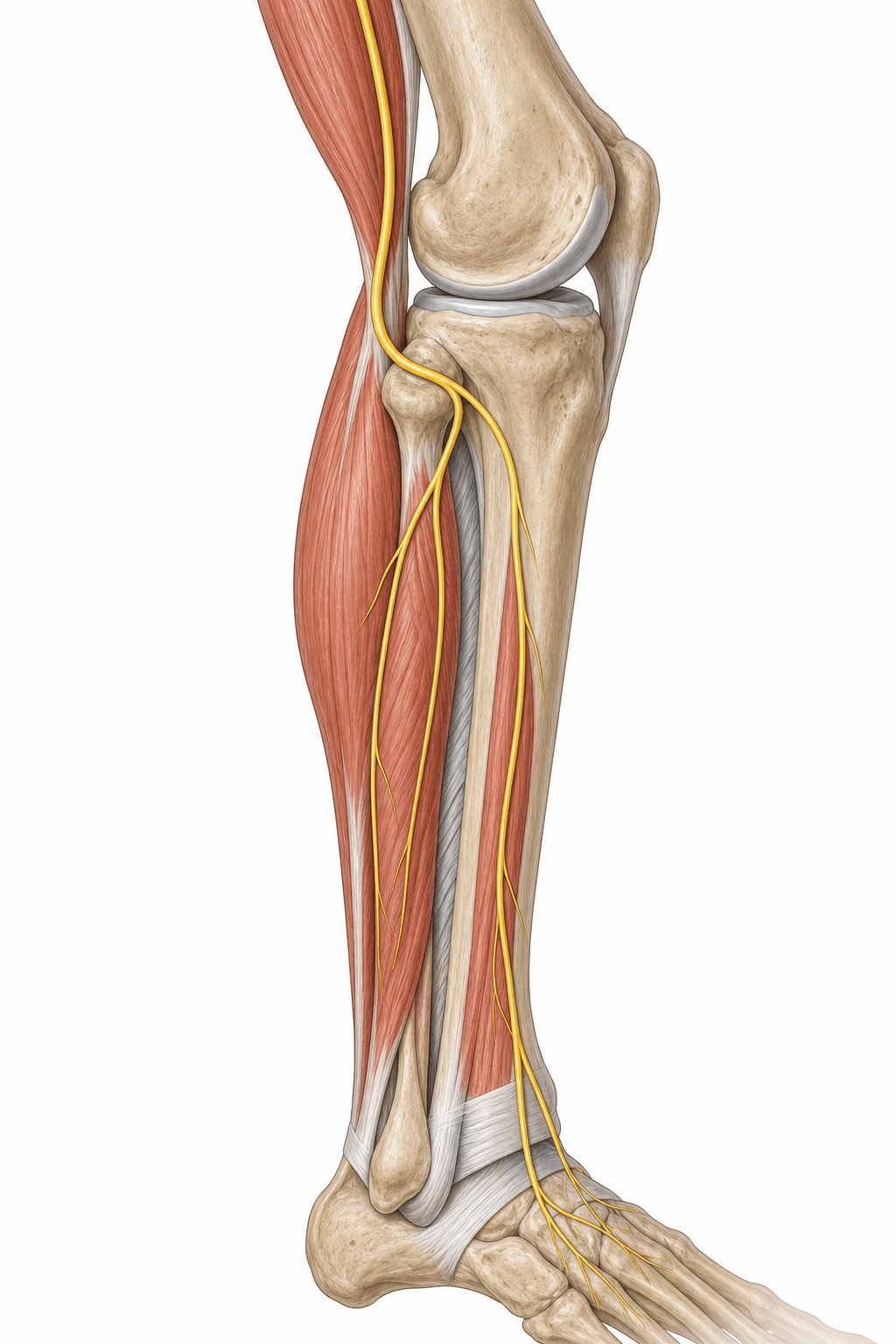
- The common peroneal (fibular) nerve is the smaller terminal branch of the sciatic nerve (L4-S2).
- It winds subcutaneously around the NECK OF THE FIBULA, where it is the most commonly injured/compressed nerve in the lower limb.
- It divides into the DEEP peroneal nerve (anterior compartment - dorsiflexion) and the SUPERFICIAL peroneal nerve (lateral compartment - eversion).
- Deep peroneal palsy → foot drop (loss of dorsiflexion/toe extension) and numbness of the FIRST web space.
- Superficial peroneal palsy → weak eversion and numbness of the dorsum of the foot (sparing the first web).
- Foot drop produces a high-steppage gait; common causes are fibular neck fracture, knee dislocation, prolonged leg-crossing/squatting, casts, and rapid weight loss.
- “Deep peroneal nerve = 'first web space' sensation (the classic small autonomous sensory zone).
- “Distinguish a peroneal palsy from an L5 radiculopathy: L5 also weakens inversion (tibialis posterior, tibial nerve) and hip abduction, which a peroneal lesion spares.
- “The fibular neck is both the danger zone and the surgical release site (deep peroneus longus fascia over the nerve).
Weak dorsiflexion and eversion; sensory loss over the dorsum of the foot / first web space. Inversion (tibialis posterior, tibial nerve) and ankle plantarflexion are PRESERVED. A positive Tinel sign at the fibular neck supports a peripheral lesion.
Also causes foot drop, but inversion is ALSO weak (tibialis posterior is L5 via the tibial nerve), as is hip abduction (gluteus medius). Back/radicular pain and a wider myotomal pattern point to the root rather than the peroneal nerve.
Origin & Course
Origin
- The common peroneal (fibular) nerve is the smaller terminal branch of the sciatic nerve, carrying fibres from the posterior divisions of L4-S2.
- It separates from the tibial nerve at (or above) the apex of the popliteal fossa and runs along the medial border of biceps femoris toward the fibular head.
Motor & Sensory Supply
The deep peroneal nerve supplies the anterior compartment - tibialis anterior, extensor hallucis longus, extensor digitorum longus, peroneus tertius (and extensor digitorum brevis in the foot) - producing ankle dorsiflexion and toe extension. The superficial peroneal nerve supplies the lateral compartment - peroneus longus and brevis - producing foot eversion.
- Sensory (deep peroneal): a small but clinically vital zone in the first dorsal web space.
- Sensory (superficial peroneal): most of the dorsum of the foot (sparing the first web space and the lateral border, which is sural territory).
- Lateral sural cutaneous nerve: upper lateral leg, contributing to the sural nerve.
Clinical Correlations
Common Peroneal Palsy
- Foot drop - loss of dorsiflexion - with a compensatory high-steppage gait and weak eversion.
- Sensory loss over the dorsum of the foot and first web space.
- Plantarflexion and inversion are preserved (tibial nerve).
DEEPPeroneal Branches
Hook:Deep = Dorsiflexion + first-web sensation; Superficial = Eversion + dorsum of foot.
Evidence Base
Common Peroneal Nerve at the Fibular Neck (Cadaveric Release)
- Cadaveric validation of common peroneal nerve release at the fibular neck in 15 specimens
- Confirms the nerve is compressed by the deep fascia of peroneus longus overlying it at the fibular neck
- Release extended to the bifurcation into the superficial and deep peroneal nerves
- No iatrogenic neurovascular or tendon injury; demonstrates the surgically relevant anatomy
Lower-Extremity Entrapment Neuropathies (incl. Common Peroneal)
- Reviews lower-limb entrapment neuropathies including common peroneal neuropathy causing foot drop
- Emphasises accurate localisation to prevent pain, sensory loss and weakness affecting mobility
- Diagnosis combines clinical examination with electrodiagnosis and imaging
- Distinguishes peripheral peroneal palsy from other causes of foot drop
Viva Scenarios
Practise clinical reasoning and management decisions out loud
“A patient develops foot drop after a below-knee cast. They cannot dorsiflex or evert the foot and have numbness over the dorsum of the foot. How do you assess and manage this, and how do you exclude an L5 radiculopathy?”
Guidelines, Registries & Global Practice
Global Practice Picture
Common peroneal neuropathy is a universal clinical problem - the leading lower-limb compressive neuropathy and the prototypical cause of foot drop. Internationally consistent teaching: protect the fibular neck (positioning, casts), localise the lesion clinically (deep vs superficial branch; peroneal vs L5), confirm with electrodiagnostics, and manage with cause removal, an AFO, and decompression or tendon transfer when indicated.
Side-by-Side Synthesis
- Compartment / muscles
- Anterior (TA, EHL, EDL, per. tertius)
- Action
- Dorsiflexion, toe extension
- Sensory
- First web space
- Compartment / muscles
- Lateral (peroneus longus/brevis)
- Action
- Eversion
- Sensory
- Dorsum of foot (spares 1st web)
- Compartment / muscles
- (parent at fibular neck)
- Action
- Both of the above
- Sensory
- Lateral leg (sural contribution)
Anatomy
- Smaller terminal branch of sciatic (L4-S2)
- Winds around the fibular neck (subcutaneous)
- Divides into deep + superficial peroneal
- Most commonly injured lower-limb nerve
Supply
- Deep: dorsiflexion + first web space
- Superficial: eversion + dorsum of foot
- Plantarflexion/inversion preserved (tibial)
- Lateral sural cutaneous → sural nerve
Clinical
- Palsy → foot drop, high-steppage gait
- Causes: cast, leg-crossing, fibular neck fracture, knee dislocation, weight loss
- L5 differs: inversion + hip abduction weak
- Mx: remove cause, AFO, decompression/transfer