A Painless Right-Sided Mid-Clavicular Lump
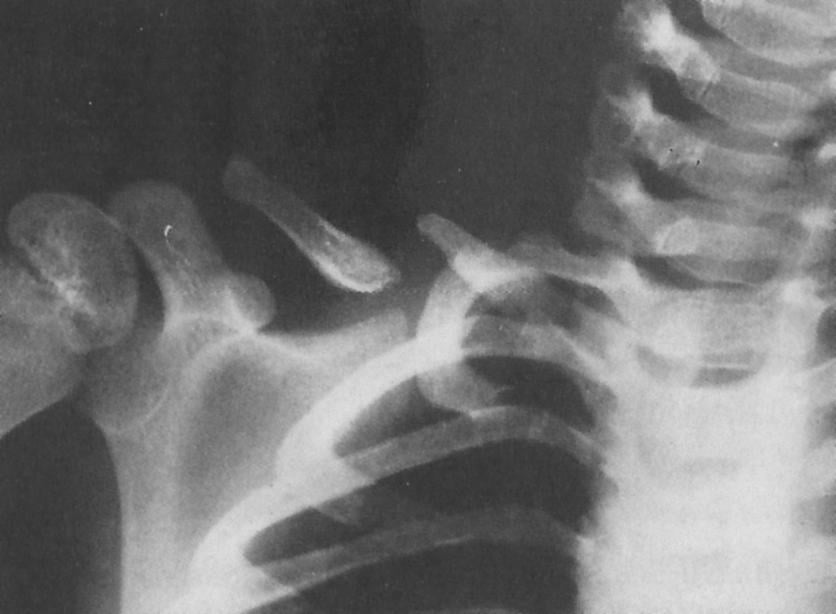
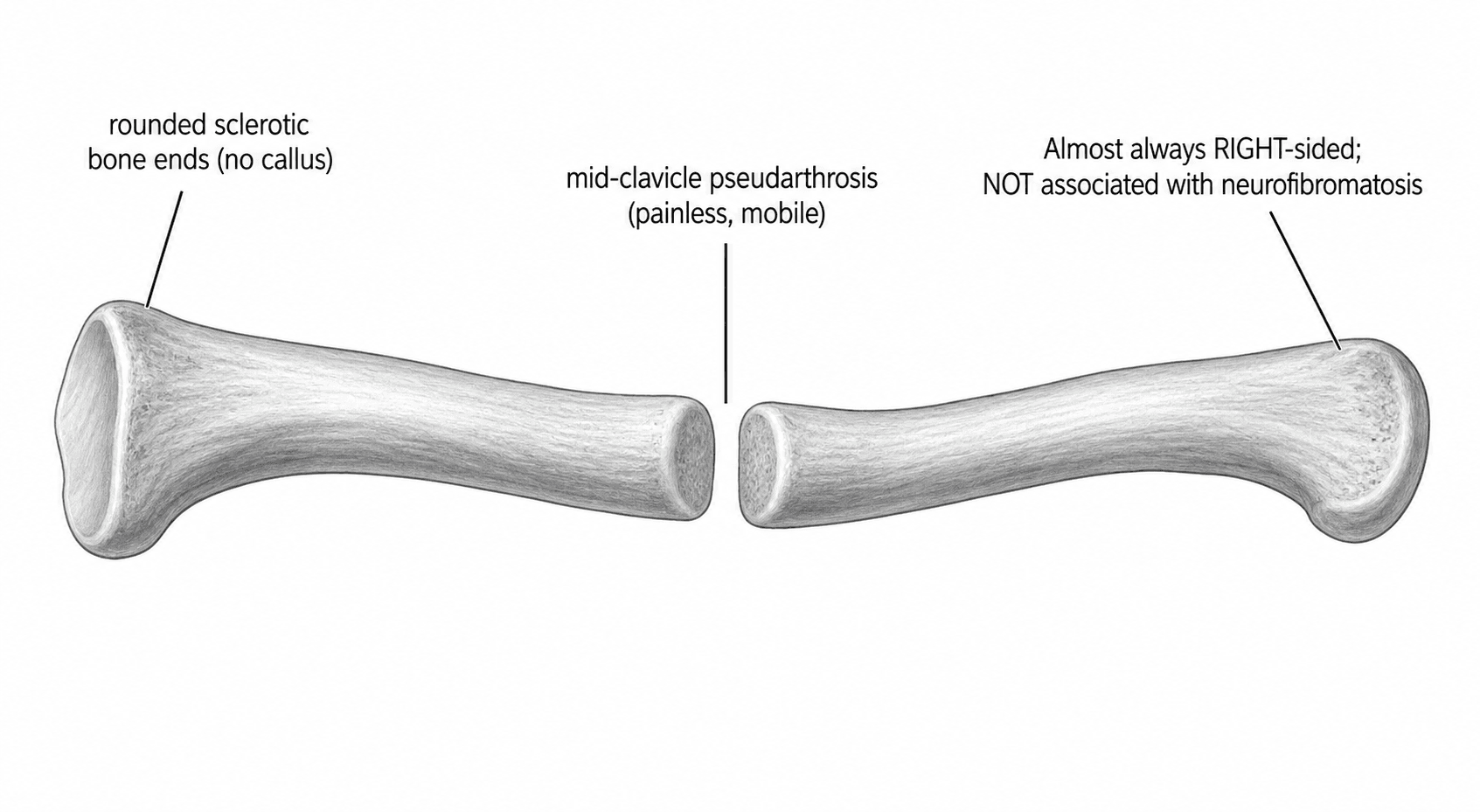
- Congenital pseudarthrosis of the clavicle (CPC) is a RARE disorder in which the medial and lateral ossification centres of the clavicle FAIL TO FUSE, producing a PAINLESS, mobile, non-tender LUMP over the MIDDLE THIRD of the clavicle, usually noticed in infancy/early childhood, with rounded SCLEROTIC non-united bone ends and NO callus on radiographs.
- It is almost ALWAYS RIGHT-SIDED; the rare LEFT-sided cases are associated with DEXTROCARDIA/situs inversus, and bilateral cases are uncommon - the right predominance is attributed to the higher position of the subclavian artery on the right (pulsation theory).
- Crucially, CPC is NOT associated with NEUROFIBROMATOSIS - this contrasts with congenital pseudarthrosis of the TIBIA, which IS strongly associated with NF1 and is far harder to treat; this distinction is a classic exam point.
- The main DIFFERENTIAL is a CLAVICLE BIRTH FRACTURE: a birth fracture follows trauma, is initially PAINFUL and HEALS with abundant CALLUS and simple immobilisation, whereas CPC is painless, present without trauma and does NOT heal spontaneously; the other differential is CLEIDOCRANIAL DYSPLASIA (BILATERAL clavicular defects with cranial/dental features, RUNX2 mutation).
- CPC is usually PAINLESS and NON-PROGRESSIVE with little or no functional deficit, so MANY cases can be simply OBSERVED; the indications for SURGERY are symptoms (pain, fatigue), cosmetic concern about the prominent lump, or functional problems, typically operated from around age 3-6 years.
- SURGICAL treatment is RESECTION of the pseudarthrosis with autologous (ILIAC CREST) bone grafting and internal FIXATION (plate, or elastic stable intramedullary nail/K-wire), which gives high union rates and good functional/cosmetic outcomes; AUTOGRAFT is preferred to allograft (allograft has higher non-union).
- “CPC = painless, mobile MID-clavicular lump from failed fusion of ossification centres; rounded sclerotic ends, no callus; almost always RIGHT-sided.
- “NOT associated with neurofibromatosis (vs congenital pseudarthrosis of the TIBIA, which IS). Left-sided CPC -> think situs inversus/dextrocardia.
- “Differentiate from birth fracture (painful, heals with callus) and cleidocranial dysplasia (bilateral + cranial/dental). Observe if asymptomatic; resection + iliac-crest autograft + fixation if symptomatic/cosmetic.
CPC is painless, present without trauma and does NOT heal; a clavicle birth fracture is initially painful, follows trauma and heals with callus on immobilisation.
CPC is NOT associated with neurofibromatosis and treats well; congenital tibial pseudarthrosis IS strongly NF1-associated and is notoriously difficult.
Presentation & Radiology
CPC results from failure of fusion of the medial and lateral clavicular ossification centres, presenting in infancy or early childhood as a painless, mobile, non-tender LUMP over the middle third of the clavicle. It is almost always RIGHT-sided; left-sided cases should prompt consideration of dextrocardia/situs inversus, and bilateral cases are rare. On radiographs the bone ends are rounded and sclerotic and non-united with NO callus - distinguishing it from a healing fracture. The condition is non-progressive with little functional deficit, and importantly it is NOT associated with neurofibromatosis (in contrast to congenital pseudarthrosis of the tibia).
Differential Diagnosis & Management
- Clavicle birth fracture: the main differential - it follows birth trauma, is initially painful, and HEALS with abundant callus and simple immobilisation; CPC is painless, atraumatic and does not heal.
- Cleidocranial dysplasia: suspect if the clavicular defects are BILATERAL or there are cranial features (delayed closure of fontanelles, Wormian bones), dental anomalies and other skeletal signs (a RUNX2 disorder).
- Neurofibromatosis: CPC is NOT associated with NF - but congenital pseudarthrosis of the tibia IS, so reserve NF concern for tibial (not clavicular) pseudarthrosis. Diagnosis is clinical (painless mid-clavicular mass) plus a radiograph showing the characteristic non-united rounded sclerotic ends.
- Observation: because CPC is usually painless, non-progressive and causes little functional deficit, many children need only reassurance and observation.
- Surgery (indications): symptoms (pain, shoulder fatigue), troublesome cosmetic prominence, or functional limitation - typically performed from around age 3-6 years.
- Technique: RESECTION of the pseudarthrosis with autologous ILIAC CREST bone grafting and internal FIXATION (a plate, or an elastic stable intramedullary nail / K-wire) - giving high union rates and good functional and cosmetic outcomes.
- Graft choice: AUTOGRAFT is preferred; allograft has a higher non-union rate. Intramedullary devices may achieve faster union and easier removal than plates.
Evidence & Key Studies
Comparison of fixation methods in the treatment of congenital pseudarthrosis of the clavicle (multicentre)
- In 15 clavicles (11 children), pseudarthrosis resection with iliac crest bone autograft and fixation (plate vs elastic stable intramedullary nail/K-wires) achieved radiological healing in almost all.
- Elastic stable intramedullary nailing or K-wires achieved a shorter union time and easier implant removal than plates, with no difference in complications or clinical outcome.
- Surgical resection with iliac crest autograft is an effective treatment, and surgery is recommended for symptomatic congenital pseudarthrosis of the clavicle.
Congenital pseudarthrosis of the clavicle: a report on 27 cases
- In 27 cases, operated patients (mostly for cosmetic appearance) were treated by pseudarthrosis resection and K-wire fixation, usually with iliac crest autograft.
- Bone healing was achieved in 74%, all operated patients were pain-free with full range of motion, and no neurovascular complications occurred.
- Use of allograft was associated with a high non-union rate, so autologous iliac crest grafting gave better functional and cosmetic outcomes.
According to PubMed, the effectiveness of resection plus iliac crest autograft and the comparison of fixation methods (intramedullary devices giving faster union) come from the cited Li multicentre study, and the good functional/cosmetic outcomes with autograft (and high non-union with allograft) from the cited Di Gennaro series. The painless right-sided presentation, the rounded non-united radiographic appearance, the non-association with neurofibromatosis and the distinction from a birth fracture and cleidocranial dysplasia are standard, well-established paediatric teaching. (See also our Clavicle Fractures, Cleidocranial Dysplasia and Congenital Tibial Pseudarthrosis topics.)
Clinical Decision Scenarios
Practise clinical reasoning and management decisions out loud
“An infant has a painless, mobile lump over the middle of the right clavicle. What is the likely diagnosis, what are the key differentials, and how would you manage it?”
“How does congenital pseudarthrosis of the clavicle differ from that of the tibia, and what does this mean for treatment and prognosis?”
Mnemonics & Memory Aids
CLAVICLE
Hook:CPC of the CLAVICLE: painless right-sided lump, not NF, observe or graft.
NOT NF
Hook:Clavicular pseudarthrosis = NOT NF (that's the tibia).
What it is
- Failure of fusion of the clavicular ossification centres
- Painless, mobile, non-tender mid-clavicular lump from infancy
- Almost always RIGHT-sided (left -> dextrocardia/situs inversus)
Radiology & associations
- Rounded, sclerotic non-united bone ends; NO callus
- NOT associated with neurofibromatosis (vs congenital tibial pseudarthrosis)
- Non-progressive; little functional deficit
Differentials
- Clavicle birth fracture (painful, trauma, heals with callus)
- Cleidocranial dysplasia (bilateral + cranial/dental, RUNX2)
- Diagnosis: clinical + radiograph
Management
- Observe if asymptomatic (usually painless, good function)
- Surgery for symptoms/cosmesis (~age 3-6): resection + iliac-crest autograft + fixation
- Autograft > allograft (lower non-union); IM device gives faster union than plate