Pure Lower Motor Neuron Disease | Flaccid Paralysis, Normal Sensation
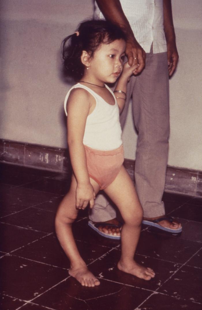
- Pure motor disease: Polio destroys anterior horn cells, causing a lower motor neuron flaccid, areflexic paralysis. Sensation is ALWAYS normal - this single fact separates polio from spina bifida and most other deformities.
- Asymmetric and patchy: Paralysis is asymmetric and skips muscles. This drives muscle imbalance, which is the engine of deformity.
- Wait for the residual phase: Reconstructive surgery is generally delayed until recovery plateaus (around 2 years) so you do not transfer or fuse a muscle that was going to recover.
- Deformity = imbalance + growth + gravity: Living muscle pulling against a dead antagonist, plus growth and posture, produces predictable deformities (equinus, calcaneus, flail joints, scoliosis, dislocated hip).
- Post-Polio Syndrome (PPS): New weakness, fatigue and pain appearing 15 or more years after the original illness in a previously stable survivor - a diagnosis of exclusion.
- “Normal sensation in a paralysed limb = think polio (or other pure LMN cause).
- “Never do a tendon transfer through a joint that cannot be made stable and plantigrade.
- “Arthrodesis is the workhorse for a flail joint with no transferable motors.
- “Polio is the classic cause of a calcaneus foot after over-zealous tendo-Achilles surgery.
The discriminator. Polio is a pure motor disease. If the candidate finds a sensory deficit, the diagnosis is NOT polio - think spina bifida, peripheral neuropathy, or spinal cord pathology instead.
Wait for the plateau. Muscle power can recover for up to ~2 years. Transferring or fusing before recovery plateaus risks sacrificing a muscle that would have recovered. Splint and stretch in the meantime.
Iatrogenic disaster. Over-lengthening or releasing the tendo-Achilles in a foot with a weak triceps surae creates an unbraceable calcaneus (heel-walking) deformity. Respect the plantarflexors.
Exclude treatable causes. A polio survivor with new weakness needs other causes excluded first (radiculopathy, entrapment, arthritis, thyroid, depression) before labelling it post-polio syndrome.
SABREGoals of Reconstructive Surgery - SABRE
Hook:A SABRE cuts the deformity down to a functional, plantigrade limb.
SPARETendon Transfer Prerequisites - SPARE
Hook:SPARE the patient a failed transfer - check the prerequisites first.
PASTEPost-Polio Syndrome Criteria - PASTE
Hook:PASTE the history together: old polio + plateau + new weakness, with nothing else to blame.
Overview and Epidemiology
What is it? Poliomyelitis is an acute viral infection (poliovirus, an enterovirus) that selectively destroys the anterior horn motor neurons of the spinal cord (and sometimes brainstem motor nuclei). The orthopaedic surgeon almost never sees the acute illness; we manage the lifelong residual paralysis and deformity it leaves behind.
- Loss of anterior horn cells produces a lower motor neuron (LMN) picture: flaccid, areflexic, wasted muscles.
- The sensory system is untouched - proprioception, light touch and pain are normal. This is the single most useful examination fact.
- Paralysis is asymmetric and patchy - the virus does not respect anatomical groups, so one muscle may be dead while its neighbour is normal.
- Near eradication: Global cases have fallen by over 99% since the Global Polio Eradication Initiative (1988). Wild poliovirus type 1 now circulates in only a small number of endemic regions; types 2 and 3 are certified eradicated.
- A huge surviving cohort: Despite eradication of transmission, millions of polio survivors remain worldwide. Survivors and their musculoskeletal sequelae will be encountered for decades to come - very much a "living history" exam topic.
- Burden shifts to limited-resource settings: The largest number of untreated residual deformities is in low- and middle-income countries, where late presentation with neglected deformity is common.
- Vaccine-derived polio: Rare circulating vaccine-derived poliovirus (cVDPV) outbreaks can still cause new paralytic cases where immunisation coverage is low.
Polio is the prototype flaccid paralysis and the classic teaching model for the principles of tendon transfer, balancing surgery, osteotomy and arthrodesis - principles that transfer directly to other LMN conditions (peripheral nerve injury, spina bifida, CMT).
Pathophysiology and Mechanisms
- Poliovirus reaches the CNS and lyses anterior horn cells.
- The number of cells destroyed determines the final deficit; surviving neurons sprout to re-innervate orphaned muscle fibres (enlarged "giant" motor units) during the convalescent phase, which is why power can recover for up to ~2 years.
Like cerebral palsy, the neurological lesion is static but the MSK deformity is progressive, driven by three forces acting over a growing skeleton:
- Muscle imbalance: A living muscle with no working antagonist pulls the joint into a fixed position (for example, intact tibialis posterior with a dead tibialis anterior leads to equinovarus).
- Growth: Imbalance acting across active physes produces progressive bony deformity and torsion. The paralysed segment also grows poorly, producing limb-length discrepancy (LLD) and atrophy (reduced muscle pump and vascularity).
- Gravity and posture: Habitual postures, gravity, and contracture of fascia/capsule convert dynamic imbalance into fixed contracture.
- Foot and ankle: Equinus, equinovarus, calcaneus, cavus, flail foot - depending on which motors survive.
- Knee: Flexion contracture (weak quadriceps, tight hamstrings/ITB), genu recurvatum (weak quadriceps with a fixed plantarflexed foot), flail knee.
- Hip: Flexion-abduction-external rotation contracture, paralytic hip subluxation/dislocation (abductor and extensor weakness), and apparent LLD from pelvic obliquity.
- Spine: Paralytic (neuromuscular) scoliosis, often a long C-shaped curve with pelvic obliquity when the trunk musculature is involved.
- Upper limb: Less common; flail shoulder, paralytic elbow, intrinsic-minus hand.
A weak/absent quadriceps forces the patient to lock the knee in hyperextension to stand. Over years, the posterior capsule and tibial plateau remodel, producing fixed genu recurvatum. A coexisting fixed equinus contributes by driving the tibia backwards on heel strike.
Classification
Clinical Phases (decides timing of surgery)
- Acute phase (days to ~2 weeks): Viraemia, fever, then asymmetric flaccid paralysis. Orthopaedic role is supportive - positioning, splinting to prevent early contracture, and respiratory support for bulbar/spinal involvement.
- Convalescent phase (up to ~2 years): Variable recovery of muscle power as surviving neurons re-innervate. Physiotherapy, dynamic splinting and contracture prevention dominate. Do NOT do definitive reconstruction yet.
- Residual phase (after ~2 years): The deficit is now fixed. This is when reconstructive surgery (transfers, osteotomy, arthrodesis) is planned.
- Late / Post-Polio Syndrome: New deterioration decades later (see dedicated tab).
Knowing the phase tells the examiner you understand WHEN to operate.
Clinical Assessment
- Age at infection, limbs affected, recovery achieved, previous surgery and bracing.
- Current function: walking distance, falls, brace use, pain, fatigue.
- New symptoms? New weakness, fatigue or pain raises the question of post-polio syndrome (but exclude other causes).
- Look: Wasting, deformity, scars, posture, brace wear, skin (trophic but usually intact because sensation is preserved), limb-length discrepancy.
- Feel: Confirm sensation is intact (the diagnostic discriminator). Palpate for fixed contractures.
- Move: Distinguish fixed vs correctable deformity. Chart power of every relevant muscle (MRC grade) - this is the single most important examination step for planning.
- Function/gait: Identify the gait pattern (for example, the hand-on-thigh / quadriceps gait of an isolated quadriceps palsy, where the patient pushes the thigh to lock the knee).
- Coleman block test: For the cavovarus foot - distinguishes a flexible (forefoot-driven) from a fixed hindfoot varus, deciding whether a calcaneal osteotomy is needed.
- Thomas test: Fixed flexion deformity of the hip.
- Limb-length measurement: True (ASIS to medial malleolus) vs apparent length; identify the contribution of pelvic obliquity.
- Trendelenburg test: Abductor competence (relevant to paralytic hip instability).
- Poliomyelitis
- LMN (anterior horn cell)
- Spina Bifida (Myelomeningocele)
- Mixed (LMN +/- UMN), level-dependent
- Cerebral Palsy
- UMN (brain)
- Poliomyelitis
- Flaccid, areflexic
- Spina Bifida (Myelomeningocele)
- Flaccid below level
- Cerebral Palsy
- Spastic, hyper-reflexic
- Poliomyelitis
- NORMAL (key clue)
- Spina Bifida (Myelomeningocele)
- Absent below level (insensate, ulcer risk)
- Cerebral Palsy
- Usually intact
- Poliomyelitis
- Asymmetric, patchy
- Spina Bifida (Myelomeningocele)
- Symmetric below a spinal level
- Cerebral Palsy
- Patterned (hemi/di/quad)
- Poliomyelitis
- Spared
- Spina Bifida (Myelomeningocele)
- Often affected (neurogenic bladder/bowel)
- Cerebral Palsy
- Usually spared
Reading the Paralytic Gait: Compensations for Dead Muscles
The topic names the "hand-on-thigh / quadriceps gait" and uses the Trendelenburg test — the point of gait analysis in polio is to read which muscle is dead from how the patient compensates.
- Quadriceps (hand-to-thigh) gait. With a weak or absent quadriceps, the patient presses a hand on the thigh at stance (or thrusts the knee into extension/recurvatum) to keep the knee from buckling — the signature of quadriceps palsy.
- Gluteus maximus lurch. With a weak gluteus maximus (hip extensor), the patient throws the trunk backwards over the hip at heel strike, moving the ground-reaction line behind the hip to lock it in extension.
- Gluteus medius (Trendelenburg) lurch. With weak abductors the pelvis drops on the swing side (Trendelenburg) and the patient lurches the trunk over the stance hip to compensate — a waddling gait if bilateral.
- Calcaneus and drop-foot patterns. A weak triceps surae gives a calcaneus gait (no push-off, a heel-only stance and crouch); weak dorsiflexors give a high-stepping foot-drop gait. Reading the gait names both the paralysed motor and what a brace or transfer must replace.
Q: A polio survivor presses a hand on the thigh in stance - what does it mean, and what other paralytic gaits should you know? A: Hand-to-thigh (quadriceps) gait = quadriceps palsy (stabilising the knee manually). Also recognise the gluteus maximus lurch (backward trunk lean = hip-extensor weakness), the Trendelenburg/abductor lurch (trunk over the stance hip = gluteus medius weakness), the calcaneus gait (no push-off = triceps surae weakness), and the high-stepping foot-drop (dorsiflexor weakness). The gait localises the dead motor and dictates the brace or transfer.
Investigations
The diagnosis of residual polio rests on the history of an acute paralytic illness in childhood and the characteristic LMN, normal-sensation examination. No single test "proves" residual polio.
- A meticulous MRC muscle chart of the whole limb is the principal "investigation" for surgical planning. It identifies expendable donors and the deformity-driving muscles.
- Plain radiographs: Document bony deformity, joint subluxation/dislocation (hip), arthritis, and limb-length discrepancy (scanogram/CT scanogram). Bones are characteristically osteopenic, slender, with a narrow medullary canal in the affected limb - important for implant selection.
- Standing alignment / scoliosis films: For paralytic spinal deformity and pelvic obliquity.
- CT: For complex bony deformity planning and assessing the narrow canal before nailing.
- Confirms a chronic neurogenic picture (large-amplitude, long-duration motor units; reduced recruitment) with normal sensory conduction.
- Especially useful in the workup of suspected PPS, to demonstrate ongoing/active denervation and to exclude alternative causes (radiculopathy, entrapment neuropathy).
The affected polio limb is osteopenic with a narrow medullary canal and a valgus neck-shaft angle - anticipate difficulty with intramedullary nailing and choose implants (smaller nails, fixed-angle plates, sliding hip screws) that allow early weight-bearing.
Genu Recurvatum: Assessment and Management
The topic explains WHY the polio knee hyperextends — here is how to assess and treat the established deformity, of which polio is the classic cause.
- Two mechanisms to distinguish. A structural (bony) recurvatum from remodelling of the posterior proximal tibia/tibial plateau, versus a ligamentous recurvatum from stretching of the posterior capsule; both are driven by a weak or absent quadriceps forcing the patient to lock the knee in hyperextension to stand, and are compounded by a fixed equinus that thrusts the tibia backwards at heel strike.
- Assess. Chart quadriceps power (the underlying cause), measure the arc of hyperextension, separate correctable from fixed, and — crucially — assess the ankle, because an uncorrected equinus will recreate the recurvatum.
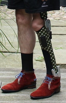
- Non-operative. A KAFO holding the knee in a few degrees (about 5-10) of flexion blocks the damaging hyperextension and is often definitive; correcting a coexisting equinus (AFO or heel raise) removes the back-thrust.
- Operative. For a disabling fixed deformity, a proximal tibial or supracondylar femoral flexion (extension-blocking) osteotomy restores a few degrees of flexion and a stable stance; the equinus must be corrected at the same sitting, and residual quadriceps weakness (or a substituting KAFO) still sets the functional ceiling.
Q: How do you manage a fixed genu recurvatum in a polio survivor? A: It is driven by quadriceps insufficiency (the patient locks the knee in hyperextension) plus a fixed equinus thrusting the tibia back. Manage with a KAFO holding a few degrees of flexion, or - if disabling and fixed - a proximal tibial/supracondylar flexion osteotomy; either way you MUST correct the coexisting equinus, or the recurvatum recurs.
Management
The Three Surgical Principles
The whole of polio reconstruction reduces to three repeatable principles, applied in a logical order:
- Correct the deformity (make the joint supple and the segment aligned): soft-tissue releases for fixed contracture, then osteotomy for residual bony/rotational malalignment.
- Stabilise the unstable (provide a stable, plantigrade base): arthrodesis for a flail joint or to create a stable foot/foundation before any transfer.
- Balance the muscles (restore a working couple): tendon transfer of an expendable, powerful donor onto the deficient action - done LAST, onto a corrected, stable limb.
Golden rule: Never balance a deformed or unstable limb. Correct and stabilise first, then transfer.
Across a whole limb these are often staged or combined; in adults, "limited surgery + external fixation" can correct multiple deformities efficiently.
Complications
- Progressive fixed contracture and joint deformity.
- Limb-length discrepancy and pelvic obliquity.
- Paralytic hip subluxation/dislocation and paralytic scoliosis.
- Osteoporosis of the affected limb with fragility fractures (low bone density, reduced muscle pull, falls).
- Genu recurvatum and degenerative joint disease from chronic abnormal loading.
- Recurrence/relapse of deformity, especially when operating before skeletal maturity or on an unbalanced limb.
- Iatrogenic calcaneus foot after over-lengthening the tendo-Achilles in a weak triceps surae.
- Failed/under-powered tendon transfer (donor too weak, transfer onto an unstable base).
- Implant difficulty: Narrow osteopenic canal, valgus neck and increased anteversion complicate nailing of femoral fractures; fixed-angle plates and sliding hip screws are often preferred, with an emphasis on constructs permitting early weight-bearing.
- New weakness, abnormal fatigue, muscle and joint pain, cold intolerance, and (less commonly) dysphagia or respiratory decline.
- Loss of previously stable function and increasing dependence on orthoses/aids.
Clinical Relevance and Exam Application
- It is the cleanest teaching model of flaccid (LMN) paralysis with intact sensation, so it tests whether you can localise a lesion from the examination alone.
- It forces you to articulate the three principles - correct, stabilise, balance - in the right order. Getting the order wrong (for example, transferring onto a deformed foot) is the classic candidate error.
- It overlaps with high-yield neighbours: tendon transfer principles (also peripheral nerve injury, CMT), neuromuscular scoliosis, paralytic hip, and limb reconstruction / external fixation.
- "Polio is a pure lower motor neuron disease with normal sensation - that is how I separate it from spina bifida."
- "I would delay reconstruction until the residual phase (around two years) once recovery has plateaued."
- "My principles are to correct deformity, stabilise the joint, and only then balance with a transfer."
- "A tendon transfer needs a supple joint, a stable base, and a grade 4-5 expendable donor."
- "New weakness decades later is post-polio syndrome - a diagnosis of exclusion."
Guidelines, Registries and Global Practice
Global epidemiology:
- Eradication trajectory: Wild poliovirus cases have fallen by more than 99% since 1988 (Global Polio Eradication Initiative). Wild type 2 and type 3 are certified eradicated; wild type 1 persists in a small number of endemic regions, with sporadic vaccine-derived (cVDPV) outbreaks elsewhere.
- Surviving cohort: Millions of polio survivors remain worldwide; large surgical databases (for example, tens of thousands of sequelae cases in single high-volume centres) confirm that an ageing survivor population will need orthopaedic care for decades.
- Geographic burden: Untreated, neglected residual deformity is concentrated in low- and middle-income settings; high-income settings now mainly manage ageing survivors and post-polio syndrome.
- Typical Presentation
- Ageing survivors; post-polio syndrome; fragility fractures
- Emphasis
- PPS clinics, energy conservation, orthotic review, fracture care with early weight-bearing
- Typical Presentation
- Neglected fixed deformity in young adults
- Emphasis
- Staged reconstruction: correct, stabilise, balance; external fixation for complex multilevel deformity
- Typical Presentation
- Prevention of new paralytic cases
- Emphasis
- Sustained immunisation (OPV/IPV) and surveillance to prevent wild and vaccine-derived polio
- WHO / Global Polio Eradication Initiative: Drives immunisation, surveillance and the endgame strategy - the upstream prevention that determines future caseload.
- March of Dimes post-polio criteria: The widely cited clinical framework for diagnosing post-polio syndrome (prior paralytic polio, stable interval, new persistent weakness, exclusion of alternatives).
- Rehabilitation principles: International rehabilitation guidance for PPS centres on energy conservation, individualised exercise that avoids overuse, orthotic optimisation, and multidisciplinary symptom management.
- No dedicated international polio-sequelae implant registry exists; outcome data come from large single-centre surgical series and national rehabilitation cohorts rather than arthroplasty registries.
Controversies and Areas of Uncertainty
- Hamstring-to-patella transfer for quadriceps palsy: Can restore active knee extension in selected, well-balanced limbs, but risks creating a flexion deformity or recurvatum and has variable, often modest, active power - patient selection is everything.
- Joint preservation vs arthrodesis: In the ageing survivor with degenerative change, when to fuse a flail joint versus continue bracing is individualised; there are no high-level trials.
- Arthroplasty in polio limbs: Hip and knee replacement can relieve pain but face instability (abductor/quadriceps insufficiency) and fixation challenges in osteopenic, deformed bone; evidence is limited to small series.
- Optimal management of PPS: No disease-modifying therapy is proven; the balance between activity (to avoid disuse) and rest (to avoid overuse) remains pragmatic and individualised.
Viva Scenarios
Clinical Decision Scenarios
Practise clinical reasoning and management decisions out loud
“How would you assess and manage this patient?”
Core Concept
- LMN: flaccid, areflexic, wasted
- Sensation NORMAL (key clue)
- Asymmetric, patchy paralysis
- Static nerve lesion, progressive MSK deformity
Timing & Phases
- Acute: support, prevent contracture
- Convalescent: recovery up to ~2 yrs
- Residual: reconstruct after plateau
- Late: post-polio syndrome
Three Principles (in order)
- 1. Correct deformity (release, osteotomy)
- 2. Stabilise joint (arthrodesis/brace)
- 3. Balance muscles (tendon transfer LAST)
- Donor: grade 4-5, loses ~1 grade
Classic Pitfalls
- Calcaneus foot from over-lengthening TA
- Transfer onto unstable/deformed base
- Operating before recovery plateau
- Calling new weakness PPS without exclusion
Evidence Base
Supracondylar Femoral Extension Osteotomy for Knee Flexion Deformity
- Fractional hamstring lengthening plus supracondylar femoral extension osteotomy was used for polio knee flexion deformity (mean deformity 65 degrees).
- At a mean 15.5-year follow-up, 22 knees achieved full extension and 26 had only a minor extension lag (-1 to -10 degrees).
- All patients were subsequently braceable in long leg orthoses with no neurovascular complications.
- Femoral shortening was recommended in severe deformities to relax the neurovascular structures.
Limited Surgery + External Fixation in Older Polio Survivors (Large Series)
- Drawn from a database of 23,310 polio sequelae cases - 629 were over 41 years old, underlining the huge ageing survivor cohort.
- 57 middle/older patients treated with limited orthopaedic surgery plus external fixation (Ilizarov or combined fixator).
- Procedures combined Achilles lengthening, supracondylar osteotomy, knee-flexion release and selective arthrodesis.
- Excellent/good results in 75% by a deformity-correction standard, with mostly minor complications (pin-track infection).