A COL2A1 Type II Collagenopathy
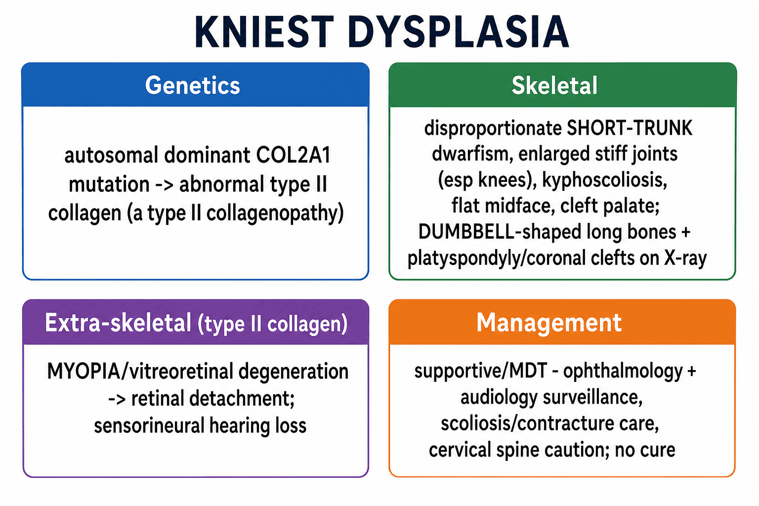
- Kniest dysplasia is an AUTOSOMAL DOMINANT skeletal dysplasia caused by a mutation in COL2A1, making it a TYPE II COLLAGENOPATHY - type II collagen is the principal collagen of cartilage, the vitreous body of the eye, the nucleus pulposus and the inner ear, which explains the combination of skeletal, ocular and auditory features.
- The SKELETAL phenotype is a disproportionate SHORT-TRUNK dwarfism with ENLARGED, STIFF joints (especially the knees) and contractures, kyphoscoliosis, a flat midface with a depressed nasal bridge, and often a CLEFT PALATE; radiographs show 'DUMBBELL'-shaped long bones (broad metaphyses), platyspondyly and coronal clefts of the vertebrae, with the histological 'Swiss-cheese' cartilage of large chondrocyte lacunae.
- The EXTRA-SKELETAL features arise because type II collagen is also in the eye and ear: high MYOPIA with vitreoretinal degeneration carries a significant risk of RETINAL DETACHMENT (requiring ophthalmology surveillance), and there is sensorineural and/or conductive HEARING LOSS - so eye and ear assessment is an essential part of care.
- Kniest dysplasia sits within the COL2A1 SPECTRUM of type II collagenopathies, which ranges (in decreasing severity) from achondrogenesis type II and hypochondrogenesis (often lethal) through spondyloepiphyseal dysplasia congenita (SEDC) and Kniest dysplasia to the milder STICKLER syndrome - radiologically it is in the 'Kniest-Stickler' group characterised by dumbbell tubular-bone deformities.
- Important ORTHOPAEDIC/ANAESTHETIC considerations include progressive kyphoscoliosis and joint contractures, early osteoarthritis, and the possibility of CERVICAL-SPINE instability (odontoid/atlantoaxial issues seen in type II collagenopathies), which - together with the midface/airway features - make anaesthesia hazardous and warrant cervical-spine assessment before any procedure.
- There is NO cure; management is SUPPORTIVE and MULTIDISCIPLINARY - regular OPHTHALMOLOGY surveillance (to prevent/treat retinal detachment), AUDIOLOGY, management of scoliosis and joint contractures, cleft palate repair, respiratory monitoring, cervical-spine assessment, and genetic counselling for the autosomal dominant inheritance.
- “Kniest dysplasia = AD COL2A1 TYPE II collagenopathy: short-trunk dwarfism, enlarged stiff joints, dumbbell long bones, platyspondyly/coronal clefts.
- “Type II collagen is in eye + ear -> high MYOPIA/RETINAL DETACHMENT and hearing loss (surveillance needed).
- “Part of the COL2A1 spectrum (achondrogenesis II -> SEDC -> Kniest -> Stickler). No cure; supportive MDT; beware cervical-spine instability/anaesthesia.
AD COL2A1 disorder: short-trunk dwarfism, enlarged stiff joints, kyphoscoliosis, dumbbell long bones and platyspondyly.
Type II collagen is in the eye and ear -> myopia/vitreoretinal degeneration with RETINAL DETACHMENT and hearing loss - surveillance is essential.
Genetics, Phenotype & Spectrum
Kniest dysplasia results from an autosomal dominant mutation in COL2A1, the gene for type II collagen - the dominant collagen of cartilage, the vitreous of the eye, the nucleus pulposus and the inner ear. This single shared collagen explains why the disorder combines a skeletal phenotype with eye and ear disease. The skeleton shows a disproportionate short-trunk dwarfism with enlarged, stiff joints (especially the knees), contractures, kyphoscoliosis, a flat midface and often a cleft palate; radiographs show 'dumbbell' long bones, platyspondyly and coronal vertebral clefts, with characteristic 'Swiss-cheese' cartilage histology. Kniest dysplasia is part of the COL2A1 spectrum (achondrogenesis type II -> hypochondrogenesis -> SEDC -> Kniest -> Stickler, in decreasing severity), and radiologically belongs to the 'Kniest-Stickler' group with dumbbell tubular-bone deformities.
Clinical Care & Management
There is no cure, so management is supportive and multidisciplinary:
- Ophthalmology surveillance: regular review for high myopia and vitreoretinal degeneration to detect and treat (or prevent) RETINAL DETACHMENT - a major preventable cause of visual loss.
- Audiology: monitor and manage sensorineural/conductive hearing loss.
- Orthopaedic care: manage scoliosis and joint contractures, anticipate early osteoarthritis, and address limb/joint problems; assess and protect the cervical spine (instability/odontoid issues).
- Airway/anaesthesia: midface hypoplasia, cleft palate and possible cervical instability make airway management and anaesthesia hazardous - plan carefully and avoid forced neck manipulation.
- Other: cleft palate repair, respiratory monitoring, and genetic counselling (autosomal dominant).
Two points are easy to miss and important: first, because Kniest dysplasia is a type II collagenopathy, the patient has high myopia and vitreoretinal degeneration with a real risk of RETINAL DETACHMENT, so lifelong ophthalmology surveillance is essential to preserve vision; second, like other type II collagenopathies it can involve the upper cervical spine with odontoid/atlantoaxial instability, which - together with the midface and airway features - makes anaesthesia and any forced neck movement hazardous, so the cervical spine should be assessed before procedures. Recognising Kniest as a systemic collagen disorder rather than a purely skeletal one is the key to safe, comprehensive care.
Evidence & Key Studies
Radiologic features of type II and type XI collagenopathies (Kniest-Stickler group)
- COL2A1 pathogenic variants cause abnormal type II collagen and a group of skeletal dysplasias (type II collagenopathies), commonly associated with vitreoretinal degeneration and hearing impairment.
- These divide radiologically into the SEDC group (pear-shaped vertebrae) and the KNIEST-STICKLER group, characterised by disordered tubular bone growth producing 'dumbbell' deformities.
- Kniest dysplasia belongs to the Kniest-Stickler group; recognising the radiographic pattern guides diagnosis and multidisciplinary care.
COL2A1 spectrum disorders (Stickler, Kniest, achondrogenesis type II)
- Heterozygous COL2A1 defects cause a continuous spectrum of disorders affecting cartilage and bone, including Kniest dysplasia, achondrogenesis type II and Stickler syndrome type 1.
- These collagenopathies share ocular, auditory, musculoskeletal and orofacial manifestations.
- The shared COL2A1 basis explains the overlapping skeletal and extra-skeletal (eye/ear) features across the spectrum.
According to PubMed, the COL2A1/type-II-collagenopathy basis, the 'dumbbell' Kniest-Stickler radiographic group and the association with vitreoretinal degeneration and hearing impairment come from the cited Handa review, and the COL2A1 spectrum (Kniest, achondrogenesis type II, Stickler) with its shared ocular/auditory/ musculoskeletal features from the cited Savasta report. The short-trunk phenotype, the histological 'Swiss- cheese' cartilage, the cervical-spine/anaesthetic considerations and the supportive multidisciplinary management are standard, well-established teaching. (See also our Skeletal Dysplasias, Spondyloepiphyseal Dysplasia and Stickler Syndrome topics.)
Clinical Decision Scenarios
Practise clinical reasoning and management decisions out loud
“What is Kniest dysplasia, and why does it affect the eyes and ears as well as the skeleton?”
“What are the key management and anaesthetic considerations in Kniest dysplasia?”
Mnemonics & Memory Aids
KNIEST
Hook:KNIEST: COL2A1 short-trunk dysplasia with stiff joints, eye and ear disease, dumbbell bones.
COL2 SPECTRUM
Hook:COL2A1 spectrum, severe to mild: Achondrogenesis II - SEDC - Kniest - Stickler.
Genetics
- Autosomal dominant COL2A1 mutation -> abnormal type II collagen
- Type II collagen: cartilage, vitreous, nucleus pulposus, inner ear
- Part of COL2A1 spectrum (achondrogenesis II - SEDC - Kniest - Stickler)
Skeletal phenotype
- Disproportionate short-trunk dwarfism; enlarged stiff joints (knees), contractures
- Kyphoscoliosis, flat midface, cleft palate
- Radiographs: dumbbell long bones, platyspondyly, coronal vertebral clefts; 'Swiss-cheese' cartilage
Extra-skeletal
- High myopia / vitreoretinal degeneration -> RETINAL DETACHMENT (surveillance)
- Sensorineural/conductive hearing loss
- Recognise as a systemic collagen disorder
Management & caution
- No cure; supportive MDT (ophthalmology + audiology surveillance)
- Scoliosis/contracture/early-OA care; cleft repair; respiratory monitoring
- Cervical-spine instability + midface -> hazardous anaesthesia (assess spine); genetic counselling