Congenital Cervical Fusion | Segmentation Failure | Classic Triad
- Definition: Congenital fusion of at least two cervical vertebrae due to failure of segmentation.
- Classic Triad: Low posterior hairline, Short neck, Limited cervical ROM (Triad seen in only less than 50% of patients).
- Associations (Hensinger, 50 patients): scoliosis in over half is the commonest, ahead of renal anomalies in about a third; Sprengel's deformity 21/50 (42%), hearing impairment 15/50 (30%), synkinesia 9/50 (18%), congenital heart disease 7/50 (14%). Being a referral series these are likely upper estimates.
- Instability: The risk is at the hyper-mobile segment adjacent to the fusion. Concern for catastrophic injury.
- Screening: Mandatory Renal US and Cardiac Check. Flexion/Extension views for stability.
- “The most consistent physical finding is Limited Cervical ROM (not the full triad).
- “Radiculopathy/myelopathy occurs in multi-level patterns (Samartzis Type II and III), not single blocks; Type I tends to cause axial neck pain.
- “Think 'VACTERL' - Klippel-Feil overlaps this spectrum of segmentation/formation defects.
- “Avoid collision sports with multi-level or high (C1-C2) fusions or any demonstrated instability.
Overview and Epidemiology
Klippel-Feil syndrome (KFS) is congenital fusion of two or more cervical vertebrae, the result of a failure of normal segmentation of the cervical somites during the 3rd to 8th weeks of gestation. The block itself is immobile; the trouble arises at the segments left mobile beside it, and in what else was affected at the same time.
Who. Prevalence is estimated at 1 in 40,000 to 42,000 births, and is likely under-reported because asymptomatic cases exist. There is a slight female predominance (60%). Most cases are sporadic; autosomal dominant and autosomal recessive forms exist, with GDF6, GDF3 and MEOX1 mutations identified.
The classic triad. The textbook description has three parts:
- Low posterior hairline
- Short neck
- Limited cervical range of motion
Fewer than 50% of patients have all three, so the absence of the triad excludes nothing. Limited cervical range of motion is the most consistent single sign.
Natural history. Many patients with limited fusion (Type I) are asymptomatic and live normal lives. Symptomatic patients present with neck pain, radiculopathy or myelopathy, usually in the 2nd or 3rd decade, as the overdriven segments adjacent to the block degenerate.
Associations. KFS overlaps the VACTERL spectrum of segmentation and formation defects, and the diagnosis obliges a deliberate search for what else is affected. Hensinger's series of 50 patients established the constellation, and because it was a referral series its frequencies are likely upper estimates. Scoliosis is the commonest association, ahead of renal anomalies.
- Incidence
- Over 50% - the commonest association, ahead of renal
- Pathology
- Congenital and/or compensatory curves
- Action
- Whole-spine radiograph at diagnosis and through growth
- Incidence
- Roughly a third; higher in referred and syndromic cohorts
- Pathology
- Congenital high scapula, often with an omovertebral bone
- Action
- Examine the scapula; X-ray shoulder girdle
- Incidence
- 25-30% (about a third in Hensinger's series)
- Pathology
- Agenesis, Horseshoe kidney, ectopic kidney
- Action
- Renal Ultrasound
- Incidence
- About 15% (7 of 50 in Hensinger's series)
- Pathology
- VSD, ASD
- Action
- Echocardiogram
- Incidence
- 30% (15 of 50 in Hensinger's series)
- Pathology
- Sensorineural or Conductive Loss
- Action
- Audiology Refer
- Incidence
- About 20% (synkinesia in 9 of 50 in Hensinger's series)
- Pathology
- Synkinesis (Mirror movements), Diastematomyelia
- Action
- MRI Spine
Pathophysiology and Mechanisms
Embryology. The vertebral column forms from the sclerotomes, and dividing them into segments requires precise regulation through the Notch signalling pathway. KFS is a failure of segmentation, to be contrasted with the hemivertebra, which is a failure of formation.
Which levels. The patterns to know:
- C2-C3 - most common; often autosomal dominant
- C5-C6 - second most common; autosomal recessive association (MEOX1)
- Occipitocervical - rare but dangerous
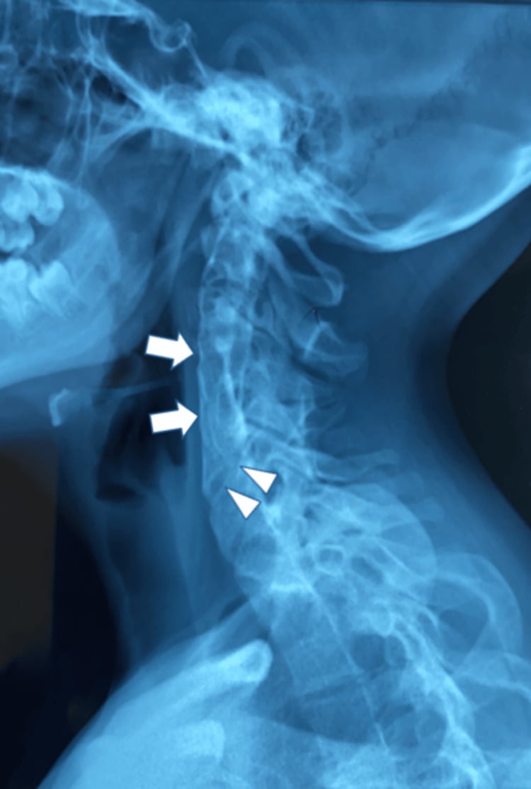
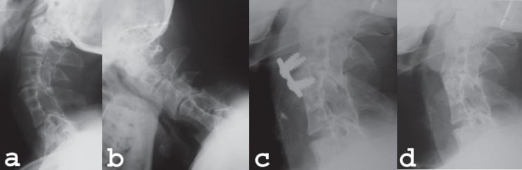
Biomechanics of the block. The fused segment is immobile, so the level above or below must compensate to maintain head motion, and that level experiences increased stress and range of motion. This hypermobility can lead to ligamentous laxity and frank instability. On flexion-extension views the segment moves excessively compared with normal, the appearance called wash-boarding.
Classification
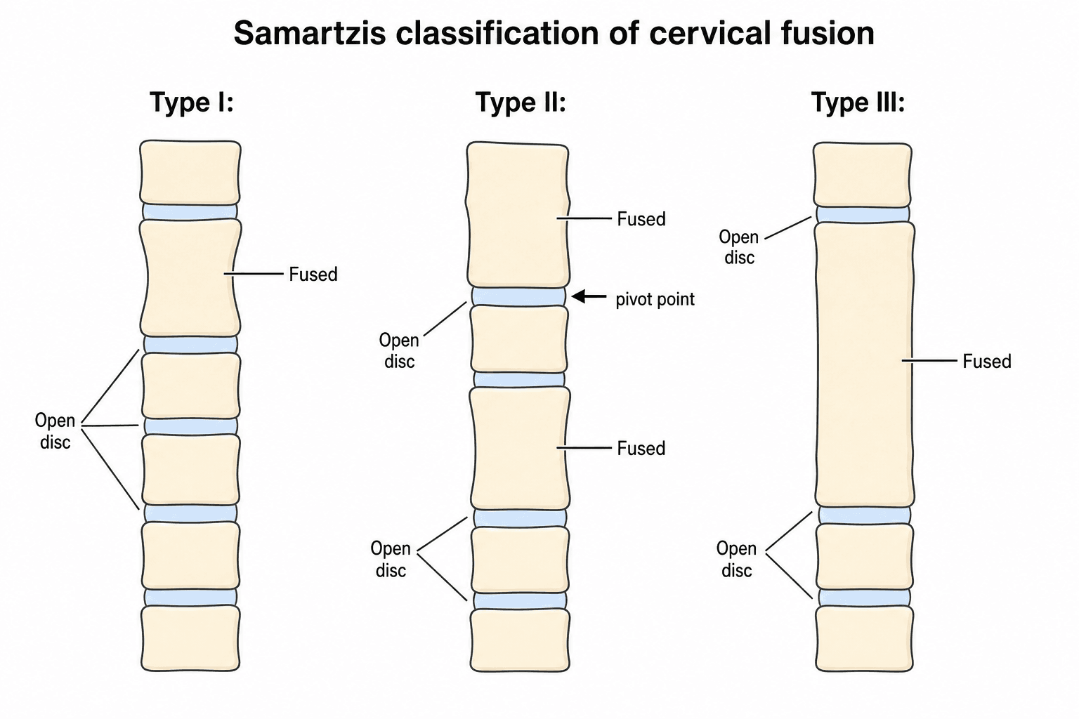
Samartzis's classification is the one to use, because the pattern of fusion predicts who gets into trouble. Feil's 1919 scheme is historical and less useful for prognosis.
- Pattern
- Single congenital block vertebra
- Example
- Risk
- Low; usually asymptomatic
- Pattern
- Multiple non-contiguous fused segments
- Example
- C2-3 fused and C5-6 fused, with an open intercalated segment
- Risk
- The intercalated open segment becomes a "pivot point" bearing concentrated stress, the classic biomechanical substrate for hypermobility and adjacent-level degeneration
- Pattern
- Multiple contiguous fused segments
- Example
- C2-3-4-5 fused into one block
- Risk
- A long stiff lever; tends to show the full clinical triad and, in the Samartzis series, carried the highest rate of radiculopathy/myelopathy
In Samartzis's own cohort, radiculopathy and myelopathy arose in Type II and Type III patients and never in Type I, which presented with axial neck pain. Both multi-level patterns warrant surveillance; do not reassure on the basis of a stable-looking film alone.
The Craniovertebral Junction: Occipitalization and Basilar Invagination
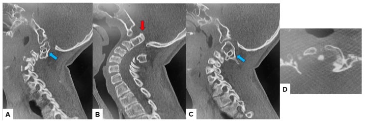
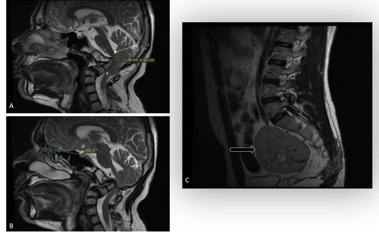
The most dangerous fusion pattern in KFS sits at the top of the spine. Occipitalization (atlanto-occipital assimilation), congenital fusion of the atlas to the occiput, concentrates motion at C1-C2 and opens the door to two linked problems.
Atlantoaxial instability. With the occiput-C1 segment fused, the C1-C2 joint becomes the compensating mobile level and can become unstable, threatening the cord at the cervicomedullary junction.
Basilar invagination. Progressive superior migration of the odontoid through the foramen magnum can compress the brainstem and upper cord and produce myelopathy, sleep apnoea or sudden death. The risk rises with the number of fused segments: four or more fused levels confer roughly a sevenfold risk of significant superior odontoid migration (more than about 4.5 mm), and occipitalization is an independent predictor.
What to do about it. In a long or high fusion, deliberately assess the craniovertebral junction for instability and invagination, because it is the lesion most likely to be catastrophic. Flexion-extension views test reducibility and occult instability, and superior odontoid migration is measured rather than described qualitatively. CT defines the bony lesion behind an abnormal dynamic study; review the canal, the facets and the C1-C2 relationships in every plane before deciding whether decompression, reduction and fixation are required. MRI supplies the neural-axis answer that CT cannot, so image the entire craniovertebral junction and cord for Chiari malformation, compression, syringomyelia and myelomalacia before treating the visible block vertebra.
Clinical Assessment
History. Parents notice a short neck or asymmetry, or that the child turns the whole body rather than the head. Ask about neck pain, which is mechanical, and radicular symptoms from nerve compression, and about weakness or clumsiness, which are signs of myelopathy.
Inspection. Look for:
- A short neck, the head appearing to sit on the shoulders
- A low hairline
- A webbed neck (pterygium colli), to be differentiated from Turner syndrome
- Torticollis or facial asymmetry
- Sprengel's deformity, a high scapula
Range of motion. Rotation and lateral bending are usually restricted; flexion and extension may be preserved if segments are open.
Neurology. Test reflexes, strength and sensation. Hyperreflexia or a positive Hoffmann's sign indicates myelopathy. Mirror movements (synkinesis), involuntary movement of one hand when the other moves, indicate failure of decussation of the corticospinal tracts.
Screening. You must examine for the other anomalies: listen for a heart murmur and check for scoliosis.
Investigations
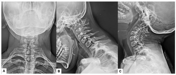
Plain radiographs. Three static views and a dynamic pair:
- AP
- Lateral
- Open-mouth odontoid
- Flexion and extension laterals, which are critical
The flexion-extension views are read for instability, translation greater than 3.5 mm or angulation greater than 11 degrees, and for hypermobility at the adjacent segments. The static films show the wasp waist, a narrowing at the fused disc space, with loss of disc height and fusion of the posterior elements (facets and laminae).
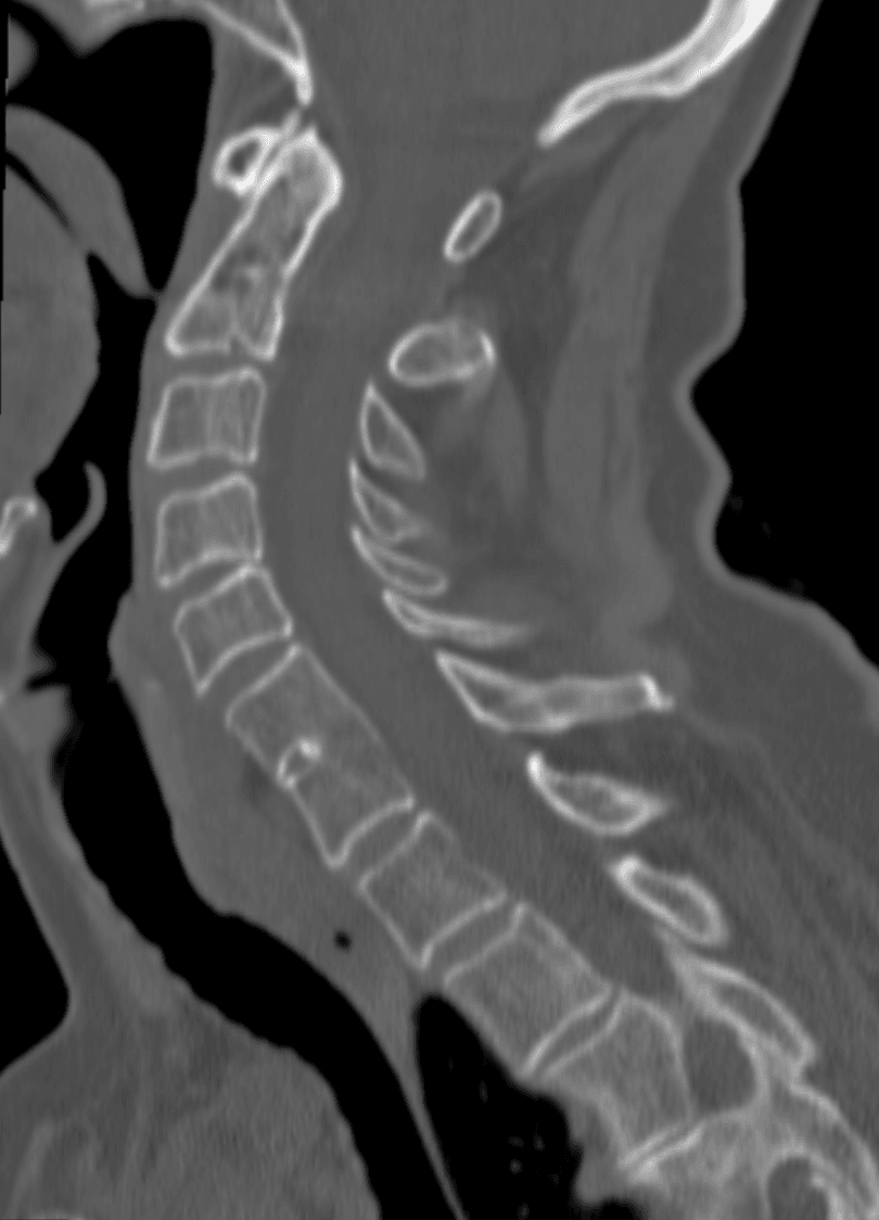
CT is best for bony architecture. It defines the extent of fusion, anterior versus posterior, and is the planning study before surgery.
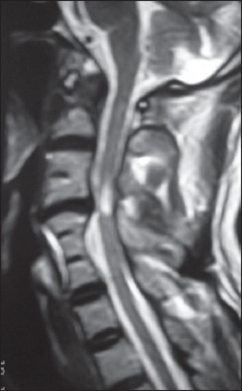
MRI of the whole spine is mandatory if there are neurological signs or before surgery. It assesses cord compression from stenosis at the adjacent levels and screens for Chiari malformation, syringomyelia and diastematomyelia.
Renal ultrasound screens for agenesis and the other renal anomalies.
Managing the Associated Sprengel's Deformity
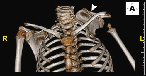
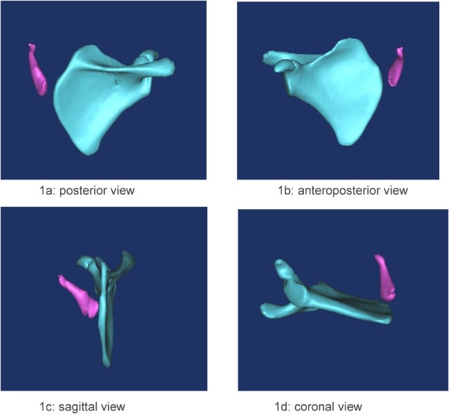
Sprengel's deformity, a congenitally high, hypoplastic, medially rotated scapula from failure of scapular descent, is often the visible clue to the diagnosis, and it deserves its own assessment and plan. The quoted frequency varies with how the cohort was assembled: Hensinger's referral series found it in 21 of 50 patients (42%), while figures nearer a quarter to a third are commonly cited from other series, so quote "roughly a third, higher in referred and syndromic cohorts" rather than a single number.
Grading. Cavendish grades clinical severity from Grade 1, very mild and invisible when dressed, to Grade 4, severe, with the superomedial scapular angle near the occiput and neck webbing.
The omovertebral bone. In a substantial minority a fibrous, cartilaginous or bony bar tethers the superomedial scapula to a lower cervical spinous process or lamina. It restricts abduction and must be excised when the scapula is released.
Surgery. Repositioning is for function and cosmesis and is usually done around age 3 to 8 years, when remodelling and functional gain are greatest. The Woodward procedure detaches the medial parascapular muscle origins and reinserts them more inferiorly; the Green procedure is an extraperiosteal muscle detachment with cable traction. Either is combined with excision of the omovertebral bone and resection of the prominent superomedial scapular angle.
The hazard. Bringing the scapula down risks brachial plexus traction injury. Many surgeons add a clavicular osteotomy (morcellation) before repositioning to reduce this risk during caudal translation, and excising the omovertebral bone and superomedial angle lets the scapula descend without over-stretching the plexus.
Differential Diagnosis
A short, stiff neck or congenital cervical fusion has several mimics. The key is to distinguish a true segmentation failure from acquired fusion and from syndromic short-neck appearances.
- Key feature
- Congenital failure of segmentation of 2+ cervical vertebrae
- Discriminator from KFS
- 'Wasp-waist' block vertebra, fused facets/posterior elements present from birth
- Key feature
- Acquired apophyseal joint ankylosis, hypoplastic vertebrae
- Discriminator from KFS
- Inflammatory history, gradual onset, normal disc height early; ankylosis develops over time
- Key feature
- Webbed neck, short stature, gonadal dysgenesis
- Discriminator from KFS
- Karyotype 45,X; cervical vertebrae are NOT fused
- Key feature
- Hemifacial microsomia, epibulbar dermoids, ear anomalies
- Discriminator from KFS
- Craniofacial asymmetry dominates; vertebral fusion may coexist (overlap spectrum)
- Key feature
- KFS + Duane retraction + sensorineural deafness
- Discriminator from KFS
- Triad with abducens/ocular involvement; almost exclusively female
- Key feature
- Acquired bony bridging
- Discriminator from KFS
- Disc space preserved or scarred; clinical history of infection or prior surgery
Management Algorithm
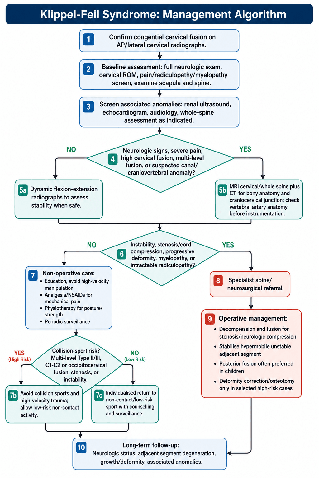
Non-operative care is the mainstay for most patients, Type I and stable Type III. Regular follow-up monitors for symptoms of degeneration. Physiotherapy maintains range of motion and strength but avoids aggressive manipulation, which risks injury, and NSAIDs treat mechanical neck pain. Always counsel on the importance of avoiding high-velocity trauma.
Sport. Collision sports are avoided, per the Torg guidelines, in any of:
- Multi-level fusion (Type II or III)
- C1-C2 involvement
- Stenosis
- Demonstrated instability
Swimming, cycling and non-contact sports are allowed.
Operative indications. Surgery is reserved for instability or neurological compression:
- Progressive myelopathy
- Intractable radiculopathy
- Demonstrated instability
- Significant deformity (rarely for cosmesis alone)
Procedures. Decompression and fusion treats instability or stenosis, and the segment fused is the hypermobile adjacent one. Posterior fusion is often preferred in children to avoid an effect on anterior growth. Deformity correction by osteotomy is high risk.
Surgical Technique
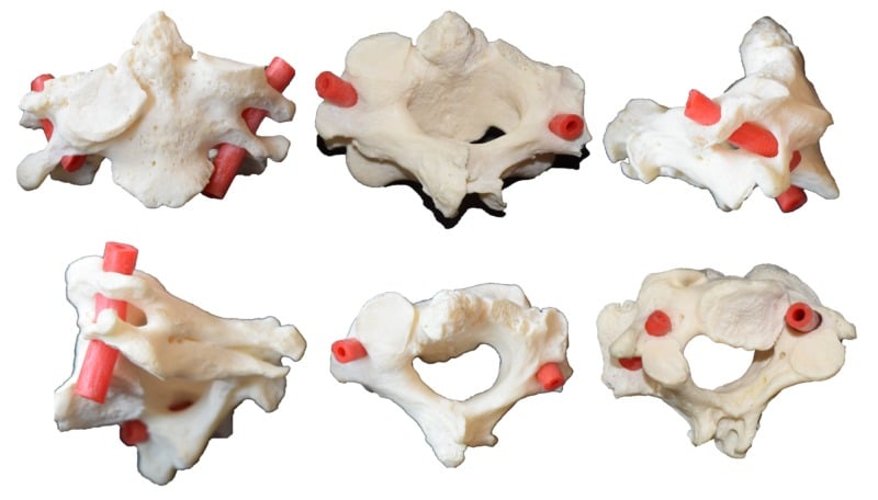
Before the incision. Anomalous vertebral arteries are common in KFS. The artery normally enters the transverse foramen at C6; in KFS it often enters higher, at C4 or C5, so pre-operative CTA or MRA is recommended to map both arteries, and they are always screened before any screw is placed. Limited neck extension means a difficult airway is anticipated; intubate with a fibreoptic scope.
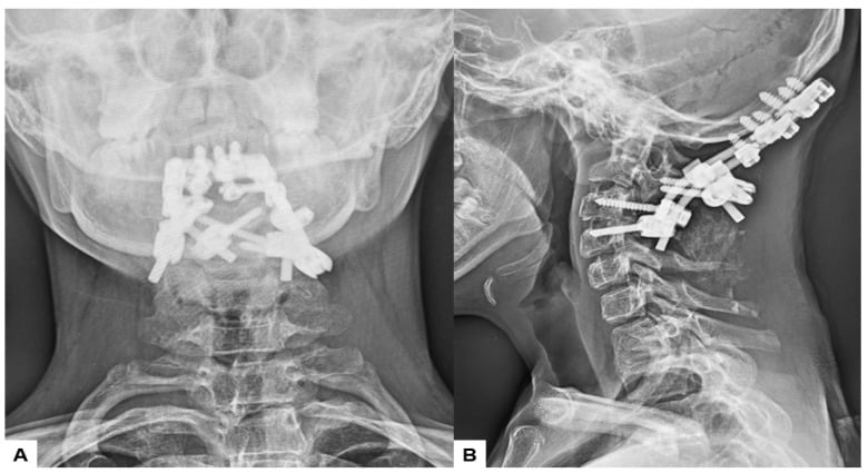
Posterior cervical fusion is the standard for stabilising an unstable segment. The patient is prone in Mayfield tongs or a halo, and neuromonitoring (SSEP and MEP) is essential. A midline posterior approach with subperiosteal dissection exposes the levels; expose carefully, with the anomalous arteries in mind.
Fixation and graft:
- Lateral mass screws at C3-C6
- Pedicle screws at C2, C7 and T1
- Wiring, the older technique, is less rigid
- Iliac crest autograft or allograft
Complications
- Risk Level
- High
- prevention
- Abnormal anatomy + Stenosis. Use Monitoring.
- Risk Level
- Moderate
- prevention
- Pre-op CTA to identify anomalous course.
- Risk Level
- Very High
- prevention
- Fusing one level stresses the next. Inevitable long-term.
- Risk Level
- Low-Mod
- prevention
- Rigid fixation required.
Postoperative Care
Fusion Protocol
- Neuro-checks.
- Collar (Rigid Miami J or Halo depending on fixation).
- Mobilise.
- X-ray check.
- Maintain collar.
- No lifting greater than 5lbs.
- Wean collar if fused.
- Isometrics.
- Lifestyle: Permanent restriction from contact sports often advised if fusion is long.
Outcomes
Prognosis follows the Samartzis type described in the classification section, and is further modified by the associated anomalies. Life expectancy is generally normal unless severe cardiac or renal anomalies are present.
Living with the block. Open segments bear increased stress and degenerate early, so accelerated adjacent segment disease is a long-term concern. Patients with stable spines who avoid contact sports generally have good function, and jobs involving heavy lifting or neck strain may need activity modification.
After surgery. Fusion rates for posterior cervical fusion are generally high, greater than 90%, with good pain relief and neurological improvement in appropriately selected patients. Adjacent segment disease can progress despite a successful fusion, and revision may be required for pseudarthrosis or adjacent-level pathology.
The team. Optimal management needs orthopaedics, paediatrics, cardiology, nephrology and genetics working together to address the full spectrum of associated anomalies.
Guidelines, Registries & Global Practice
Global epidemiology
- Estimated prevalence approximately 1 in 40,000-42,000 live births, with under-reporting because many single-level (Type I) blocks are asymptomatic and found incidentally.
- Slight female predominance reported across series. Most cases are sporadic; familial autosomal dominant (often C2-C3, GDF6) and autosomal recessive (often C5-C6, MEOX1) forms are described.
- KFS overlaps the VACTERL spectrum and syndromes such as fetal alcohol syndrome, Goldenhar (oculo-auriculo-vertebral) and Wildervanck syndrome.
Side-by-side guidance (no single-country frame)
- Focus
- Collision-sport clearance
- Practical recommendation
- Single sub-C3 fusion with full ROM, no stenosis/instability = generally permitted; multi-level, C1-C2 or unstable patterns = relative/absolute contraindication
- Focus
- Screening + surveillance
- Practical recommendation
- Renal ultrasound and echocardiography at diagnosis; dynamic flexion-extension films for stability; MRI if any neurologic sign
- Focus
- MDT pathway
- Practical recommendation
- Tertiary paediatric spine referral; audiology and renal/cardiac screening; counsel on activity restriction
- Focus
- Craniocervical risk
- Practical recommendation
- Heightened vigilance for occipitalization, basilar invagination and superior odontoid migration in long fusions
Registry context
- There is no disease-specific KFS implant registry; KFS is rare and surgery is uncommon. Cervical instrumentation outcomes are extrapolated from national spine and arthroplasty registries and from paediatric deformity databases rather than KFS-specific data.
High- vs limited-resource practice variation
- Well-resourced settings: routine MRI of the whole neuraxis, CT angiography/MRA for vertebral artery mapping, intra-operative neuromonitoring, and panel genetic testing (GDF6, GDF3, MEOX1, RIPPLY2).
- Limited-resource settings: diagnosis often rests on plain radiographs and a careful clinical examination; renal ultrasound (cheap, high-yield) remains the single most valuable screening test; advanced imaging and genetics may be unavailable, raising the importance of clinical screening for cardiac murmurs, hearing loss and scoliosis.
Controversies & Areas of Uncertainty
Which fusion pattern is truly "most dangerous"? Classic teaching emphasises the Type II intercalated open segment as the biomechanical risk for instability. However, the Samartzis 2006 cohort found radiculopathy/myelopathy in both Type II and Type III, with Type III carrying the highest neurologic rate. The honest answer for examiners: neurologic risk lives in multi-level patterns (Type II and III), and a single radiograph cannot fully predict an individual's trajectory.
Sports clearance thresholds are consensus, not trial-based. The Torg framework is built from registry data and expert opinion; there is no randomised evidence defining a safe level of collision-sport exposure in KFS. Practice therefore errs toward caution, particularly for high (C1-C2) or long fusions.
Prophylactic stabilisation is not established. There is no high-level evidence supporting prophylactic fusion of an asymptomatic hypermobile adjacent segment. Surgery remains reserved for instability, progressive deformity, or neurologic compromise.
Genetic heterogeneity. GDF6, GDF3, MEOX1 and RIPPLY2 explain only a minority of cases; many patients have no identified mutation, and newer panels implicate additional candidate genes. Genotype-phenotype correlation remains weak, limiting prognostic use of genetics.
Optimal screening cadence is undefined. While renal ultrasound and echocardiography at diagnosis are widely accepted, the frequency and duration of subsequent cervical surveillance imaging are not standardised across societies.
MCQ Practice Points
Q: What is the most common associated anomaly in KFS? A: Musculoskeletal (Scoliosis, Sprengel's). Extraspinal: Renal (30%).
Q: Which Samartzis types develop radiculopathy/myelopathy? A: Type II and Type III - never Type I (which causes axial neck pain). In the Samartzis 2006 series, Type III carried the highest rate of radiculopathy/myelopathy, while Type II's intercalated open segment is the classic source of hypermobility/instability.
Q: What is the Torg Ratio significance? A: Used to assess cervical stenosis. Ratio of canal diameter to vertebral body diameter. Less than 0.8 indicates significant stenosis.
Q: What is the 'Wasp-Waist' sign involved in KFS? A: It refers to the narrowing of the vertebral body at the level of the fused disc space, seen on AP/Lateral X-rays. A classic radiographic sign of congenital fusion.
Q: What is the significance of synkinesis (mirror movements)? A: It indicates a failure of decussation of the corticospinal tracts. Often seen in KFS and other midline defects.
Exam Viva Scenarios
Practise clinical reasoning and management decisions out loud
“Describe your approach to this patient.”
“Classify this lesion and predict the natural history.”
“What is the connection between this and the cervical fusion?”
Classification (Samartzis)
- Type I: Single Block (axial pain, C2-3 most common)
- Type II: Non-Contiguous (pivot-segment instability)
- Type III: Contiguous (full triad; highest neuro risk)
- Neuro symptoms only in Type II/III, never Type I
Triad (less than 50%)
- Short Neck (Head on shoulders)
- Low Posterior Hairline
- Limited ROM (Most consistent)
- Webbed Neck (Pterygium)
Associations (VACTERL)
- Renal (30%) - URGENT US
- Cardiac (15%) - Echo required
- Sprengel's - roughly a third (42% in Hensinger) - omovertebral bone
- Hearing Loss (30%) - Audiology
- Scoliosis (60%)
Management
- Observe (Type I / Asymptomatic)
- Modify Activity (No Rugby)
- Fusion (Instability greater than 3.5mm)
- Decompression (Myelopathy)
- Osteotomy (Rare, High Risk)
Evidence Base
Samartzis Radiographic Classification and Symptom Risk
- Single-institution radiographic and clinical review of 28 KFS patients (mean clinical follow-up 8.5 years).
- Defined Type I (single fused segment, 25%), Type II (multiple non-contiguous, 50%) and Type III (multiple contiguous, 25%).
- 64% had no cervical complaints; axial neck symptoms were most strongly associated with Type I.
- Radiculopathy and myelopathy occurred only in Type II and Type III patients, with Type III carrying the highest risk of radiculopathy/myelopathy.
- Mean age at onset of cervical symptoms was 11.9 years; symptomatic neurologic cases diagnosed at a mean of 17.9 years.
Associated Anomalies - the Original Constellation
- Classic series of 50 KFS patients establishing the systemic anomaly constellation.
- Fewer than half had the complete classic clinical triad.
- More than half had scoliosis; approximately one-third had renal anomalies.
- Sprengel's deformity in 21, hearing impairment in 15, synkinesis in 9 and congenital heart disease in 7 patients.
Return-to-Play Guidelines for Cervical Anomalies
- Consensus management guidelines built from over 1,200 cervical lesions in the National Football Head and Neck Injury Registry.
- Stratifies congenital, developmental and post-injury cervical lesions into no, relative, or absolute contraindication to collision sport.
- A single-level fusion below C3 with full motion and no stenosis or instability is generally no contraindication.
- Multi-level fusion, fusion involving C1-C2, occipitocervical fusion, instability or stenosis represent relative or absolute contraindications.
Superior Odontoid Migration Risk
- Prospective radiographic and retrospective clinical review of 27 KFS patients (mean age 13.5 years).
- Mean superior odontoid migration 5.0 mm; C2-C3 was the most commonly fused segment (74%).
- Four or more fused segments conferred an approximately sevenfold relative risk of migration greater than 4.5 mm.
- Occipitalization and increasing number of fused segments were the strongest predictors of migration.
Genetic Basis - GDF6 Mutations
- Identified mutations at the GDF6 locus in familial and sporadic KFS, including a recurrent p.Leu289Pro missense change.
- GDF6 is expressed at developing vertebral, carpal and tarsal boundaries and within the adult disc.
- GDF6 knockdown in Xenopus produced anterior axial (vertebral) defects, supporting a causal role.
- Implicates a bone morphogenetic protein family member in segmentation failure.
Genotype-Phenotype and the Symptomatic Type III Pattern
- Cohort of 25 KFS patients with multigene panel sequencing.
- Limited cervical range of motion was the single most common clinical feature (48%).
- Type III (multiple contiguous) patients were significantly more likely to manifest short neck and limited ROM than Type I or II.
- Novel candidate variants (e.g. COL6A1, COL6A2, CDAN1, GLI3, FLNB) detected in 8 of 25 patients, broadening the known mutational spectrum
- THE NEGATIVE RESULT IS THE STRIKING ONE: no pathogenic mutation was found in ANY of the five previously reported KFS genes - GDF6, MEOX1, GDF3, MYO18B or RIPPLY2 - including the GDF6 locus carded immediately above