Pediatric Spine | Vertebral Anomalies | Early Onset | VACTERL Association
- Definition: Spinal deformity caused by abnormal vertebral development in utero (first 6 weeks)
- Worst Prognosis: Unilateral unsegmented bar with contralateral hemivertebra (5-10 degrees progression per year)
- Associations: 55% have at least one extraspinal defect - Intraspinal 37%, CARDIAC 26% (more common than renal, and often quoted far too low), Urogenital 21%
- MRI Mandatory: Must screen for intraspinal anomalies (tethered cord, syrinx, diastematomyelia) before surgery
- Renal Ultrasound: Mandatory screening for all patients
- “Unilateral Bar + Contralateral Hemivertebra = 'Malignant' curve (rapid progression)
- “Always check kidneys (Renal US) and heart (Echo)
- “Thoracic Insufficiency Syndrome is the main mortality risk in early onset severe curves
- “Growth potential: T1-S1 grows 10cm from age 0-5, and 10cm from 10-maturity. Fusing too early creates short trunk.
Overview and Epidemiology
Congenital scoliosis is a lateral curvature of the spine caused by vertebral malformations present at birth. Unlike adolescent idiopathic scoliosis the deformity is structural from the outset and often rigid, and unlike neuromuscular scoliosis the problem is in the bone rather than the muscle. Where it presents as a severe curve in a small child it becomes one of the early-onset scoliosis problems, with the growth-friendly options set out in VEPTR and growing rods.
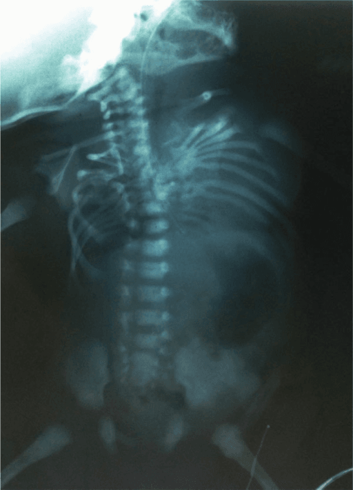
Who. The incidence is 0.5-1 per 1000 live births, slightly more girls than boys. Most cases are sporadic with no strong genetic pattern, though familial clusters exist, and maternal diabetes and valproic acid exposure are risk factors.
Natural history. Without treatment 75% progress, and how fast depends on the balance of growth across the anomaly: the type of defect, whether it is unilateral (unilateral anomalies progress faster), its location (thoracolumbar curves progress most), an intraspinal tether (which worsens progression) and the rapid growth phases, the first five years and the adolescent spurt. The life-threatening complication of a severe early-onset deformity is thoracic insufficiency syndrome (TIS), the inability of the thorax to support normal respiration or lung growth.
Pathophysiology and Mechanisms
Embryology. The axial skeleton forms in the mesenchymal stage, weeks 4-6. Somites form along the notochord and then re-segment: the caudal half of one somite fuses with the cranial half of the next to form a vertebra. A failure of formation leaves a hemivertebra or wedge; a failure of segmentation leaves an unsegmented bar or block.
Formation failure is not only a hemivertebra. A butterfly vertebra is a midline sagittal cleft, two alae of body that never fused. It is a formation defect, but a balanced one, so it does not act as a growing wedge. The hemivertebra is the unbalanced cousin that drives the curve.
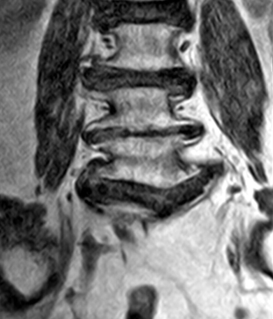
Growth potential. T1-S1 lengthens by 10cm in the first five years and by another 10cm in the adolescent growth spurt. Fusing the thoracic spine early stops that vertical growth, and the loss is lung volume rather than correction: adequate adult pulmonary function needs roughly 18-22cm of T1-T12 height.
Most candidates answer this from intuition and get it wrong: "fusing too early damages the lungs" is only half right. In the reference study of 28 children fused before age nine and followed to a mean of 14.6 years (PMID 18519321), the damage was severe: mean FVC 57.8% of age-matched normal, mean FEV1 54.7%, FVC below 50% in 12 of the 28, and two needing respiratory support. What predicted it was not age.
- Age at fusion: no significant correlation, and nor did length of follow-up
- Extent of thoracic spine fused: significant (p = 0.01, r = -0.46); the average child had 58.7% of the thoracic spine fused
- Proximal level: the strongest signal (p = 0.0004, r = 0.62). Eight of twelve children whose fusion started at T1 or T2 had an FVC below 50%, against four of sixteen whose fusion began below T2
So the operative variables are how much and how high, not how young. A short caudal thoracic fusion in a three-year-old is a very different proposition from a long fusion starting at T1. The often-quoted "don't fuse more than four segments" threshold does not come from this study, which reports extent as a proportion of the thoracic spine rather than a segment count; treat four segments as a conventional rule of thumb and, if pressed, quote the proportion and the proximal level.
The Genetics: TBX6 Compound Inheritance
Most congenital scoliosis is sporadic, but a recognised genetic mechanism now explains a meaningful minority and is increasingly examined.
The compound-inheritance model. In roughly 10% of patients with isolated congenital scoliosis (vertebral malformation without a syndrome) the cause is a rare loss-of-function TBX6 variant, commonly a 16p11.2 microdeletion, on one allele combined with a common hypomorphic TBX6 risk haplotype on the other. This compound heterozygosity drops TBX6 dosage below the threshold needed for normal somite segmentation, and because neither allele alone causes disease the inheritance looks sporadic.
The pathway. TBX6 acts in the Notch / segmentation-clock machinery that drives somitogenesis, the same pathway whose disruption (DLL3, MESP2, LFNG, HES7) causes spondylocostal dysostosis.
Why it matters. Identifying a 16p11.2 deletion has counselling implications, since it also carries developmental and neuropsychiatric associations, and it frames congenital scoliosis as a dosage-sensitive developmental disorder rather than a purely random event.
Classification
Winter's classification is read from the radiograph and sorts anomalies by what went wrong in embryology: failure of formation (Type I), failure of segmentation (Type II) or both (Type III). Its point is urgency. The type tells you how the growth is unbalanced, and therefore how fast the curve is likely to move.
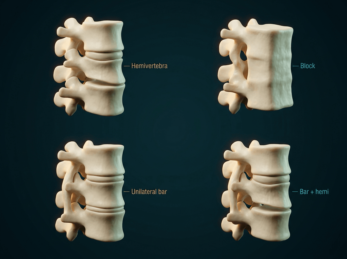
Type I, failure of formation. The subtypes differ in how the hemivertebra is joined to its neighbours:
- Fully segmented hemivertebra: a disc space above and below, so it acts as a growing wedge. Rapid progression
- Semi-segmented: fused to one adjacent vertebra, still a wedge
- Incarcerated hemivertebra: only the pedicles are deformed and the body is set into the spine. Slow progression
- Wedge vertebra: partial formation failure
Type II, failure of segmentation. A block vertebra is a bilateral failure: stable, a short segment, and usually needs no treatment. A unilateral unsegmented bar is the dangerous one, because the fused side cannot grow while the open side does, and the spine bends towards the bar.
Type III, mixed. A combination of both. A unilateral bar with a contralateral hemivertebra is the "malignant curve": the bar tethers one side while the hemivertebra pushes growth on the other, and progression is rapid and relentless.
- Progression Rate
- Less than 1° / year
- Severity
- Benign
- Management Action
- Observation
- Progression Rate
- 1-2° / year
- Severity
- Mild
- Management Action
- Observation
- Progression Rate
- 2-5° / year
- Severity
- Moderate
- Management Action
- Early surgery often required
- Progression Rate
- 5-6° / year
- Severity
- Severe
- Management Action
- Fusion of bar typically needed
- Progression Rate
- 5-10° / year
- Severity
- Malignant
- Management Action
- URGENT Surgery
Two cautions about those numbers. They are thoracic rates from McMaster's series, and the same anomaly progresses more slowly in the lumbar spine, so the site modifies the figure. And no abstract is indexed for the 1982 paper, so the per-year rates rest on the full text and decades of secondary citation: the ranking is not in doubt, the decimal precision is. In a viva, give the order and the approximate rate and say "in McMaster's series" rather than implying a figure you could look up in seconds.
What triggers the operation is the observed rate, not the predicted one. The classification tells you which child to watch closely and how alarmed to be at presentation; serial radiographs at consistent intervals tell you what the curve is really doing. A fully segmented hemivertebra behaving like a bar is treated like a bar.
Clinical Assessment
Look. Start with the skin over the spine (the alert below), then shoulder balance and trunk shift, the face for asymmetry (Goldenhar) and the limbs for a radial clubhand (VACTERL).
Always look at the skin over the spine. A hairy patch (faun's beard) or deep dimple is highly suggestive of underlying spinal dysraphism (spina bifida occulta, diastematomyelia); a sinus or haemangioma is another cutaneous marker.
Feel. A detailed lower-limb neurological examination and the reflexes: abdominal reflexes are often absent in an extensive syrinx, and ankle clonus suggests tethering.
Ask and listen. Auscultate the heart for murmurs, ask about renal problems or urinary tract infections, and confirm anal patency.
Investigations
Radiographs. Whole-spine films are the first line. Count the vertebrae, identify bars and hemivertebrae, and measure the Cobb angle.
MRI of the whole spine is mandatory before any corrective surgery, looking for a tethered cord, syrinx, diastematomyelia or Chiari malformation. See tethered cord syndrome and Chiari malformation for the other neural-axis findings the scan is looking for.
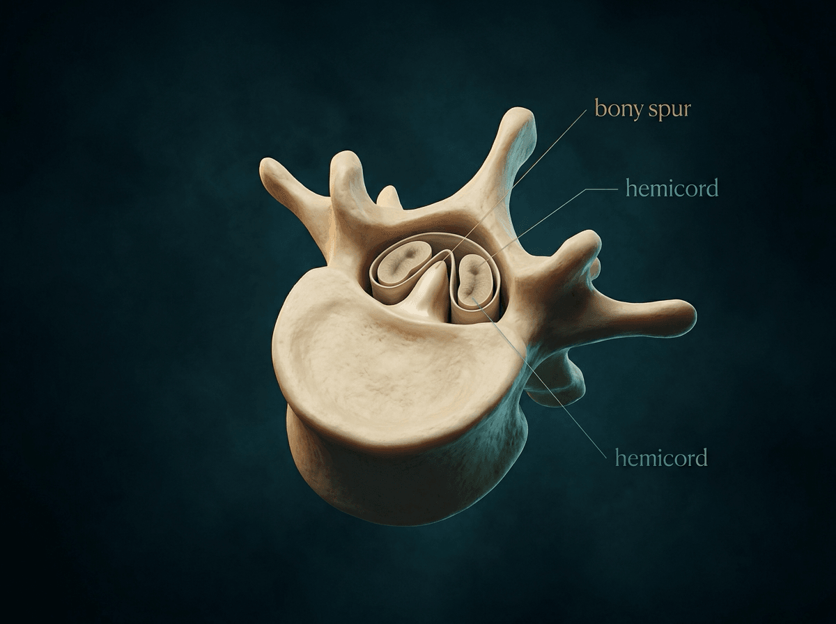
The spur must go first. If a bony spur is present you cannot distract or straighten the spine until it is excised. Distraction against a spur splits the cord, a neurological catastrophe.
Everyone gets the MRI, so this does not change who is scanned. It changes how hard you look, how alarmed you are before correction and what you say to the family, and it is the level of detail that separates a good viva answer from a recited percentage. In the 126-patient reference series (PMID 12394903), intraspinal anomalies were significantly concentrated:
- Congenital kyphosis carried significantly more intraspinal anomalies than scoliosis (p = 0.0048)
- Mixed and segmentation defects carried more than pure formation failures
- Cervical and thoracic hemivertebrae had significantly more intraspinal anomalies than lumbar hemivertebrae (p = 0.0253), so the level of the anomaly predicts the neural-axis risk
- Extraspinal organ defects were likewise commonest in mixed defects (p = 0.002)
The one-line version: a high thoracic or cervical anomaly, a segmentation or mixed defect, or any congenital kyphosis is the child in whom you should genuinely expect to find something on the MRI, not the isolated lumbar hemivertebra.
Renal ultrasound and echocardiography are mandatory for every child; the yield, and what each is looking for, is set out under associated anomalies.
CT with 3D reconstruction is for planning once you have already decided to operate; the MRI still comes first for the cord. It shows the complex bony anatomy and the pedicle morphology for screws: count the pedicles, see whether there is a disc above and below the hemivertebra (fully segmented) and plan the screws on it.
Differential Diagnosis
The task is to separate congenital scoliosis, a structural vertebral malformation present at birth, from the other early-onset curves, because management and prognosis differ sharply. The defining feature is an identifiable vertebral anomaly (hemivertebra, bar, block) on plain films, not simply an early or rigid curve.
- Distinguishing feature
- Discrete vertebral anomaly (hemivertebra/bar/block) on radiograph
- Response to bracing
- Poor (rigid, structural)
- Pitfall
- Missing intraspinal anomaly on MRI before correction
- Distinguishing feature
- Normal vertebral morphology; rib-vertebra angle difference (Mehta) guides risk
- Response to bracing
- Many resolve; serial casting effective if progressive
- Pitfall
- Labelling a progressive curve idiopathic without ruling out a subtle hemivertebra
- Distinguishing feature
- Long C-shaped curve, pelvic obliquity, underlying CP/SMA/DMD
- Response to bracing
- Limited; postural seating support
- Pitfall
- Attributing a syndromic child's curve solely to vertebral anomaly
- Distinguishing feature
- Cafe-au-lait spots, short sharp dystrophic curve, rib penciling
- Response to bracing
- Poor in dystrophic curves
- Pitfall
- Treating an NF-1 dystrophic curve as routine congenital deformity
- Distinguishing feature
- Multiple, generalized segmentation defects with rib fusions (TBX6/DLL3)
- Response to bracing
- Poor; high TIS risk
- Pitfall
- Calling diffuse multi-level segmentation a single congenital curve
Management Algorithm
Observation. Non-progressive curves, block vertebrae and balanced deformities are watched, with a radiograph every 6-12 months during growth. Observation requires strict radiographic intervals.
Bracing. Its role is limited to none. Congenital curves are rigid and do not respond to a brace. The one exception is the compensatory curve above or below the congenital defect, which is occasionally braced to delay surgery.
Surgical strategy. Treatment is a race between curve control and lung growth, and the strategy depends on how much growth remains. The goals are to stop progression, first of all, while keeping the thoracic height that lung volume needs, preventing thoracic insufficiency syndrome and restoring the sagittal profile.
The options:
- In situ fusion: fuse the curve as it is to stop progression. Best for an unsegmented bar (posterior fusion of the bar). Safe and simple, but no correction, and a short segment
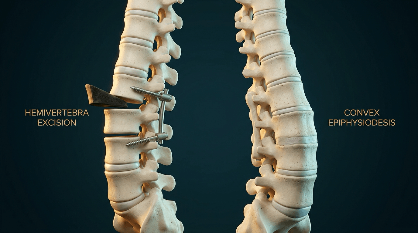
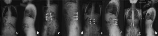
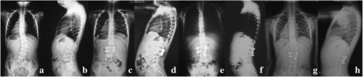
- Hemivertebra excision: remove the wedge to straighten the spine, the gold standard for an L4 or L5 hemivertebra causing trunk shift. Posterior-only (eggshell) or combined anterior and posterior. Excellent correction with a short fusion, but technically demanding
- Growth-friendly surgery for the long early-onset curve and for thoracic insufficiency, not for a single lumbar hemivertebra:
- Growing rods: distract the spine and lengthen every 6 months
- MAGEC rods: magnetically controlled lengthening, with no trips to theatre
- VEPTR: rib-to-rib expansion thoracoplasty for TIS and fused ribs
- Shilla: guided growth, the rods sliding at their ends
- Spinal osteotomy, vertebral column resection (VCR), for the severe rigid deformity in the older child
Surgical Technique
Convex epiphysiodesis for the unilateral bar. The logic is in the anatomy: the bar has no growth plates, the contralateral side has normal ones, and the difference is what bends the spine. Stopping growth on the convex (healthy) side matches it to the concave (bar) side, and early intervention prevents the severe deformity. Through a posterior or combined anterior and posterior approach:
- Ablate the growth plates and discs on the convex side
- Apply a fusion mass
- Expect arrest of progression, and perhaps some auto-correction
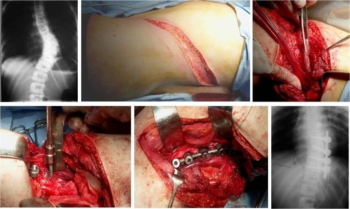
Hemivertebra excision. The indication is a lumbosacral hemivertebra causing a severe list. The logic is Ruf and Harms': take the wedge, compress a short segment and leave the rest of the spine to grow.
Steps
- Posterior approach, subperiosteal dissection
- Identify the hemivertebra, often by its widely separated pedicles
- Pedicle screws in the vertebrae above and below
- No screw in the hemivertebra
- Remove the lamina and facet of the hemivertebra
- Isolate the nerve root (crucial)
- Decancellation: drill out the body of the hemivertebra through the pedicle (transpedicular subtractive osteotomy)
- Remove the discs above and below
- Compress the osteotomy site
- Watch the nerve root for kinking
- Confirm that lordosis is restored
Complications
- Risk Factor
- Osteotomy, Diastematomyelia distraction
- Management
- IOM monitoring. Wake up test. Remove spurs first.
- Risk Factor
- Posterior fusion with open anterior growth plates in young child
- Management
- Add anterior fusion or exclude growth plates.
- Risk Factor
- Long revisions, syndromes
- Management
- Washout, antibiotics.
- Risk Factor
- Growing rods, poor bone stock
- Management
- Revision.
- Risk Factor
- Proximal Junctional Kyphosis
- Management
- Extension of fusion.
Postoperative Care and Rehabilitation
Protocol
- ICU observation (neuromonitoring considerations)
- Pain control
- Mobilise as tolerated; no brace usually needed with rigid fixation
- Wound checks
- Return to school
- No impact sports
- X-ray to assess arthrodesis
- Gradually increase activity
- Monitor for crankshaft (if young)
- Monitor for PJK
- Lung function tests
Outcomes
Hemivertebrae. Excision yields excellent correction and balance.
Unsegmented bars. Early convex epiphysiodesis can halt progression but rarely corrects the deformity substantially.
Untreated. The curve can reach severe deformity (greater than 100 degrees), cor pulmonale, and early death from respiratory failure.
Associated Anomalies and Screening
The reference series. Basu, Elsebaie and Noordeen screened 126 consecutive patients with congenital spinal deformity with MRI, echocardiography and renal ultrasound (PMID 12394903). 55% had at least one extraspinal defect: intraspinal anomalies in 37%, cardiac in 26% and urogenital in 21%. Cardiac is more common than urogenital, not less, and is often quoted far too low. The authors concluded that MRI and echocardiography "should be an essential part in the evaluation".
So every child is screened, not the selected few. A quarter of these children have a cardiac defect and auscultation will not find them all, so echocardiography is for every child at presentation, not for the one with a murmur. Renal ultrasound is ordered on diagnosis, and a whole-spine MRI before any corrective surgery. What each test is looking for:
- Intraspinal: diastematomyelia, tethered cord, syrinx, Chiari
- Cardiac: VSD, ASD, tetralogy of Fallot
- Renal: renal agenesis, horseshoe kidney
- VACTERL features on examination: imperforate anus, thumb hypoplasia (radial deficiency)
VACTERLVACTERL Association
Hook:A child with a bad back needs checking from top (TEF) to bottom (Anus) and limbs!
Named associations. Beyond VACTERL, the syndromes worth recognising at the bedside:
- Klippel-Feil syndrome: fusion of cervical vertebrae, with the triad of low hairline, short neck and limited neck movement
- Goldenhar syndrome: hemifacial microsomia, auricular tags and congenital scoliosis
- Diastematomyelia: a split cord malformation in which a bony or fibrous spur divides the cord
The Sagittal Counterpart: Congenital Kyphosis
Congenital vertebral anomalies also deform the spine in the sagittal plane. Congenital kyphosis is graded by the same formation-versus-segmentation logic but carries a distinct and dangerous complication: it is the commonest congenital cause of paraplegia. It also has the highest intraspinal anomaly rate of any congenital spinal deformity (the MRI section below gives the figures), so the two risks compound.
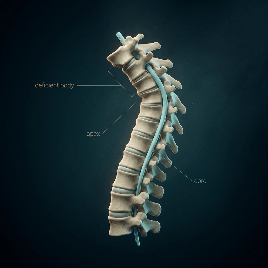
- Anomaly
- Failure of FORMATION (posterior/posterolateral vertebral-body defect)
- Behaviour / risk
- Most progressive AND highest risk of paraplegia - the apex angulates sharply over the cord
- Anomaly
- Failure of SEGMENTATION (anterior unsegmented bar)
- Behaviour / risk
- Slower, more rounded progression; lower (but not zero) paralysis risk
- Anomaly
- Mixed
- Behaviour / risk
- Variable
Why Type I is dangerous. The deficient vertebral body lets the kyphosis angulate acutely at the apex and drapes the cord over the deformity, so a progressive Type I kyphosis can produce a gradual spastic paraparesis. Type II, an anterior bar, is slower and rounder.
Treatment. Early posterior fusion can arrest a small Type I curve in a very young child; an established or larger deformity needs combined anterior and posterior fusion, with anterior decompression if neurology is present. Bracing does not work.
Guidelines, Registries & Global Practice
Global epidemiology:
- Incidence approximately 0.5-1 per 1000 live births worldwide; one of the commonest causes of structural scoliosis presenting in infancy.
- Largely sporadic; environmental contributors (maternal pregestational diabetes, valproate, gestational hypoxia/carbon monoxide in experimental models) and genes affecting somitogenesis (e.g. the Notch/segmentation-clock pathway, TBX6 in spondylocostal/multiple-vertebral-segmentation phenotypes) are implicated.
- Slight female predominance; high rate of associated VACTERL, intraspinal, cardiac and renal anomalies across all populations.
Side-by-side guidance (no single national standard exists for this rare condition; principles are shared):
- Screening emphasis
- Whole-spine MRI, echo and renal US routine at diagnosis; PFTs before/after early surgery
- Treatment philosophy
- Growth-sparing constructs favoured under age 8-10; early short-segment resection for progressive hemivertebrae
- Screening emphasis
- MRI plus cardiac and renal imaging; multidisciplinary tertiary pathway
- Treatment philosophy
- Document progression before fusing; MAGEC and casting in early-onset disease
- Screening emphasis
- MRI mandatory before any correction; CT 3D for bony planning
- Treatment philosophy
- Posterior-only hemivertebra resection well established; VCR reserved for rigid late deformity
- Screening emphasis
- Plain radiographs and clinical screening; MRI/echo where available
- Treatment philosophy
- Later, often larger presentations; in-situ fusion and convex epiphysiodesis when implants/MRI scarce
- There is no implant-survival registry specific to congenital scoliosis the way NJR/AJRR/AOANJRR exist for arthroplasty; growth-friendly implant data come from collaborative early-onset-scoliosis databases (e.g. the Pediatric Spine Study Group / Growing Spine Study Group), which track rod fractures, anchor failure, infection and the law of diminishing returns with repeated lengthening.
- These collaboratives, not national joint registries, are the main source of comparative MAGEC vs traditional growing-rod and VEPTR outcome data.
- High-resource: MRI/echo/renal US at diagnosis, intra-operative neuromonitoring, 3D-CT planning, MAGEC rods to reduce repeat anaesthetics.
- Limited-resource: later presentation with larger rigid curves, restricted MRI access (raising the threshold before any distraction because diastematomyelia may be undetected), and greater reliance on in-situ fusion or convex hemiepiphysiodesis. Where MRI is unavailable, intra-operative neuromonitoring and a low threshold for the Stagnara wake-up test become even more important.
every child with confirmed congenital scoliosis should be managed by a paediatric spine service. A generalist may appropriately initiate screening imaging (whole-spine radiographs, renal ultrasound) but should not undertake correction.
Controversies & Areas of Uncertainty
When to operate on a hemivertebra. Early excision at age 2-4 gives a short fusion and lets the compensatory curves self-correct, but it carries the anaesthetic and neurological risk of operating on tiny pedicles, and some advocate watchful waiting until progression is documented. The balance shifts toward earlier surgery for the fully segmented lumbosacral hemivertebra causing trunk decompensation.
MAGEC versus traditional growing rods. Magnetically controlled rods avoid repeated open lengthenings and anaesthetics, but face metallosis, titanium wear debris, lock-pin failure and the same law of diminishing returns. Whether they truly reduce total complications is still debated, and the question is tracked by early-onset-scoliosis collaboratives rather than by randomised trials.
Prophylactic anterior fusion for crankshaft. Whether routine anterior fusion is needed to prevent crankshaft after posterior fusion in a skeletally immature child is contested; pedicle-screw constructs with a robust posterior arthrodesis may reduce the phenomenon, but not abolish it.
Tethered cord release before correction. Whether an asymptomatic low-lying conus or filum lipoma must be released before deformity correction, or simply monitored, is unsettled. A bony diastematomyelia spur is a different matter: excising it before any distraction is not controversial.
MCQ Practice Points
Q: What is the most common intraspinal anomaly associated with congenital scoliosis? A: Diastematomyelia (Split cord malformation), followed by tethered cord and syrinx.
Q: Rank the following from worst to best prognosis: Block Vertebra, Fully Segmented Hemivertebra, Unilateral Bar + Contralateral Hemi. A: Worst: Bar + Contralateral Hemi. Middle: Fully Segmented Hemi. Best: Block Vertebra.
Q: How many features are needed to diagnose VACTERL association? A: Typically at least 3 of the 6 features (Vertebral, Anal, Cardiac, TE, Renal, Limb).
Q: Why is an MRI mandatory before casting or surgery in congenital scoliosis? A: To rule out intraspinal anomalies (e.g. syrinx, tethering). Correcting the curve stretches the spinal canal; if the cord is tethered, this stretch causes ischemia and paraplegia.
Q: What is the classic triad of Klippel-Feil syndrome? A: 1. Low posterior hairline. 2. Short neck ("Webbed"). 3. Limited cervical range of motion.
Q: A child with scoliosis has ear tags and facial asymmetry. What is the diagnosis? A: Goldenhar Syndrome (Oculo-auriculo-vertebral spectrum).
Clinical Decision Scenarios
Practise clinical reasoning and management decisions out loud
“A newborn baby is noted to have a spinal deformity. X-ray shows a hemivertebra at L1. The parents ask if surgery is needed now.”
“A 6-year-old boy presents with a worsening thoracolumbar curve. X-rays show a unilateral unsegmented bar on the left from T10-L2 with multiple hemivertebrae on the right.”
“You are performing a hemivertebra excision. During the osteotomy, the neuromonitoring signals (MEPs) drop bilaterally.”
VACTERL Checklist
- Vertebral (X-ray)
- Anal (Exam)
- Cardiac (Echo)
- TE (History/Swallow)
- Renal (Ultrasound)
- Limb (Radius exam)
McMaster Prognosis
- Block: Benign
- Wedge: Mild
- Hemi: Moderate
- Bar: Severe
- Bar+Hemi: Malignant
Key Principle
- MRI before Surgery/Bracing
- Renal US AND Echo for everyone - cardiac 26% is MORE common than urogenital 21%, and a murmur will not find them all
- Intraspinal 37%, but concentrated in KYPHOSIS, segmentation/mixed defects, and CERVICAL/THORACIC (not lumbar) hemivertebrae
- Preserve lung volume (TIS risk)
- Lungs are threatened by HOW MUCH thoracic spine and HOW PROXIMAL - fusion starting at T1/T2 is the danger; age at fusion did NOT correlate
- 'Don't fuse more than 4 segments' is a rule of thumb, not a finding from the pulmonary evidence
- Check for Diastematomyelia spur
Evidence Base
Natural History of Congenital Scoliosis
- Seminal study of 251 patients with congenital scoliosis followed over time
- Defined the hierarchy of progression by anomaly type
- Worst prognosis: unilateral unsegmented bar with a contralateral hemivertebra (the 'malignant' curve)
- Best prognosis: block vertebra (essentially non-progressive)
Posterior Hemivertebra Resection (Innovative Technique)
- 21 consecutive young children: posterior-only hemivertebra resection with transpedicular instrumentation
- Mean main-curve Cobb angle improved from 41 degrees pre-op to 14 degrees post-op (maintained at 15 degrees)
- Mean kyphosis improved from 24 degrees to 11 degrees
- Complications limited to one infection, one pedicle fracture, two early wire-instrument failures; no permanent neurological injury
Thoracic Insufficiency Syndrome (TIS)
- Defined TIS as the inability of the thorax to support normal respiration or lung growth
- Described the three-dimensional 'windswept' thoracic deformity of fused ribs plus congenital scoliosis
- Introduced the thumb-excursion test and 'space available for lung' as clinical/radiographic markers
- Treatment goal: acutely increase thoracic volume and maintain it during growth, ideally without spine fusion (rationale for VEPTR)