Control the deformity while preserving spinal, thoracic and pulmonary growth
- Early definitive fusion in a young child stunts thoracic height and lung development — thoracic insufficiency syndrome (Campbell): the inability of the thorax to support normal respiration or lung growth
- Serial Mehta (elongation-derotation-flexion) casting can be curative in progressive infantile idiopathic scoliosis if started young — RVAD greater than 20 degrees and phase 2 rib overlap predict progression (Mehta)
- Law of diminishing returns (Sankar): each successive TGR lengthening yields progressively less length as the spine stiffens and autofuses
- VEPTR was designed by Campbell for thoracic insufficiency syndrome — fused ribs and constricted hemithorax (Jeune, Jarcho-Levin, congenital rib fusion with scoliosis) — via opening-wedge expansion thoracostomy
- Aetiology drives strategy: congenital, neuromuscular, syndromic and idiopathic infantile EOS behave differently and fail differently
- “MCGR avoids repeated anaesthetics but does not avoid revisions — metallosis around the actuator, failure to lengthen and actuator pin fracture are recognised
- “Graduation is a decision, not an event: definitive fusion, in-situ retention of the construct, or removal — each has advocates and trade-offs
- “Outcome measures the examiner wants: T1-T12 and T1-S1 height gain, space-available-for-lung ratio, EOSQ-24 quality of life, complications per patient
Definitive fusion of the young spine arrests T1-T12 growth, producing a short, stiff thorax and restrictive lung disease. Quote Karol precisely, because the usual paraphrase is wrong. In 28 children fused before age NINE and tested at a mean age of 14.6 years, mean FVC was 57.8 per cent of predicted and 12 of 28 were below 50 per cent. Age at fusion did NOT correlate with pulmonary function; what did was the extent fused (p = 0.01) and above all the proximal level — 8 of 12 fused from T1 or T2 had an FVC under 50 per cent, against 4 of 16 starting below T2 (p = 0.0004). So the surgeon-controlled variables are how many levels and how proximal, not the birthday. The entire growth-friendly strategy exists to avoid this.
A progressive infantile idiopathic curve (RVAD greater than 20 degrees, phase 2 rib) in a child under 2 years should have serial derotational casting before any implant — it is the only potentially curative growth-friendly treatment.
Across all systems, unplanned returns to theatre are the rule: anchor pull-out, rod fracture, deep infection, proximal junctional kyphosis, wound breakdown in thin children. Bess reported complication risk rises with each additional procedure — consent families honestly at the outset.
Rib cradles placed too high, or excessive distraction elevating the first rib, can cause brachial plexus traction injury. Neuromonitoring during implantation and lengthening, and anchor placement at the second rib or below, mitigate this.
Rationale: Why Growth Matters More Than Correction
The child with early-onset scoliosis has two coupled problems: a progressive spinal deformity and a growing respiratory system that depends on a growing thorax.
- Spinal growth: T1-S1 gains roughly 2 cm per year from birth to age 5, slows to about 1 cm per year from 5 to 10, then accelerates again in the adolescent spurt (Dimeglio). Around half of final sitting height is achieved by age 5.
- Thoracic growth: the thorax reaches about 30 percent of adult volume by age 5 and about 50 percent by age 10. An adult T1-T12 height of approximately 18 to 22 cm is regarded as the threshold for adequate lung volume; a fused short thorax cannot reach it.
- Lung growth: alveolar multiplication is most rapid in the first 2 years and largely complete by around age 8; thereafter lungs enlarge mainly by alveolar hypertrophy. Deformity control before age 8 therefore protects irreplaceable alveolar development — this is the biological deadline that frames every decision.
- Thoracic insufficiency syndrome (Campbell): the inability of the thorax to support normal respiration or lung growth — seen with fused ribs, constricted hemithorax, severe early scoliosis, and hypoplastic thorax syndromes (Jeune asphyxiating thoracic dystrophy, Jarcho-Levin spondylothoracic dysostosis). The windswept, rotated thorax reduces the space available for lung on the concavity.
Space-available-for-lung (SAL) ratio: on an upright PA radiograph, measure the height of the concave hemithorax from the apex of the most cephalad rib to the centre of the ipsilateral hemidiaphragm, and divide by the same measurement on the convex side. A ratio below approximately 0.8 to 0.9 indicates significant concave hemithorax constriction and supports thorax-based (VEPTR) treatment.
Untreated progressive EOS carries excess cardiorespiratory mortality in adulthood (Pehrsson's Swedish natural history series), which is why observation of a clearly progressive curve is not an option — but neither is early fusion. Hence: control the curve while the spine and thorax keep growing.
Aetiology Groups and How They Change Strategy
- Typical behaviour
- Onset under 3 years; many resolve (RVAD less than 20 degrees, phase 1 rib); progressive curves respond well to casting
- Strategic implications
- Serial Mehta casting first — potentially curative; implants reserved for casting failures or late presentation
- Typical behaviour
- Failure of formation or segmentation; unilateral unsegmented bar with contralateral hemivertebra is the most malignant pattern; often rib fusions
- Strategic implications
- Casting largely ineffective for structural anomalies; hemivertebra excision for isolated anomalies; VEPTR where fused ribs constrict the hemithorax; see the congenital scoliosis topic for anomaly-specific surgery
- Typical behaviour
- Long sweeping collapsing curves, pelvic obliquity, poor bone, poor soft tissue envelope, high infection risk
- Strategic implications
- Distraction constructs frequently extended to pelvis (rib-to-pelvis VEPTR or growing rods with iliac/S2AI anchors); higher wound and infection burden; unit rod definitive constructs at maturity — see the neuromuscular scoliosis topic
- Typical behaviour
- Marfan, neurofibromatosis, skeletal dysplasias, VACTERL; dystrophic curves progress rapidly; anaesthetic and airway comorbidity
- Strategic implications
- Multidisciplinary work-up mandatory; MCGR attractive to reduce anaesthetic exposure; anchor fixation compromised by dysplastic bone (NF1 dural ectasia, rib penciling)
All EOS patients need whole-spine MRI before instrumentation: neural axis anomalies (syrinx, Chiari, tethered cord, diastematomyelia) occur in roughly one in five infantile idiopathic curves and more frequently in congenital scoliosis.
Non-Operative Growth-Friendly Treatment First
- 1Step 1 — Characterise the curve
Infantile idiopathic curve on PA film: measure Cobb angle, rib-vertebra angle difference (RVAD) at the apical vertebra (perpendicular to endplate versus rib neck line, convex angle subtracted from concave), and note rib phase.
RVAD and rib phase separate the curves that resolve from those that will progress.
- 2Step 2 — Observe
RVAD less than 20 degrees, phase 1 rib (no overlap of convex rib head on apical body), curve under 25 degrees.
The majority resolve spontaneously; reassess if progression is documented.
- 3Step 3 — Serial Mehta casting
Documented progression, RVAD greater than 20 degrees or phase 2 rib, age under 2 to 3 years, curve roughly 25 to 60 degrees.
Started before age 2 with curves under approximately 60 degrees, full resolution is achievable (Mehta's series). In non-idiopathic EOS casting rarely cures but is a legitimate delay tactic, buying spinal height and postponing the first implant.
- 4Step 4 — Bracing
Thoraco-lumbo-sacral orthoses hold rather than correct in EOS.
Poorly tolerated and less effective in neuromuscular and syndromic curves. A bridge, not a destination: it defers when, not whether, to instrument.
- 5Step 5 — Flip to surgery
Progression despite casting or bracing beyond approximately 50 to 60 degrees; congenital anomalies with predicted relentless progression; thoracic insufficiency syndrome with declining respiratory function or deteriorating space available for lung.
Proceed to implant selection on anatomy and anaesthetic burden.
- 6Step 6 — Select the system
Fused ribs and a constricted hemithorax push towards VEPTR; the wish to avoid repeated anaesthetics pushes towards magnetically controlled growing rods (MCGR); hyperkyphosis or very small stature limits MCGR through actuator length and rod contouring constraints; neuromuscular pelvic obliquity mandates pelvic anchors.
Traditional growing rods remain the fallback where MCGR geometry cannot be accommodated.
Operative Systems: The Exam Comparison
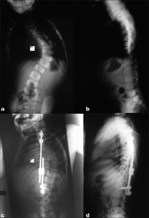
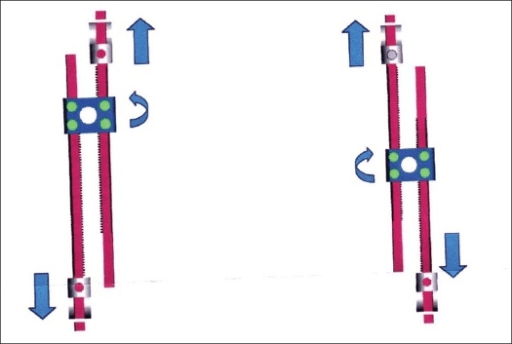
- Working principle and anchors
- Distraction-based. Dual rods with proximal foundation (pedicle screws or hooks, T2-T4) and distal foundation, spanning the curve without fusing intervening levels; tandem connectors
- Lengthening regime
- Open surgical lengthening every 6 months under anaesthesia
- Advantages
- Powerful correction; longest track record; dual rods superior to single (Thompson)
- Characteristic failures
- Repeated anaesthetics and incisions; law of diminishing returns (Sankar, Spine 2011) with progressive stiffening and autofusion; rod fracture; anchor pull-out; infection; proximal junctional kyphosis
- Working principle and anchors
- Distraction-based. Same proximal and distal foundations as TGR, but the rod houses a magnetically driven internal actuator
- Lengthening regime
- Outpatient external remote-controller distraction, typically every 1 to 3 months in small increments; no anaesthetic
- Advantages
- Avoids repeated surgical lengthenings; more frequent, more physiological distraction; reduced family and anaesthetic burden
- Characteristic failures
- Metallosis and titanium debris in surrounding tissue (Teoh); failure to lengthen or rod slippage; actuator pin fracture; MRI compatibility caveats; regulatory scrutiny episodes; planned exchange once actuator stroke is exhausted, plus unplanned revisions
- Working principle and anchors
- Chest-wall based. Vertical expandable prosthetic titanium rib (Campbell); rib-to-rib, rib-to-spine or rib-to-pelvis cradles; opening-wedge thoracostomy for fused ribs
- Lengthening regime
- Surgical expansion every 6 months
- Advantages
- Only device designed for thoracic insufficiency syndrome; treats the thorax and lung volume directly; rib-based constructs spare the spine of anchors
- Characteristic failures
- Rib anchor migration and rib erosion; brachial plexus injury with cephalad anchors; wound breakdown over prominent implants in thin children; modest scoliosis correction versus spine-based systems
- Working principle and anchors
- Guided growth, not distraction. Limited apical fusion with corrective apical screws; proximal and distal non-locked sliding screws let the rods run through as the spine grows
- Lengthening regime
- None scheduled — growth drives elongation along the rods
- Advantages
- Fewest planned operations; apex controlled directly at index surgery
- Characteristic failures
- Metal-on-metal wear debris at sliding interfaces; unpredictable guidance and add-on deformity; rod exchange when growth outruns rod length
- Principle and anchors
- Dual rods; proximal (pedicle screws or hooks, T2-T4) and distal foundations spanning the curve without fusing intervening levels; tandem connectors; surgical lengthening every 6 months
- Advantages
- Powerful correction; long track record; dual rods superior to single (Thompson)
- Key problems
- Repeated anaesthetics and incisions; law of diminishing returns; rod fracture, anchor pull-out, infection, PJK
- Principle and anchors
- Same foundations as TGR but rod contains a magnetically driven actuator; outpatient external remote-controller lengthenings, typically every 1 to 3 months in small increments
- Advantages
- Avoids repeated surgical lengthenings and anaesthetics; more frequent, more physiological distraction; family burden reduced
- Key problems
- Metallosis and tissue titanium debris around actuator (Teoh); failure to lengthen or rod slippage; actuator pin fracture; MRI compatibility caveats; regulatory scrutiny episodes; still needs planned exchanges when actuator stroke is exhausted, plus unplanned revisions
- Principle and anchors
- Vertical expandable prosthetic titanium rib (Campbell); rib-to-rib, rib-to-spine or rib-to-pelvis cradles; opening-wedge expansion thoracostomy for fused ribs; surgical expansions every 6 months
- Advantages
- Only device designed for thoracic insufficiency syndrome; treats the thorax directly; spares the spine of anchors in rib-based constructs
- Key problems
- Rib anchor migration and rib erosion; brachial plexus risk with cephalad anchors; wound breakdown over prominent implants in thin children; modest scoliosis correction compared with spine-based systems
- Principle and anchors
- Limited apical fusion with corrective apical screws; proximal and distal non-locked sliding screws guide growth along the rods
- Advantages
- No scheduled lengthenings — fewer planned operations; apex controlled directly
- Key problems
- Metal-on-metal wear debris at sliding interfaces; unpredictable guidance and add-on deformity; rod exchange when growth outruns rod length
Sankar and colleagues (Spine, 2011; 38 children, five centres) showed that with dual growing rods the T1-S1 length gained per lengthening falls significantly with each successive procedure (p = 0.007) and with time (p = 0.014) — attributed by the authors, as a hypothesis, to autofusion under a rigid immobilising construct. Two figures make it usable. The Cobb angle fell 74° to 36° at the primary implantation and then did not change at all with repeated lengthenings (p = 0.96) — the correction is won once. And the returns diminish but do not reverse: average annual T1-S1 gain was still 1.76 ± 0.71 cm/year. Practical consequences: obtain the best correction at index surgery, do not delay the index procedure expecting to "catch up" later, and use the falling per-lengthening yield to judge when the programme has stopped earning its complication rate.
Operative Technique
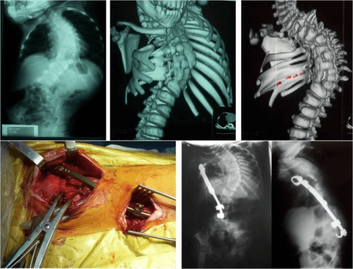
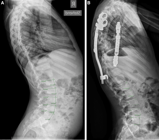
Indication: progressive EOS (typically greater than 50 to 60 degrees) with substantial growth remaining, failed or unsuitable for casting, without fused ribs demanding thorax-based treatment. Contraindication: active infection, insufficient bone stock for foundations, kyphosis beyond actuator tolerance (MCGR), very small stature precluding actuator (MCGR). Why this over alternatives: spine-based distraction gives more powerful curve correction than VEPTR; MCGR over TGR when reducing anaesthetic exposure is a priority.
- Position: prone on a radiolucent table, chest and pelvic bolsters, abdomen free, head in a padded holder or Mayfield in the very small; all pressure points padded.
- Imaging/equipment: fluoroscopy; multimodal neuromonitoring (SSEP and MEP) mandatory; cell salvage in small children; MCGR external remote controller checked and rods templated for actuator length.
- Preparation: prophylactic antibiotics; tranexamic acid per protocol; whole-spine MRI reviewed preoperatively.
- Approach: two limited midline (or paramedian muscle-splitting) incisions over the planned proximal and distal foundations only — do not expose the intervening spine, to limit autofusion.
- Dissection: subperiosteal exposure confined to foundation levels; proximal foundation typically T2-T4 with 4 anchor points (pedicle screws, or hooks/rib anchors in poor bone — hooks or rib-based proximal anchors may reduce PJK by preserving the posterior tension band); distal foundation 2 levels of pedicle screws below the end vertebra; iliac or S2-alar-iliac screws for pelvic obliquity.
- Reduction/Reconstruction: rods contoured to sagittal profile (limit contour near the MCGR actuator — it must remain straight over the actuator segment), tunnelled submuscularly (submuscular passage has lower infection and rod fracture rates than subcutaneous), connected via tandem connectors (TGR) or as single actuator rods (MCGR); gentle distraction with monitoring stable.
- At-risk structures: spinal cord (monitor throughout, especially during distraction), pleura at proximal thoracic anchors, great vessels with mal-directed screws, skin envelope in thin children.
- Closure: layered, watertight; local antibiotic powder used by many units.
- Aftercare: TGR — surgical lengthening every 6 months, day-case where feasible; MCGR — outpatient magnetic lengthenings every 1 to 3 months, small increments, distraction confirmed by ultrasound or radiograph; brace rarely needed with dual rods.
- Pitfalls: exposing the mid-spine (accelerates autofusion); over-distraction causing PJK or neurological signal change — distract to soft endpoint, never force; MCGR actuator placed where kyphosis contour is needed.
- Salvage of failure: rod fracture — exchange; anchor pull-out — augment or extend foundation; deep infection — debridement with implant retention early, staged exchange if chronic; construct exhausted — proceed to graduation strategy.
Complications Across All Systems
- Where it bites
- Proximal rib anchors and at every distraction
- Mechanism
- Traction on the plexus and cord as the construct is lengthened — distraction is the moment of neurological risk
- Prevention
- Multimodal MEP and SSEP monitoring at index surgery and every surgical lengthening; small, frequent increments for unmonitored MCGR outpatient lengthenings
- Salvage
- Release distraction immediately, restore blood pressure, re-check signals before proceeding
- Where it bites
- VEPTR rib cradles, proximal foundation
- Mechanism
- Cradle cuts through rib under repetitive distraction load; thin soft tissue envelope offers no cushion
- Prevention
- Multiple ribs captured, proactive soft tissue planning in thin children, avoid over-distraction
- Salvage
- Revise cradle to a fresh rib level, convert to hooks or spine anchors, extend the foundation
- Where it bites
- Traditional growing rods — commonest at tandem connectors
- Mechanism
- Cyclical loading and stress risers at connectors; subcutaneous placement raises risk (Bess, JBJS Am 2010)
- Prevention
- Dual rods, avoid connectors at the apex or deformity crossing, submuscular rather than subcutaneous placement
- Salvage
- Exchange the fractured rod or connector at the next scheduled lengthening; revise pulled-out anchors
- Where it bites
- Any incision, risk compounding with each reopening
- Mechanism
- Repeated exposure, neuromuscular hosts, poor soft tissue coverage over prominent implants
- Prevention
- Meticulous closure, staged skin planning, minimise unnecessary reopenings
- Salvage
- Early deep: debride and retain implants with targeted antibiotics. Chronic or recurrent: staged removal and exchange, sometimes forcing early graduation to fusion
- Where it bites
- Proximal foundation and the level above
- Mechanism
- Distraction force concentrates at the top of the construct; posterior tension band violated and sagittal contour lost
- Prevention
- Preserve interspinous ligaments and facets above the construct, avoid over-distraction, consider hooks or rib anchors proximally, restore sagittal contour
- Salvage
- Extend the construct proximally, correct the kyphosis, address anchor pull-out
- Where it bites
- MCGR actuator; Shilla sliding interface
- Mechanism
- Wear at the actuator and at gliding metal-on-metal interfaces raises local tissue titanium
- Prevention
- Recognise failure to lengthen or actuator pin fracture early; account for MRI artefact and rod behaviour caveats in surveillance
- Salvage
- Exchange the affected implant, debride stained tissue; note regulatory scrutiny and temporary market suspension of MCGR in some jurisdictions
- Where it bites
- Whole construct over years of distraction
- Mechanism
- Prolonged distraction and immobilisation cause spontaneous fusion; continued anterior growth around a posterior-only fusion produces crankshaft in the very young
- Prevention
- Maintain lengthening intervals, avoid early definitive posterior-only fusion
- Salvage
- Accept reduced late correction, plan osteotomies at graduation, or convert to definitive fusion
The rule, not the exception. Bess (JBJS Am, 2010, Growing Spine Study Group — 140 patients, 897 procedures) found 81 of 140 (58 per cent) had at least one complication. Two dose-response figures are the ones to quote at consent: risk fell 13 per cent for every year of increased age at initiation and rose 24 per cent for every additional procedure. Construct matters too — unplanned procedures for implant problems in 27 per cent of single rods against 10 per cent of dual, and wound complications in 26 per cent of subcutaneous against 10 per cent of submuscular. Consent at the outset must frame this as a multi-year surgical programme, not a single operation.
Distraction forces concentrate at the proximal foundation. Prevention: preserve the posterior tension band and interspinous ligaments above the construct, avoid over-distraction, consider hooks or rib anchors proximally, restore sagittal contour. Management: extend proximally, restore kyphosis correction, address pull-out.
Higher in neuromuscular patients and with each reopening. Early deep infection: debride, retain implants, targeted antibiotics. Chronic/recurrent: staged removal and exchange — sometimes forcing early graduation. Thin children with VEPTR need proactive soft tissue planning.
Prolonged distraction and immobilisation cause spontaneous fusion, limiting late lengthening and final correction. Historically, crankshaft phenomenon (continued anterior growth rotating around a posterior-only fusion) complicated isolated posterior fusion in the very young — one more reason early definitive fusion fails.
Implant-specific additions: TGR — rod fracture (commonest at tandem connectors), anchor pull-out. MCGR — metallosis and elevated tissue titanium around the actuator, failure to lengthen, actuator pin fracture, MRI compatibility caveats (image quality artefact and rod behaviour concerns), and episodes of regulatory scrutiny including temporary market suspension in some jurisdictions. VEPTR — rib cradle migration, rib erosion, brachial plexus traction. Shilla — sliding-interface metal debris, adding-on deformity.
Distraction is the moment of neurological risk. Multimodal monitoring (MEP and SSEP) during index surgery and surgical lengthenings; if signals deteriorate, release distraction immediately, restore blood pressure, and re-check before proceeding. MCGR outpatient lengthenings are unmonitored — hence small, frequent increments.
Outcome Measures
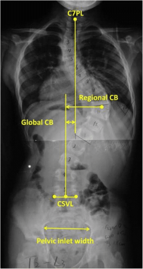
- T1-T12 height: proxy for thoracic and lung volume growth; target trajectory towards 18 to 22 cm at maturity; normal growth about 1.4 cm per year in this age range (Dimeglio) — compare achieved gain against this benchmark.
- T1-S1 height: total spinal growth achieved by the programme.
- Cobb angle: control, not necessarily cure; expect the best correction at index surgery.
- Space-available-for-lung ratio: thorax-specific measure, particularly for VEPTR.
- EOSQ-24: validated parent-reported quality-of-life instrument for EOS — increasingly the headline outcome, since radiographs do not capture the burden of repeated surgery.
- Complications per patient and unplanned returns to theatre: report honestly.
RVAD 20 / Phase 2Predictors of infantile curve progression
Hook:Twenty and two: RVAD over twenty or a phase two rib — treat, do not observe.
CASTGrowth-friendly strategy choice
Hook:CAST — because casting really is the first move.
Lengthening Programme and Surveillance
Growth-friendly programme: index surgery to graduation
Best correction is obtained at implantation — the law of diminishing returns means every subsequent lengthening adds progressively less. Document baseline T1-T12 and T1-S1 length, major Cobb angle, space available for lung (SAL) and EOSQ-24 before leaving theatre.
Performed as a day case where possible; distract to a soft endpoint under neuromonitoring. Each return to theatre carries cumulative infection, wound and anaesthetic risk, so batch procedures and treat every incision as a full aseptic case.
External remote controller in clinic, no anaesthetic. Confirm achieved distraction with ultrasound (avoids repeated radiation) or radiograph. Investigate repeated failure to lengthen: rod slippage, actuator failure or spontaneous autofusion.
MCGR actuator stroke is finite at approximately 28 to 48 mm depending on rod length. Exchange electively once the stroke is consumed rather than waiting for failure, and inspect retrieved implants for metallosis and titanium wear debris.
Serial coronal and sagittal radiographs for Cobb, thoracic height and proximal junctional kyphosis; monitor anchor migration and rod fracture; track EOSQ-24 and respiratory function alongside radiographic gain.
Choose between definitive posterior spinal fusion, retention of the construct in situ, or implant removal — guided by residual growth, curve behaviour, autofusion and family preference.
Best correction is obtained at implantation — the law of diminishing returns means later lengthenings add less. Document T1-T12, T1-S1, Cobb, SAL and EOSQ-24 baseline.
Day-case where possible; distract to soft endpoint under neuromonitoring; each return to theatre adds infection and anaesthetic risk.
External remote controller; confirm achieved distraction with ultrasound (avoids radiation) or radiograph; investigate repeated failure to lengthen (rod slippage, actuator failure, autofusion).
MCGR actuator stroke is finite (approximately 28 to 48 mm depending on rod); exchange when consumed, inspect for metallosis.
Definitive fusion, retention in situ, or removal — see below.
Graduation: What To Do at the End of the Programme
When the child approaches skeletal maturity (or the construct is exhausted and the spine autofused), three options exist:
- Definitive posterior fusion: traditional default — formal exposure, implant exchange to a definitive construct, correction of residual deformity. Reality check: the autofused, stiff, previously distracted spine yields limited additional correction, and complication rates at final fusion are meaningful (Flynn's Growing Spine Study Group data).
- Retention in situ ("graduate without fusion"): leave the mature construct undisturbed if alignment is acceptable and the spine is effectively autofused — avoids a large final operation; risk of late rod fracture and slow curve creep requires surveillance.
- Implant removal without fusion: occasionally chosen for infection or prominence; risk of deformity progression after removal even in apparently stiff spines — least favoured for structural curves.
The decision weighs residual deformity, sagittal balance, autofusion extent, implant integrity, infection history and family priorities. There is no consensus — this is a legitimate "discuss the options" viva answer.
Guidelines, Registries & Global Practice
- Global epidemiology: EOS is uncommon relative to adolescent idiopathic scoliosis but disproportionately morbid; congenital and syndromic causes dominate surgical caseloads worldwide, while presentation is later and curves larger in resource-limited settings where casting infrastructure and device access are constrained.
- Society guidance: the Scoliosis Research Society Growing Spine Committee consensus defines EOS as onset before age 10 and endorses growth-friendly strategies over early fusion; the SRS-endorsed EOS classification (Williams — aetiology, Cobb, kyphosis, progression modifier) standardises communication. POSNA and BSCOS practice statements support serial casting as first-line for progressive infantile idiopathic curves. AAOS and NICE have no dedicated EOS implant guidance; NICE and MHRA involvement has centred on MCGR device safety review, including a period of restricted use pending investigation of metallosis and actuator concerns — candidates should know MCGR has faced genuine regulatory scrutiny.
- Registry and study-group evidence: national arthroplasty-style registries do not capture EOS; the field's registry equivalents are the Growing Spine Study Group and the Children's Spine Study Group / Paediatric Spine Study Group, whose pooled multicentre data underpin most complication and outcome figures quoted above.
- Practice variation by resource setting: MCGR cost and controller availability limit use in many health systems; TGR and Shilla (no scheduled lengthenings) remain workhorses where repeated theatre access or magnetic controllers are unavailable; casting programmes are highly cost-effective and exportable, and several global outreach programmes prioritise establishing EDF casting before implant services.
Controversies & Areas of Uncertainty
- MCGR versus TGR overall benefit: MCGR clearly reduces anaesthetic episodes, but total revision burden, metallosis significance, and cost-effectiveness remain debated; randomised data are sparse.
- Graduation strategy: definitive fusion versus retention in situ versus removal — no consensus; observational data suggest many "graduates" do acceptably without formal final fusion, but selection bias is heavy.
- Optimal proximal anchor: screws versus hooks versus rib anchors for PJK prevention — biomechanically argued, clinically unresolved.
- Distraction frequency: more frequent small MCGR distractions are argued to be more physiological; the ideal interval is unknown.
- How much correction matters: whether aggressive Cobb correction improves pulmonary outcomes beyond height and SAL gains is unproven — thoracic growth, not radiographic cosmesis, is the goal.
MCQ Practice Points
A: Approximately age 8. Alveolar number increases most rapidly in the first 2 years of life; thereafter lung growth is mainly by alveolar enlargement. This is the biological rationale for controlling thoracic deformity early rather than fusing early.
A: Approximately 18 to 22 cm. Karol's work linked thoracic height after early fusion to adult pulmonary function, underpinning growth-friendly strategy.
A: Sankar's observation that each successive traditional growing-rod lengthening yields progressively less T1-S1 length, due to progressive spinal stiffening and autofusion under the construct. Implication: maximise correction at index surgery.
A: Rib-vertebra angle difference greater than 20 degrees at the apical vertebra, and phase 2 rib (convex rib head overlapping the apical vertebral body) — Mehta, 1972.
A: The brachial plexus — from cradles placed too high (first rib) or over-distraction elevating the first rib. Prevention: superior cradle at the second rib or below, upper-limb neuromonitoring, distraction to a soft endpoint.
A: Metallosis — titanium wear debris in periprosthetic tissues around the actuator (Teoh and others), one of the drivers of regulatory scrutiny of the device. It has not been shown to cause systemic harm but should be documented and debrided at exchange.
Exam Viva Scenarios
Practise clinical reasoning and management decisions out loud
“An 18-month-old presents with a 45 degree left thoracic curve. RVAD is 28 degrees and the convex rib head overlaps the apical vertebral body. The parents ask whether she needs rods.”
“A 4-year-old with Jarcho-Levin spondylothoracic dysostosis has multiple fused ribs on the concavity, a 70 degree congenital curve, a space-available-for-lung ratio of 0.7 and recurrent chest infections. Which device and why?”
“A 7-year-old, 3 years into MCGR treatment, has had no measurable length gain on ultrasound over the last three clinic lengthening attempts. Cobb angle is stable. The family ask what is wrong.”
“A 12-year-old girl, post-menarchal, has completed 7 years of dual traditional growing rods. Residual curve is 38 degrees, sagittal profile acceptable, implants intact, spine radiographically stiff on bending films. What now?”
Core numbers
- EOS: onset before age 10 (SRS)
- Alveolar multiplication largely complete by age 8
- T1-T12 target approximately 18 to 22 cm at maturity
- SAL ratio below about 0.8 indicates constricted hemithorax
- RVAD greater than 20 degrees or phase 2 rib predicts infantile curve progression
- TGR/VEPTR lengthened surgically every 6 months; MCGR magnetically every 1 to 3 months
Strategy
- Never early definitive fusion in the young thorax — thoracic insufficiency and restrictive lung disease
- Cast first: Mehta EDF casting potentially curative under age 2 in infantile idiopathic curves
- Whole-spine MRI before any instrumentation
- Fused ribs / constricted hemithorax: VEPTR with expansion thoracostomy
- Anaesthetic-averse or frequent-distraction rationale: MCGR
- No lengthening infrastructure or family logistics: consider Shilla
- Neuromuscular pelvic obliquity: extend distal anchors to pelvis
Technique essentials
- Dual rods, submuscular passage, expose foundations only — spare the mid-spine
- Neuromonitoring at index surgery and every surgical lengthening; release distraction if signals change
- VEPTR superior cradle at second rib or below — brachial plexus protection
- Maximise correction at index surgery — law of diminishing returns (Sankar)
- Preserve the posterior tension band proximally to reduce PJK
Complications and graduation
- Unplanned returns to theatre are the rule — consent as a multi-year programme (Bess)
- TGR: rod fracture, anchor pull-out, infection; MCGR: metallosis, failure to lengthen, pin fracture; VEPTR: cradle migration, wound breakdown; Shilla: debris, adding-on
- Autofusion limits late lengthening and final correction
- Graduation options: definitive fusion vs retention in situ vs removal — no consensus, shared decision
- Outcomes: T1-T12 and T1-S1 gain, SAL, Cobb, EOSQ-24, complications per patient
Evidence Base
The Rib-Vertebra Angle in the Early Diagnosis Between Resolving and Progressive Infantile Scoliosis
- The paper that introduced the rib-vertebra angle difference (RVAD) as the radiographic discriminator between a curve that will resolve and one that will progress
- An RVAD greater than 20 degrees on the initial film predicts progression; below that, most infantile curves resolve spontaneously
- Phase 2 rib head appearance - the convex rib head overlapping the vertebral body - indicates progression regardless of the RVAD value
- The MeSH indexing covers growth, differential diagnosis and radiography of the ribs and thoracic spine in infants, consistent with that content
Growth as a Corrective Force in the Early Treatment of Progressive Infantile Scoliosis
- PROSPECTIVE study of 136 children with progressive infantile scoliosis treated under four years of age with serial corrective plaster jackets, followed for nine years
- GROUP 1 - referred EARLY, 94 children, mean age 1 year 7 months, mean Cobb 32 degrees: the scoliosis RESOLVED by a mean age of 3 years 6 months and needed no further treatment
- GROUP 2 - referred LATE, 42 children, mean age 2 years 6 months, mean Cobb 52 degrees: treatment could REDUCE but not reverse the deformity
- By last follow-up 15 of those 42 late-referred children (35.7 per cent) had already undergone spinal fusion - and the author writes 'as may all the rest eventually'
- The mechanism is the point of the title: the infant's own vigorous growth is harnessed as the corrective force, which is why the window closes
The Characteristics of Thoracic Insufficiency Syndrome Associated with Fused Ribs and Congenital Scoliosis
- THE PAPER THAT DEFINED THE SYNDROME: thoracic insufficiency syndrome is 'the inability of the thorax to support normal respiration or lung growth'
- The normal thorax has two properties - a stable VOLUME (width and depth from the rib cage, height from the thoracic spine) and the ability to CHANGE that volume, which is thoracic function, supplied by the diaphragm and secondary muscles of respiration
- Radiographic measure: loss of vertical lung height in the concave restricted hemithorax, expressed as the percentage SPACE AVAILABLE FOR THE LUNG
- Spinal rotation produces a WINDSWEPT thorax - restricting the volume of the CONVEX hemithorax and the motion of its ribs, so both sides are affected
- Progression is diagnosed from clinical respiratory signs, loss of chest wall mobility on the THUMB EXCURSION TEST, worsening three-dimensional indices on radiograph and CT, or a relative decline in percent predicted vital capacity - thoracic 'failure to thrive'
- The treatment principle stated here: acutely increase thoracic volume, stabilise flail chest-wall defects, and maintain that as the child grows WITHOUT spine fusion
The Effect of Opening Wedge Thoracostomy on Thoracic Insufficiency Syndrome Associated with Fused Ribs and Congenital Scoliosis
- 27 patients with congenital scoliosis and fused ribs of the concave hemithorax treated by opening wedge thoracostomy with a vertical expandable prosthetic titanium rib, lengthened every four to six months. Mean age 3.2 years, mean follow-up 5.7 years
- All had PROGRESSIVE disease, deteriorating a mean 15 degrees per year before surgery; scoliosis fell from a mean 74 degrees to 49 degrees WITHOUT spinal fusion
- Thoracic spine height increased by a mean 0.71 cm per year, and both the interpedicular line ratio and space available for the lung improved significantly
- THE PULMONARY RESULT IS THE ONE TO READ CAREFULLY: absolute vital capacity VOLUME increased significantly (p less than 0.0001), but the PERCENTAGE OF PREDICTED vital capacity did NOT change - the lungs grew with the child rather than catching up
- Percent predicted vital capacity at last follow-up was 58 per cent in those operated under two years of age, 44 per cent in those over two (p less than 0.001), and 36 per cent in those over two who had had previous spine surgery
- FIFTY-TWO complications occurred in 22 of the 27 patients; the commonest was asymptomatic proximal migration of the device through the ribs, in seven
Lengthening of Dual Growing Rods and the Law of Diminishing Returns
- Retrospective MULTICENTRE study, five centres, 38 children with dual growing rods, at least three lengthenings and minimum two-year follow-up. Mean age 5.7 years, mean follow-up 3.3 years, mean interval between lengthenings 6.8 months
- Cobb angle fell from 74 degrees to 36 degrees AFTER THE PRIMARY IMPLANTATION and then did NOT change significantly with repeated lengthenings (p = 0.96) - the correction is won at the index operation
- Average annual T1-S1 gain after implantation was 1.76 plus or minus 0.71 cm per year
- The T1-S1 gain from each individual lengthening DECREASED significantly with successive procedures (p = 0.007), and also decreased with time (p = 0.014)
- Repeated lengthenings still produce a NET increase in T1-S1 - the returns diminish, they do not reverse
- The authors' proposed mechanism is autofusion of the spine from prolonged immobilisation by a rigid device
Complications of Growing-Rod Treatment for Early-Onset Scoliosis: Analysis of One Hundred and Forty Patients
- Multicentre Growing Spine Study Group data: 140 patients undergoing a total of 897 growing-rod procedures between 1987 and 2005, mean age six years at first surgery, mean follow-up five years
- 81 of 140 patients (58 PER CENT) had at least one complication
- SINGLE rods: 19 of 71 (27 per cent) needed unplanned procedures for implant complications, against 7 of 69 (10 per cent) with DUAL rods (p at or below 0.05)
- SUBCUTANEOUS placement: 13 of 51 (26 per cent) had wound complications, against 9 of 88 (10 per cent) SUBMUSCULAR (p at or below 0.05)
- The two dose-response findings that drive practice: complication risk FELL by 13 per cent for each year of increased age at initiation, and ROSE by 24 per cent for each additional surgical procedure
- The recommendations follow directly - delay the index implantation if it is safe to, use dual rods, place them submuscularly, and limit the number of lengthenings
Pulmonary Function Following Early Thoracic Fusion in Non-Neuromuscular Scoliosis
- 28 patients who had thoracic spinal fusion BEFORE THE AGE OF NINE, with minimum five-year follow-up; mean age 3.3 years at surgery and 14.6 years at testing, with a mean 58.7 per cent of the thoracic spine fused
- Mean forced vital capacity was 57.8 PER CENT of age-matched normal, and mean FEV1 54.7 per cent
- FVC was below 50 per cent of normal in 12 of the 28, and two children required respiratory support
- WHAT DID NOT PREDICT IT: with these numbers, NO significant correlation was found between pulmonary function and the AGE at fusion, or the length of follow-up
- WHAT DID: the EXTENT of spine fused correlated with FVC (p = 0.01, r = minus 0.46), and the PROXIMAL LEVEL mattered most - 8 of 12 children fused from T1 or T2 had an FVC below 50 per cent, against 4 of 16 fused caudad to T2 (p = 0.0004, r = 0.62)
- The authors conclude that proximal thoracic deformity requiring fusion of more than four segments, especially with rib anomalies, carries the highest risk of restrictive lung disease, and that alternatives to early fusion are merited