Crowe Classification of Developmental Hip Dysplasia
A Crowe grade of III or above means the hip is high-riding and the examiner expects you to name subtrochanteric shortening osteotomy as part of the THA plan. State all four grade thresholds from memory, describe how to measure the migration percentage on an AP pelvis radiograph, and explain why each grade changes the operative strategy. The interteardrop line, the femoral head-neck junction, and the normal head diameter are the three measurement landmarks you must know.
The Crowe Classification System
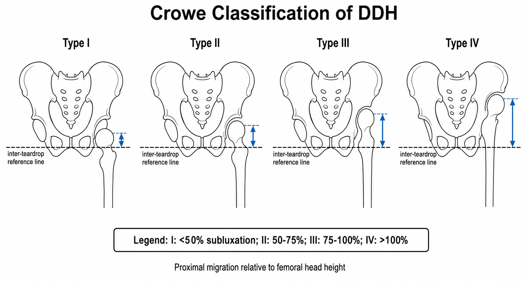
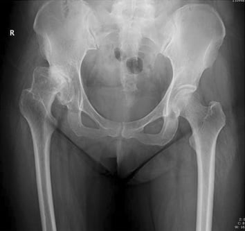
The Crowe classification quantifies the severity of developmental dysplasia of the hip by measuring how far the femoral head has migrated superiorly out of the true acetabulum. It was described in 1979 for planning total hip replacement in congenital dislocation of the hip and remains the reference grading system worldwide.
- Femoral head migration
- Less than 50 percent of the femoral head is proximal to the acetabular roof
- Radiographic appearance
- Mild subluxation; femoral head partially seated in a dysplastic but recognisable acetabulum; Shenton line disrupted but the head remains partially contained
- Typical surgical approach at THA
- Standard or slightly modified THA; small acetabular component or lateralised socket; no femoral shortening usually needed
- Femoral head migration
- 50 to 75 percent of the femoral head is proximal to the roof
- Radiographic appearance
- Moderate subluxation; the femoral head sits at the level of the superior acetabular rim; the true acetabulum is shallow and deficient superiorly
- Typical surgical approach at THA
- Modified THA; small or oblong cup, possible structural bone graft; femoral shortening may be considered if the leg length discrepancy is marked
- Femoral head migration
- 75 to 100 percent of the femoral head is proximal to the roof
- Radiographic appearance
- Severe subluxation; the femoral head is above the acetabular rim and lies adjacent to the ilium; the true acetabulum is small, anteverted, and deficient
- Typical surgical approach at THA
- Complex THA; subtrochanteric shortening osteotomy, acetabular reconstruction with structural graft or oblong cup; trochanteric osteotomy may be required
- Femoral head migration
- Greater than 100 percent of the femoral head is proximal to the roof (fully dislocated)
- Radiographic appearance
- Complete dislocation; the femoral head has migrated above the acetabulum entirely; a false acetabulum may form on the ilium; the true socket is severely deficient
- Typical surgical approach at THA
- Complex THA; subtrochanteric shortening osteotomy mandatory, major acetabular reconstruction; high risk of nerve palsy and dislocation; consider cementless reconstruction or custom implants
Fifty, Three-Quarters, Full, AboveThe four grade thresholds
Hook:Fifty Three-Quarters Full Above: think of stacking femoral heads — once the head has risen more than one full head-height above the roof, it is Crowe IV
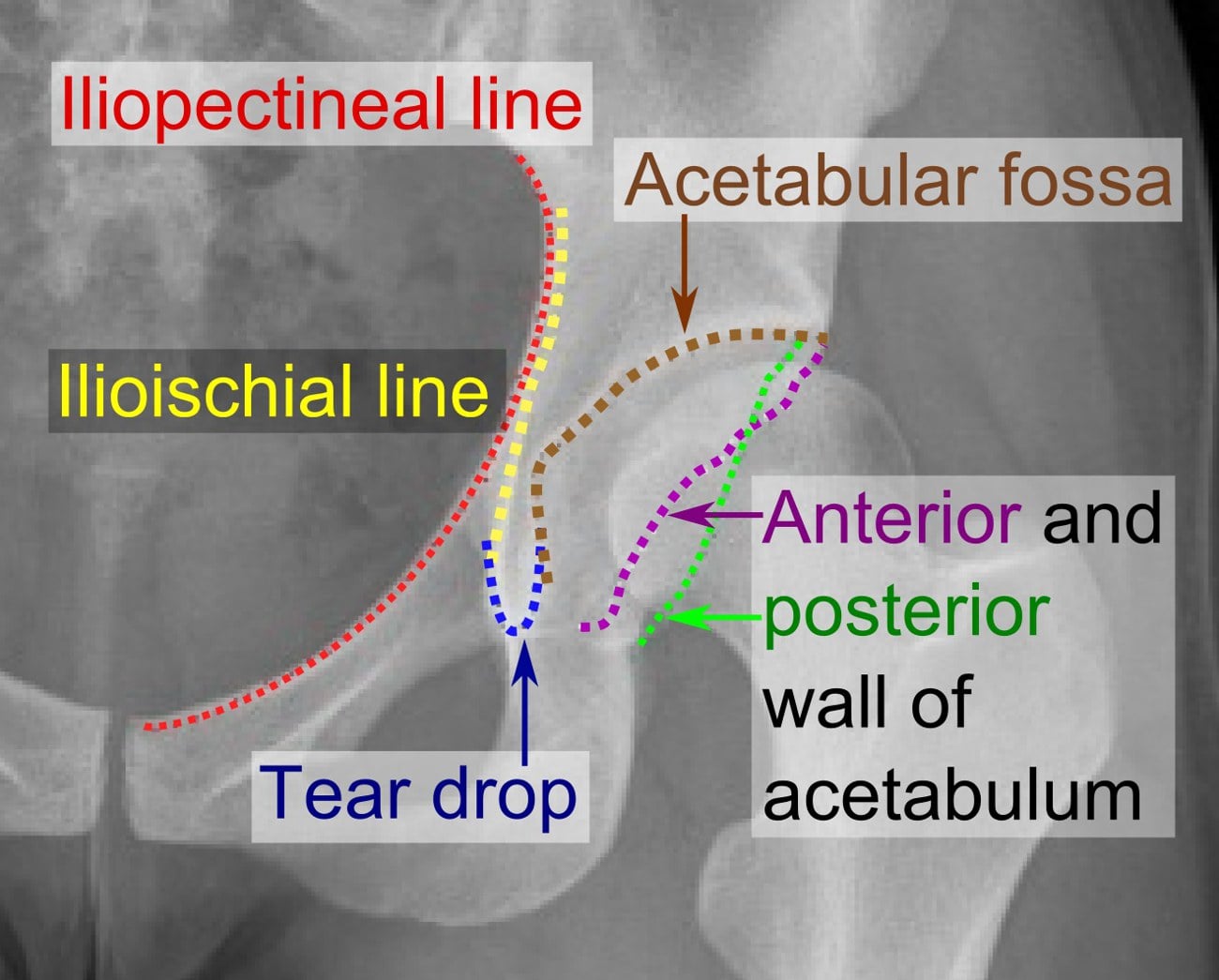
The measurement reference line is the interteardrop line — a horizontal line drawn between the inferior margins of both acetabular teardrops on a standing AP pelvis radiograph. In bilateral high dislocations, where no teardrop may be recognisable, use the inferior margin of the sacroiliac joint or estimate. The migration distance is measured to the junction of the femoral head and neck (the head-neck junction), and it is divided by the vertical diameter (height) of the normal femoral head.
Measurement Method on the AP Pelvis Radiograph
Accurate measurement requires a well-positioned standing AP pelvis radiograph with adequate exposure of both hips. The technique is reproducible but requires attention to landmarks.
- Action
- Draw the interteardrop line
- Landmark or Principle
- Connect the inferior margins of both acetabular teardrops with a horizontal line; this is the reference baseline
- Action
- Identify the femoral head-neck junction
- Landmark or Principle
- The point where the femoral head meets the neck on the dysplastic side; this is where migration is measured from
- Action
- Measure the vertical distance
- Landmark or Principle
- Draw a perpendicular line from the interteardrop line to the femoral head-neck junction; this is the migration distance
- Action
- Measure the normal femoral head height
- Landmark or Principle
- The vertical diameter of the femoral head on the normal contralateral side; if bilateral, estimate the head diameter from the femoral shaft width or pelvic proportions
- Action
- Calculate the migration percentage
- Landmark or Principle
- Migration distance divided by femoral head height, multiplied by 100; round to the nearest integer and assign the Crowe grade
Line — Mark — Measure — Divide — GradeHow to measure
Hook:Line Mark Measure Divide Grade: five steps from drawing the baseline to stating the grade — do it in this order every time you describe it in the exam
In bilateral DDH there is no normal contralateral head to measure. Estimate the head diameter using the contralateral side if only one hip is severely dislocated, or use the rule that the femoral head diameter is roughly one-fifth of the pelvic height from the superior border of the symphysis pubis to the sacral promontory. Some surgeons use the transverse diameter of the femoral condyles as a surrogate (the femoral head is approximately 80 percent of the condylar width).
Surgical Implications for Total Hip Arthroplasty
The Crowe grade is the single most important pre-operative classification for planning THA in the dysplastic hip because it predicts acetabular deficiency, femoral anatomy, leg length discrepancy, and the need for femoral shortening.
- Crowe I
- Small, anterolaterally deficient; usually reconstructable in the true socket
- Crowe II
- Shallow, deficient superiorly; may need a small cup with lateralised centre of rotation or structural graft
- Crowe III
- Severely deficient; small and anteverted; often needs structural bone graft, oblong socket, or reinforcement ring
- Crowe IV
- Very small, thin bone stock; may need medial wall protrusio technique, bulk graft, custom triflange cup, or cage
- Crowe I
- Not required
- Crowe II
- Consider if leg length discrepancy exceeds 2 to 3 cm
- Crowe III
- Usually required: subtrochanteric shortening osteotomy of 2 to 4 cm
- Crowe IV
- Mandatory: subtrochanteric shortening osteotomy of 3 to 6 cm or more
- Crowe I
- May be narrow; choose an appropriately sized stem
- Crowe II
- Narrower and straighter than normal; a tapered or modular stem is useful
- Crowe III
- Narrow, straight, often with excessive anteversion; a slim straight or modular stem; previous childhood surgery may have produced scarred soft tissues
- Crowe IV
- Very narrow, straight; anterior bow may be present; small diameter cementless stems or custom stems; the greater trochanter may override the femoral shaft
- Crowe I
- Low
- Crowe II
- Moderate if the hip is reduced without shortening
- Crowe III
- High if forced reduction; mitigated by subtrochanteric osteotomy and controlled, gradual intraoperative lengthening
- Crowe IV
- Highest risk; aggressive reduction without shortening almost guarantees nerve stretch; mandatory osteotomy
- Crowe I
- Standard
- Crowe II
- Slightly elevated; careful component positioning and soft-tissue repair
- Crowe III
- Elevated; abductor mechanism is deficient and tensioning is difficult
- Crowe IV
- Highest; abductors are severely attenuated; large head, constrained liner, or dual-mobility may be considered
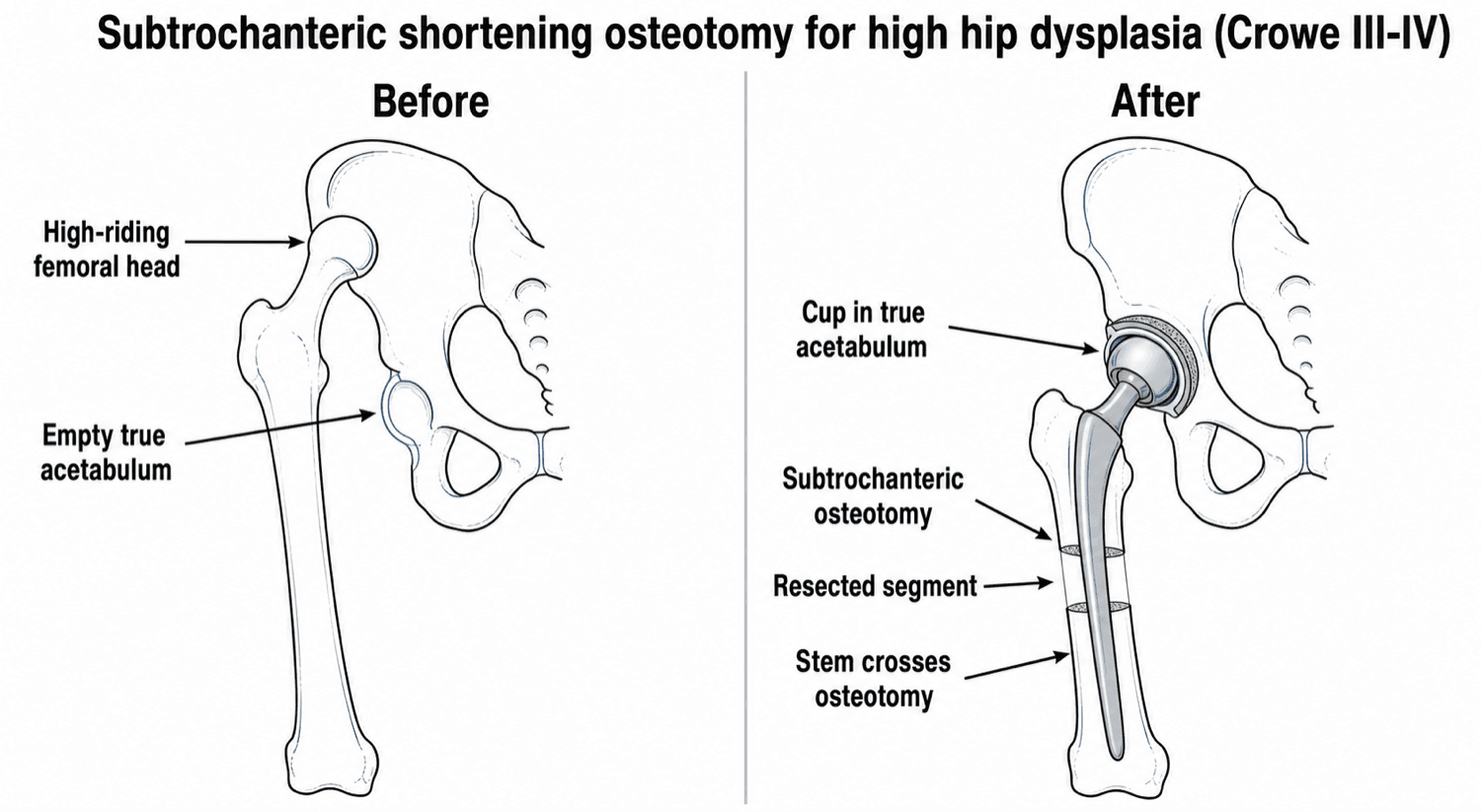
Subtrochanteric shortening osteotomy is the key safety procedure for Crowe III and IV hips. It allows the femoral head to be brought down to the true acetabulum without stretching the sciatic nerve or over-tensioning the soft tissues. Without it, attempted forced reduction carries a significant risk of permanent sciatic nerve palsy (reported up to 5 to 10 percent in historical series). The osteotomy also improves exposure and allows correct femoral component version. At the examination, if you describe a THA for a Crowe III or IV hip without mentioning subtrochanteric shortening, you will lose marks.
Socket — Shorten — NerveSurgical priorities by grade
Hook:Socket Shorten Nerve: every dysplastic THA follows these three priorities in order — rebuild the socket first, shorten the femur to fit, and protect the nerve throughout
The dysplastic femur is abnormal in a predictable way, which dictates implant choice:
- Excessive femoral anteversion — often markedly increased (commonly in the region of 40 to 60 degrees, and higher in high dislocation), with a valgus neck.
- A narrow, straight, hypoplastic medullary canal with thin cortices, a small femoral head and a short neck.
- A posteriorly positioned greater trochanter that may override the shaft.
Because the native anteversion is excessive and unreliable, a stem that simply follows the metaphysis would be left grossly anteverted (a dislocation risk). The solution is a small-diameter cementless stem that lets you set version independently — a modular stem (e.g. an S-ROM-type design, where the metaphyseal sleeve fits the canal while the stem is rotated to the correct version) or a conical/tapered fluted stem (e.g. a Wagner Cone) that gains diaphyseal fixation regardless of the deformed metaphysis. This is a further reason the subtrochanteric osteotomy is convenient — it allows simultaneous correction of the rotational (version) deformity.
The topic stresses that you shorten; examiners also want how:
- Transverse osteotomy — the simplest and most common (the commonest technique in the Casciaro review) and the easiest for shortening and de-rotation, but it has poor inherent rotational stability. It is therefore augmented: the cementless diaphyseal-fixing stem crosses the osteotomy as an intramedullary splint, and the resected segment is split longitudinally and applied as an onlay cortical strut with cerclage cables to control rotation and add bone.
- Step-cut (and Z, oblique, or double-chevron/V) osteotomies — give a larger contact area and better rotational stability (less reliant on the implant), at the cost of being more technically demanding.
- Union and pitfalls — non-union is low overall but is higher with a transverse cut combined with a cemented stem (Casciaro); achieve solid distal-fragment fixation, preserve the periosteal blood supply, and protect weight-bearing until union.
Limitations and Alternative Classifications
- Measurement variability. The interteardrop line is not always clearly visible, and the femoral head-neck junction can be difficult to identify in severely deformed or previously operated hips. CT-based measurements improve reproducibility but are not required for routine clinical use.
- It does not classify acetabular deficiency directly. The Crowe system grades femoral migration but not the shape or bone stock of the acetabulum itself. For acetabular planning, supplement Crowe with the Hartofilakidis classification (low dislocation, high dislocation, true dislocation) or the AAOS acetabular bone deficiency classification (type I to VII) for bone stock quantification.
- Leg length discrepancy is not captured. A Crowe III hip with a moderate leg length discrepancy may be easier to manage than a Crowe II hip with a severe discrepancy that requires lengthening of the contralateral limb. Clinical leg length measurement complements the radiographic grade.
- It does not account for previous surgery. Patients who had open reduction, Salter osteotomy, or pelvic osteotomy in childhood have scarring and distorted anatomy that increase surgical difficulty regardless of Crowe grade. Previous surgery should be noted separately.
- Bilateral disease doubles the planning burden. When both hips are affected, staging, leg length balance, and acetabular bone stock become more complex than the Crowe grade alone predicts.
- The Hartofilakidis classification (dysplastic, low dislocation, high dislocation) is more commonly used by European surgeons and focuses on the relationship between the femoral head and the true acetabulum rather than a percentage measurement.
- The Eftekhar classification describes three types (A, B, C) based on the degree of subluxation and the adequacy of the acetabular roof, offering a simpler system for quick clinical use.
Exam Viva
Practise clinical reasoning and management decisions out loud
“A 52-year-old woman presents with end-stage osteoarthritis of the left hip. She had untreated developmental dysplasia. Radiographs show the left femoral head sitting above the acetabular roof, with the femoral head-neck junction approximately 3.5 cm above the interteardrop line. The left femoral head measures approximately 4.4 cm in diameter. The right hip is normal. How would you classify this and how would you plan her total hip arthroplasty?”
“A 38-year-old man with bilateral Crowe IV developmental dysplasia presents with bilateral hip pain. Both femoral heads are fully dislocated and lie against the lateral ilium. He has a marked waddling gait and a leg length discrepancy of approximately 5 cm on each side. How would you approach his surgical management?”
The four grade thresholds
- Crowe I: less than 50 percent of the femoral head proximal to the acetabular roof
- Crowe II: 50 to 75 percent migration
- Crowe III: 75 to 100 percent migration
- Crowe IV: greater than 100 percent migration (fully dislocated, head completely above the acetabulum)
Measurement method
- Reference line: interteardrop line (horizontal line between inferior teardrop margins on AP pelvis)
- Migration distance: vertical distance from interteardrop line to femoral head-neck junction
- Normalise by: vertical diameter of the normal contralateral femoral head; estimate if bilateral
- Migration percentage equals migration distance divided by head height, multiplied by 100
Surgical implications at THA
- Crowe I: standard or modified THA, small cup, no femoral shortening
- Crowe II: modified THA, small or oblong cup, structural graft, consider shortening for leg length
- Crowe III: complex THA, subtrochanteric shortening osteotomy (2 to 4 cm), acetabular reconstruction
- Crowe IV: complex THA, mandatory subtrochanteric shortening (3 to 6 cm), major acetabular reconstruction, custom components possible
- Nerve protection: limit lengthening to approximately 2 cm; perform subtrochanteric osteotomy for Crowe III and IV
Limitations and alternatives
- Crowe grades femoral migration only — does not directly classify acetabular bone stock or shape
- Hartofilakidis classification (dysplastic, low dislocation, high dislocation) is an alternative widely used in Europe
- Previous childhood surgery (open reduction, Salter, pelvic osteotomy) increases complexity independent of Crowe grade
- Bilateral disease requires staged surgery and careful leg length planning
Evidence Base
Every citation below has been checked against its source record in PubMed. Crowe 1979 is the original classification, Hartofilakidis 1996 the main alternative (and acetabular-focused) system, Yiannakopoulos 2008 the reliability study (both systems are substantially-to-almost-perfectly reproducible), and Eskelinen/Casciaro the modern operative evidence — both confirming the central role of subtrochanteric shortening osteotomy in high (Crowe IV) dislocation.
Total hip replacement in congenital dislocation and dysplasia of the hip
- The paper that introduced the four-grade classification of proximal femoral head migration in congenital dysplasia/dislocation
- Reported 31 total hip replacements in 24 patients (mean 4-year follow-up): excellent or good in 27, with a 19% major-complication rate
- 27 of 31 hips needed smaller, straighter femoral components and 6 required superolateral bone grafts to increase acetabular coverage — establishing the link between migration severity and reconstructive difficulty
Congenital hip disease in adults. Classification of acetabular deficiencies and operative treatment with acetabuloplasty combined with total hip arthroplasty
- Proposed an alternative classification: dysplastic (head in true acetabulum), low dislocation (head in contact with superior acetabulum), and high dislocation (head against ilium)
- Demonstrated that acetabular reconstruction in the true anatomic position was feasible and durable in the majority of hips
- Reported that femoral shortening osteotomy was essential for high dislocations to allow safe reduction
Inter- and intra-observer variability of the Crowe and Hartofilakidis classification systems for congenital hip disease in adults
- AP pelvic radiographs of 145 patients (209 hips) were rated twice by three experienced European hip surgeons using both classifications
- Both systems were substantially-to-almost-perfectly reliable — Crowe inter-observer kappa 0.90-0.92 and intra-observer 0.86-0.95; Hartofilakidis inter-observer 0.85-0.93 and intra-observer 0.80-0.93
- The two classifications are comparably reproducible, although they convey different information (femoral migration versus acetabular/dislocation type)