Delbet Classification of Paediatric Hip Fractures
The avascular necrosis rate is the single most important fact about the Delbet classification. Types I and II carry the highest risk because the fracture passes through or very near the vascular watershed of the femoral head. Examiners expect you to state the AVN risk by type and to explain the anatomical reason. Capsular decompression within 24 hours is the intervention that most reduces AVN — examiners will push you on this. Do not delay: admit, decompress, fix, and protect.
The Delbet Classification
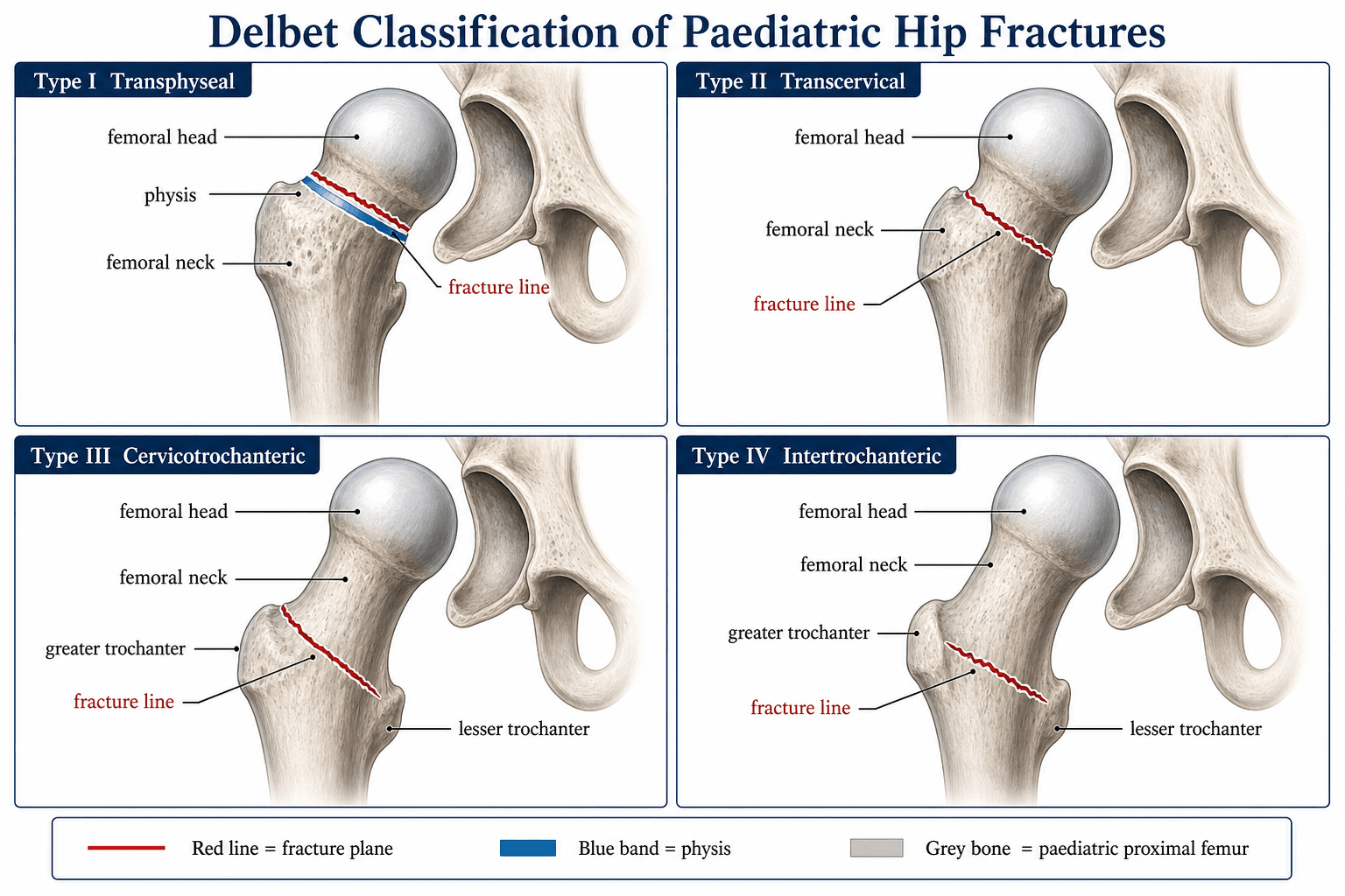
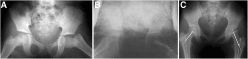
The Delbet classification, originally described in 1907, separates paediatric proximal femoral fractures into four types based on the fracture line's relationship to the proximal femoral physis and the greater trochanter. The type predicts the risk of avascular necrosis, which remains the most important determinant of long-term outcome.
- Fracture Line
- Through the proximal femoral physis; epiphysis separates from metaphysis
- AVN Risk
- Up to 100 percent (displaced)
- Frequency
- Rare (5–10 percent)
- Mechanism
- Low energy in young children; may be pathological (child abuse screening)
- Fracture Line
- Through the femoral neck, between physis and intertrochanteric line
- AVN Risk
- Approximately 50 percent
- Frequency
- Most common (45–50 percent)
- Mechanism
- High-energy trauma; falls, road traffic collisions in older children
- Fracture Line
- At the junction of the femoral neck and the intertrochanteric region
- AVN Risk
- Approximately 25 percent
- Frequency
- Uncommon (25–35 percent)
- Mechanism
- Moderate to high energy
- Fracture Line
- Between the greater and lesser trochanters, below the neck
- AVN Risk
- Near 0 percent
- Frequency
- Uncommon (20–25 percent)
- Mechanism
- High-energy trauma; similar to adult intertrochanteric fractures
Physis · Neck · Junction · TrochantericThe four Delbet types and AVN risk
Hook:The closer the fracture is to the physis and femoral head, the higher the AVN risk — it falls from ~100% (I) to near zero (IV).
Why does Type I carry the highest AVN rate? The transphyseal fracture tears the retinacular vessels (the ascending cervical branches of the medial and lateral femoral circumflex arteries) as they enter the femoral head through the posterosuperior portion of the physis. In a child whose capital femoral epiphysis is not yet fused, there is no collateral intraosseous supply — the retinacular vessels are effectively the sole blood supply to the ossifying epiphysis. When these are disrupted, the femoral head is ischaemic.
Blood Supply and the Basis of Avascular Necrosis Risk
Understanding why the Delbet types differ in AVN risk requires understanding the paediatric femoral head blood supply:
- The primary supply comes from the lateral epiphyseal arteries (posterosuperior retinacular vessels), which are terminal branches of the medial femoral circumflex artery. These enter posterosuperiorly and are at greatest risk in types I and II.
- The secondary supply comes from the ligamentum teres (foveolar artery), a branch of the obturator artery. This vessel is insignificant in young children and does not compensate when retinacular vessels are torn.
- Metaphyseal vessels contribute from below but cannot cross the physis to reach the epiphysis in children with an open growth plate.
- Type I disrupts the lateral epiphyseal vessels at their entry point through the physis — the proximal fragment is almost entirely devascularised.
- Type II passes through the neck, potentially sparing some retinacular vessels posteriorly if the fracture is undisplaced, but displaced type II fractures tear the same vessels.
- Type III is distal to the main retinacular entry, so more vessels are preserved, but displaced fractures can still damage them.
- Type IV is well distal to the femoral head circulation — AVN is essentially not a concern.
- Lateral Epiphyseal Arteries
- Almost always disrupted (vessels torn at physis)
- Ligamentum Teres
- Inadequate alone in children
- Resulting AVN Risk
- Extremely high (approaching 100 percent if displaced)
- Lateral Epiphyseal Arteries
- Often disrupted; depends on displacement and fracture level
- Ligamentum Teres
- Inadequate alone
- Resulting AVN Risk
- High (approximately 50 percent; lower if undisplaced)
- Lateral Epiphyseal Arteries
- Usually preserved; fracture is distal to vascular entry
- Ligamentum Teres
- Irrelevant to outcome
- Resulting AVN Risk
- Moderate (approximately 25 percent)
- Lateral Epiphyseal Arteries
- Preserved — fracture is well distal to vascular supply
- Ligamentum Teres
- Irrelevant
- Resulting AVN Risk
- Near 0 percent
Proximity = PerilWhy AVN risk drops from type I to IV
Hook:The retinacular vessels are the effectively sole supply to the open-physis epiphysis — the ligamentum teres does not compensate in young children.
Capsular tamponade is an emergency. A haemarthrosis in the confined paediatric hip capsule raises intracapsular pressure above femoral head perfusion pressure, causing ischaemia in addition to any direct vascular injury. This is a supracondylar-humerus equivalent at the hip: the tamponade effect compounds the mechanical vascular disruption. Urgent capsular decompression (aspiration of haematoma or open capsulotomy) within 24 hours significantly reduces AVN rates and is the single most important modifiable factor.
Management by Type
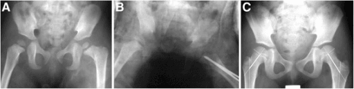
Management is guided by the Delbet type, the age of the child, the degree of displacement, and the status of the physis. The overarching principles are: urgent capsular decompression, anatomic reduction, stable fixation, and protection of the proximal femoral physis.
- Reduction
- Urgent closed or open
- Fixation
- Smooth K-wires or cannulated screws across the physis (avoid thread crossing the physis if possible)
- Capsular Decompression
- Mandatory — aspiration or capsulotomy
- Key Considerations
- High AVN regardless. In children under 2 years, consider pathology (non-accidental injury screening). Prophylactic contralateral spica may be considered in very young children.
- Reduction
- Closed reduction first; open if unreduced
- Fixation
- Cannulated screws (parallel, 2 or 3) — avoid crossing the physis in young children; use smooth K-wires in the very young
- Capsular Decompression
- Mandatory — capsulotomy or aspiration
- Key Considerations
- Anatomic reduction is critical. In displaced fractures, consider open reduction (Watson-Jones or Smith-Petersen approach). AVN remains the dominant long-term problem.
- Reduction
- Usually closed reduction achievable
- Fixation
- Cannulated screws or paediatric hip plate and screws
- Capsular Decompression
- Recommended but less critical than types I and II
- Key Considerations
- Better prognosis than types I and II. Ensure adequate screw purchase in the proximal fragment.
- Reduction
- Closed reduction
- Fixation
- Paediatric hip plate and screws (fixed-angle device) or cannulated screws
- Capsular Decompression
- Not routinely required
- Key Considerations
- Similar to adult intertrochanteric fixation principles. AVN is not a concern. Excellent prognosis with stable fixation.
CRISPThe five treatment pillars
Hook:Every paediatric hip fracture needs CRISP management — the five pillars apply to all Delbet types, but the urgency of each varies.
In children under about 8 to 10 years, avoid placing screw threads across the proximal femoral physis if possible — use smooth K-wires or place cannulated screws with the threads entirely in the metaphysis and the smooth shaft across the physis. In older children approaching physeal closure, crossing the physis with cannulated screws is acceptable. Growth arrest of the proximal femoral physis leads to progressive coxa vara and limb-length discrepancy.
The tables say "closed reduction, open if it fails, Watson-Jones or Smith-Petersen" — examiners want the how.
- Gentle, single, anatomic reduction. Reduce once, gently, under general anaesthesia and image intensifier. Repeated forceful manipulation is harmful — each attempt can further tear the already-tenuous retinacular vessels and worsen AVN. If a closed reduction is not anatomic, proceed to open reduction rather than re-manipulating.
- The capsulotomy does double duty. Whichever anterior/anterolateral approach you use, the capsulotomy both decompresses the haemarthrosis (the AVN-reducing step) and lets you reduce the fracture under direct vision — so a displaced fracture needing open reduction and the need to decompress are solved by the same exposure.
- Watson-Jones (anterolateral) — interval between gluteus medius and tensor fasciae latae (both superior gluteal nerve); a workhorse giving access to the anterolateral neck for reduction and screw placement.
- Smith-Petersen (anterior) — a true internervous approach: superficial sartorius (femoral nerve) / tensor fasciae latae (superior gluteal nerve), deep rectus femoris (femoral nerve) / gluteus medius (superior gluteal nerve) — excellent anterior exposure of the neck and capsule.
- Fixation, age-tailored: two or three parallel cannulated screws up the neck — in the young child keep the threads in the metaphysis (smooth shaft across the physis) or use smooth K-wires; nearer maturity, crossing the physis with screws is acceptable for stability. For type IV (and unstable III) use a paediatric hip plate / fixed-angle device. Augment with a hip spica in very young children or when the construct (e.g. K-wires only) is not robust, because small bones limit implant purchase.
Complications and Long-Term Sequelae
Paediatric hip fractures carry one of the highest complication rates of any paediatric fracture. Long-term follow-up to skeletal maturity is mandatory.
- Avascular necrosis of the femoral head is the most common and most devastating complication. It presents clinically with progressive pain, limp, and limited hip movement, typically within the first 1 to 2 years but sometimes later. Radiographic findings include sclerosis, collapse of the femoral head, and secondary osteoarthritis changes. Management of established AVN ranges from containment (bracing, orthoses) in early stages to valgus osteotomy, shelf acetabuloplasty, or ultimately hip arthroplasty in young adulthood.
- Coxa vara occurs from growth arrest of the greater trochanteric apophysis or from malunion of the fracture with a valgus collapse pattern. The neck-shaft angle drops below 110 to 120 degrees, producing a Trendelenburg limp and abductor weakness. A valgus subtrochanteric osteotomy corrects the mechanical alignment and improves abductor function.
- Premature physeal closure of the proximal femoral physis causes progressive limb-length discrepancy and coxa vara. The growth plate contributes approximately 15 percent of total limb length; early closure can produce a significant discrepancy.
- Non-union is seen more often in types I and II and in older children. Treatment is with valgus osteotomy combined with internal fixation and bone grafting.
- Chondrolysis (loss of articular cartilage of the hip) is a rare but serious complication that presents with progressive hip stiffness, pain, and radiographic joint-space narrowing. It is associated with internal fixation penetration into the joint and with prolonged immobilisation.
- Infection after surgical fixation is uncommon but catastrophic in the paediatric hip.
- Pathological fractures in children under 2 years — always screen for underlying pathology, including non-accidental injury, osteogenesis imperfecta, and metabolic bone disease. A transphyseal fracture in a non-ambulating child should raise immediate safeguarding concerns.
The topic's evidence cards grade outcome by "Ratliff" — examiners expect you to actually know it. Ratliff (1962) described both the pattern of post-traumatic AVN and an outcome grade:
- Ratliff AVN type I — whole head. Diffuse involvement and collapse of the entire capital epiphysis — the commonest and worst prognosis.
- Ratliff AVN type II — partial / localised. A segmental area of the head — intermediate.
- Ratliff AVN type III — from the fracture line to the physis (neck/metaphyseal, the epiphysis largely spared) — the best prognosis.
- Ratliff outcome grade (the scale the studies use): good / fair / poor, judged on pain, hip function and movement, and radiographic change.
Managing established AVN in the child is a stepwise ladder, by stage and extent:
- Pre-collapse / early: protect — restricted weight-bearing and abduction bracing/containment, with close surveillance; bisphosphonates have been tried to slow collapse but the paediatric evidence is limited.
- Segmental head involvement: a realignment/containment osteotomy (valgus or varus proximal femoral osteotomy to rotate healthy cartilage into the weight-bearing zone) and/or shelf or Chiari acetabuloplasty for containment.
- Salvage in the young adult once the head has failed: historically arthrodesis; in practice total hip arthroplasty, accepting the longevity/revision burden of a prosthesis in a young patient.
The point that scores marks: AVN is predicted by Delbet type but, once present, is classified by Ratliff and managed by its stage and extent — not by the original fracture level.
Long-term follow-up is non-negotiable. Children with a Delbet type I, II, or III fracture must be followed to skeletal maturity with serial radiographs to detect AVN, growth arrest, coxa vara, and limb-length discrepancy. A child who appears well at 1 year can develop AVN at 3 years or coxa vara at skeletal maturity. Plan follow-up for a minimum of 2 years and ideally until the physis closes.
Viva practice
Exam Viva
Practise clinical reasoning and management decisions out loud
“A 9-year-old girl falls from her bicycle and presents with a painful left hip, unable to weight-bear. Radiographs show a displaced transcervical fracture of the left femoral neck. How would you classify this, and what is your management plan?”
“An 18-month-old boy is brought in by ambulance with a swollen, painful left thigh and will not move the leg. Radiographs show a transphyseal separation of the left proximal femur. What are your immediate concerns and how would you proceed?”
Exam cheat sheet
The four types
- Type I — Transphyseal: through the physis, epiphysis separates from metaphysis — highest AVN risk (up to 100 percent)
- Type II — Transcervical: through the femoral neck — most common type, AVN approximately 50 percent
- Type III — Cervicotrochanteric: at the neck-trochanteric junction — AVN approximately 25 percent
- Type IV — Intertrochanteric: below the neck — AVN near 0 percent, excellent prognosis
Blood supply and AVN
- Lateral epiphyseal (retinacular) arteries are the primary supply to the paediatric femoral head
- These vessels enter posterosuperiorly near the physis — types I and II disrupt them directly
- Ligamentum teres (obturator branch) is insignificant in children and does not compensate
- Metaphyseal vessels cannot cross the physis to reach the epiphysis
Management principles (CRISP)
- Capsular decompression: aspirate haematoma or open capsule within 24 hours — reduces AVN
- Reduction: anatomic closed reduction; open (Watson-Jones or Smith-Petersen) if closed fails
- Internal fixation: cannulated screws (type II/III), paediatric hip plate (type IV), smooth K-wires (type I in young children)
- Salvage planning: counsel about AVN risk, plan follow-up to skeletal maturity
- Physis protection: avoid screw threads across the physis in young children
Complications
- AVN: most devastating, highest in types I and II, may present years later — long-term follow-up mandatory
- Coxa vara: growth arrest of greater trochanteric apophysis or malunion — treat with valgus osteotomy
- Premature physeal closure: progressive limb-length discrepancy and deformity
- Non-union: more common in types I and II — valgus osteotomy with bone grafting
- Chondrolysis: rare, associated with intra-articular hardware penetration
- Non-accidental injury: always consider in children under 2 years with a transphyseal fracture
Evidence
Risk factors for avascular necrosis after femoral neck fractures in children: 25 Cincinnati cases and meta-analysis of 360 cases
- Meta-analysis of 360 paediatric femoral neck fractures (plus 25 Cincinnati cases) — the classic source of the AVN-rate-by-Delbet-type figures.
- AVN rate by Delbet class: type I 38%, type II 28%, type III 18%, type IV 5% — risk falls as the fracture moves distally away from the femoral-head blood supply.
- On logistic regression, fracture type and age were the only significant independent predictors; type I-III were 15x/6x/4x more likely to develop AVN than type IV.
According to PubMed, the classic AVN-rate-by-Delbet-type data (I 38%, II 28%, III 18%, IV 5%; type and age the key predictors) come from Moon & Mehlman 2006 (DOI 10.1097/00005131-200605000-00005); the benefit of early treatment (less AVN and growth disturbance with presentation under 48 hours; type II commonest) is from Regmi et al. 2024 (DOI 10.1016/j.otsr.2024.103840), and the 15-year risk-factor analysis (high-velocity trauma and — unexpectedly — type IV associated with AVN; delayed diagnosis in neurologically impaired children) is from Mueller et al. 2025 (DOI 10.1007/s00068-024-02728-z).