IWGDF/IDSA severity classification guides treatment | Probe-to-bone test diagnoses osteomyelitis | Multidisciplinary care reduces amputation rates
- Acute limb-threatening infection requires emergency surgical debridement within 24 hours
- Empiric antibiotics must cover MRSA in moderate-severe infections (vancomycin or linezolid)
- Duration: 1-2 weeks for soft tissue only, 4-6 weeks for osteomyelitis with debridement, 12 weeks if conservative
- Systemic signs may be absent despite severe infection due to neuropathy and vasculopathy
- Multidisciplinary diabetic foot team reduces amputation rates by 50% compared to standard care
- “Probe-to-bone positive in deep ulcer (greater than 2cm²) has 89% PPV for osteomyelitis
- “Severe (PEDIS 4) infection with SIRS requires emergency debridement within 24 hours
- “MRI triad for osteomyelitis: low T1, high T2/STIR, cortical destruction
- “Deep tissue cultures (curettage) essential; superficial swabs misleading
- Failure to recognise a limb-threatening infection that needs emergency surgery.
- Not understanding the probe-to-bone test as the bedside diagnostic for osteomyelitis.
- Inadequate empiric antibiotic cover, missing MRSA or anaerobes in moderate to severe infection.
- Not appreciating the need for deep tissue cultures rather than superficial swabs.
- Underestimating vascular assessment and revascularisation, without which healing is not possible.
Pathophysiology and Microbiology
The diabetic foot triad. Neuropathy, peripheral arterial disease and immune dysfunction run together in the diabetic foot, and between them they impair recognition of the ulcer, its healing and the control of infection. Read them as a chain rather than a list of risk factors: deformity creates the pressure point, autonomic dry skin breaks the barrier, sensory loss means nobody notices, impaired immunity lets the infection spread with few systemic signs, and poor perfusion stops it healing.
Neuropathy. Sensory loss removes protective sensation, so the ulcer forms undetected, the infection is recognised late, and the patient keeps walking on it. Motor neuropathy wastes the intrinsic muscles, producing claw toes, prominent metatarsal heads, abnormal pressure points and altered biomechanics. Autonomic dysfunction brings anhidrosis with dry, cracked skin, loss of thermoregulation and an impaired inflammatory response.
Peripheral arterial disease. The macrovascular lesion is atherosclerosis of the tibial vessels, often calcified (Monckeberg's sclerosis), which reduces perfusion to the foot and impairs both wound healing and antibiotic delivery to the wound. Microvascular disease adds basement membrane thickening, endothelial dysfunction, reduced oxygen delivery and impaired neutrophil function. The clinical result is delayed healing, a higher infection risk and higher amputation rates.
Immune dysfunction. Neutrophil chemotaxis, phagocytosis and bactericidal activity are all impaired and the inflammatory response is delayed. Humoral immunity is blunted by glycosylation of immunoglobulins, impaired antibody function and reduced complement activity. Hyperglycaemia itself impairs immunity, and advanced glycation end products add to the susceptibility to infection.
Microbiology
The flora changes with the age of the wound and after surgery, which is what the empiric regimens in Management are built on.
- Common Organisms
- Staphylococcus aureus (MSSA), Streptococcus spp (Group A, B), occasionally Gram-negative bacilli
- Frequency
- Monomicrobial in 70% of cases. MSSA most common single organism (40-60%).
- Antibiotic Implications
- First-generation cephalosporin (cefazolin) usually adequate for mild infection. Add MRSA coverage if risk factors.
- Common Organisms
- Polymicrobial: S. aureus, Streptococcus, Enterococcus, Enterobacteriaceae, Pseudomonas, Anaerobes (Bacteroides, Peptostreptococcus)
- Frequency
- Polymicrobial in 50-75%. Average 3-4 organisms isolated. MRSA in 20-30% of chronic wounds.
- Antibiotic Implications
- Broad-spectrum coverage required: piperacillin-tazobactam or carbapenem. Must include anaerobic coverage in deep infections.
- Common Organisms
- MRSA (healthcare-associated), Pseudomonas aeruginosa, Enterococcus (including VRE), resistant Gram-negatives
- Frequency
- Higher proportion of resistant organisms. MRSA 30-40%. Multi-drug resistant organisms common.
- Antibiotic Implications
- Vancomycin or linezolid for MRSA. Antipseudomonal coverage essential. Culture-directed therapy critical.
- Common Organisms
- Similar to chronic wounds but S. aureus predominates in bone (60%). Polymicrobial in 30% of bone specimens.
- Frequency
- Bone culture often shows different organisms than superficial swab. Deep culture essential.
- Antibiotic Implications
- Bone-penetrating antibiotics required: fluoroquinolones, clindamycin, linezolid, rifampin. Prolonged duration 4-6 weeks minimum.
IWGDF/IDSA Classification
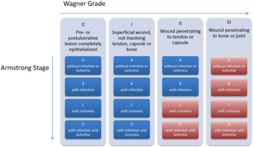
The International Working Group on the Diabetic Foot (IWGDF) system is the most widely used classification for diabetic foot infection, endorsed by the Infectious Diseases Society of America (IDSA). It grades severity as uninfected, mild, moderate or severe, and the grade sets both the antibiotic choice and the surgical urgency. The PEDIS grading (Perfusion, Extent, Depth, Infection, Sensation) integrates infection severity with the other wound characteristics for a comprehensive assessment.
- Clinical Findings
- Wound present but no purulence or inflammation. No signs of infection.
- Systemic Response
- None
- Treatment
- No antibiotics needed. Focus on wound care, off-loading, glycaemic control, and addressing ischaemia if present.
- Clinical Findings
- Local infection: erythema 0.5-2cm around ulcer, induration, warmth, tenderness. Superficial tissues only.
- Systemic Response
- No systemic signs. Cellulitis less than 2cm from wound edge.
- Treatment
- Oral antibiotics targeting S. aureus and Streptococcus. First-generation cephalosporin or amoxicillin-clavulanate. Outpatient management.
- Clinical Findings
- Erythema greater than 2cm, lymphangitis, deep tissue involvement (fascia, muscle, tendon), abscess formation. No systemic toxicity.
- Systemic Response
- Minimal systemic response. May have low-grade fever. No haemodynamic instability.
- Treatment
- IV antibiotics with MRSA coverage (vancomycin or linezolid) plus Gram-negative coverage. Surgical debridement if abscess or necrotic tissue. Hospital admission.
- Clinical Findings
- Limb-threatening infection: extensive cellulitis, necrotising fasciitis, wet gangrene, gas in tissues.
- Systemic Response
- Two or more SIRS criteria (listed below the table). Sepsis or septic shock.
- Treatment
- Urgent surgical debridement within 24 hours. Broad-spectrum IV antibiotics: vancomycin PLUS piperacillin-tazobactam or carbapenem. ICU if haemodynamically unstable. Often requires amputation.
SIRS is what makes an infection severe. Two or more of the criteria below convert a moderate infection into a severe (PEDIS 4) one, and move it from a clinic problem to emergency debridement within 24 hours, with ICU input considered:
- Temperature above 38°C or below 36°C
- Heart rate above 90 per minute
- Respiratory rate above 20 per minute, or PaCO2 below 32 mmHg
- White cell count above 12,000 or below 4,000
The trap runs in both directions. A limb-threatening infection can present without fever, while a low temperature and a low white count are themselves SIRS criteria, so the sickest patients may look deceptively quiet.
Clinical Presentation
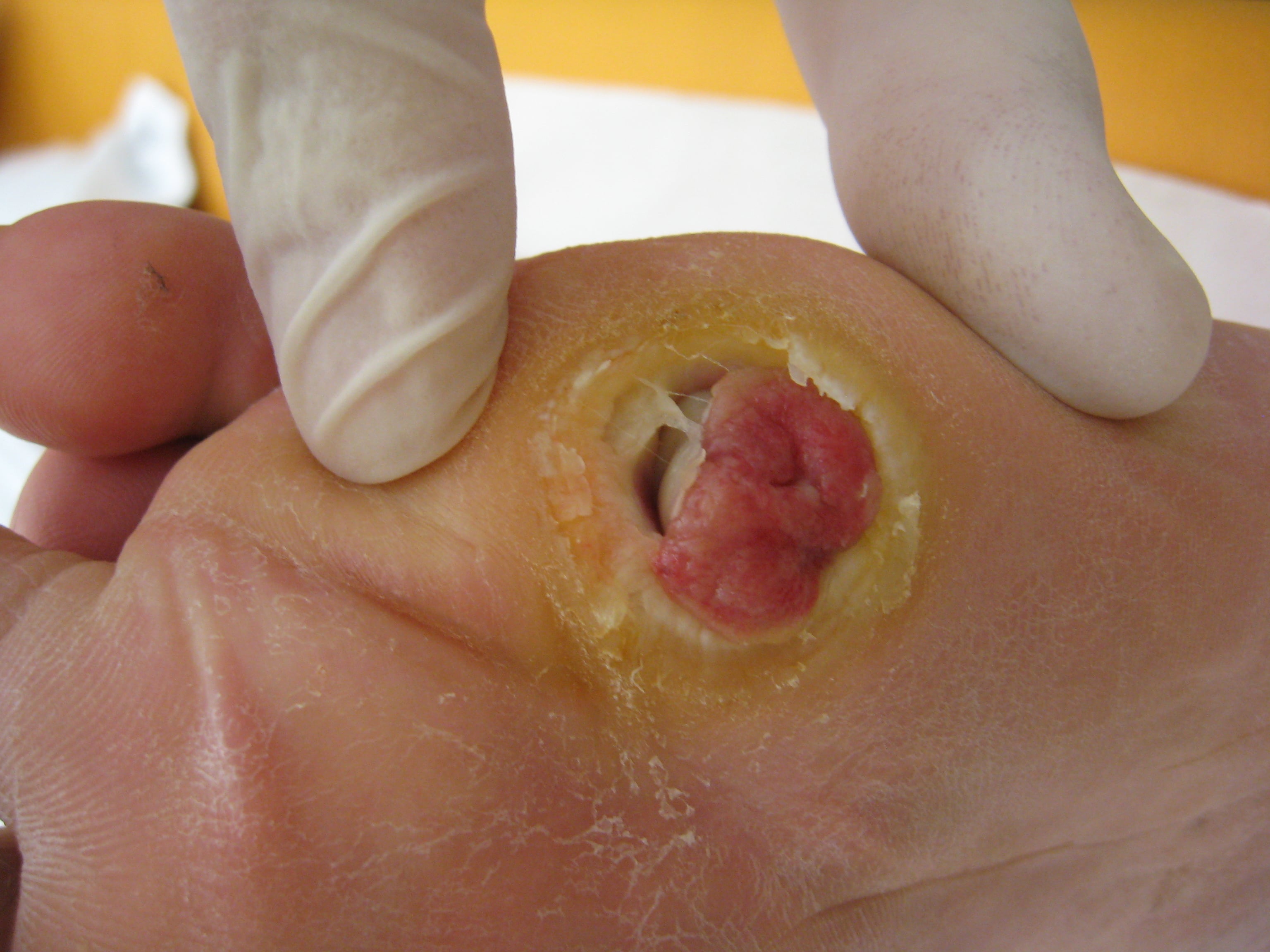
A 67-year-old man with poorly controlled type 2 diabetes (HbA1c 9.2%) presents with three days of worsening right foot swelling, erythema and purulent drainage from a long-standing plantar ulcer under the first metatarsal head. He noticed the ulcer four months ago but did not seek treatment because it did not hurt. He is now unable to walk because of the swelling. He has no fever but feels unwell.
Two features of that history do the diagnostic work. Painlessness explains the four-month delay - neuropathy removes the symptom that would have brought anyone else in early, so late presentation is the rule rather than negligence. And the absence of fever does not downgrade the severity: systemic signs are blunted in diabetes, so a patient who is afebrile but unwell, with a rapidly progressing foot, is still a candidate for urgent debridement.
Who is at risk. The high-risk patient has:
- Diabetes for more than 10 years
- HbA1c above 8% (poor glycaemic control)
- Peripheral arterial disease (absent pedal pulses, ABI below 0.7)
- A previous foot ulcer or amputation
- Severe neuropathy (unable to feel the 10g monofilament)
- Renal insufficiency (eGFR below 60)
- Immunosuppression (steroids, chemotherapy)
- Poor footwear or barefoot walking
Red flags for severe infection. Any of these should raise the alarm:
- Rapid progression over 24-48 hours
- Crepitus, suggesting gas in the tissues (necrotising infection)
- Foul-smelling discharge (anaerobic infection)
- Systemic signs: fever, tachycardia, hypotension
- Mental status changes in the elderly
- Wet gangrene with tissue necrosis
- Bullae (necrotising fasciitis)
Examination
The wound. Measure length, width and depth in centimetres and record the location, since pressure points (metatarsal heads, heel) are high-risk sites. Note the base (healthy granulation versus necrotic tissue), the drainage (serous in an uninfected wound, purulent in an infected one) and the odour, because a foul smell suggests anaerobes. Then turn to the surrounding tissue: measure the extent of erythema from the wound edge, palpate for induration and fluctuance (abscess), look for lymphangitic streaking, feel for crepitus (gas in the soft tissues), and document any exposed bone, tendon or joint.
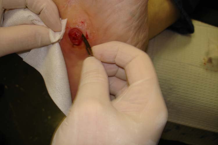
Probe-to-bone. Perform the test in every ulcer larger than 2cm² or deeper than 3mm. Pass a sterile metal probe or a cotton swab gently into the ulcer to its base; the test is positive when hard, gritty bone is felt. It is most accurate in ulcers larger than 2cm² and deeper than 3mm, and a positive test in the appropriate clinical context is highly predictive of osteomyelitis and may be sufficient to start treatment without confirmatory imaging, particularly in a large or deep ulcer or when inflammatory markers are high.
Reading the numbers. The test's predictive value depends on who is being probed. In Grayson's hospitalised patients, where two-thirds of ulcers had osteomyelitis, a positive probe had a positive predictive value of 89% (sensitivity 66%, specificity 85%); in Lavery's outpatient cohort, where 12% did, sensitivity was 87% and specificity 91%, but the positive predictive value fell to 57% while the negative predictive value was 98%. A positive probe in a high-risk inpatient therefore strongly suggests osteomyelitis, and a negative probe in an outpatient helps to rule it out. The test is most useful for ruling osteomyelitis in; a negative probe alone does not exclude it, and equivocal cases proceed to MRI and, ultimately, bone culture.
Perfusion. Infection will not resolve without adequate perfusion, so arterial assessment is part of every examination. Look for the skin changes of ischaemia (hairless, shiny, atrophic), palpate the dorsalis pedis and posterior tibial pulses (graded 0-2), check capillary refill (normal under 2 seconds), look for dependent rubor and elevation pallor (Buerger's test), and compare foot temperature between the two sides. Then measure the ankle-brachial index:
- 0.9-1.3 is normal; below 0.9 is peripheral arterial disease
- Above 1.4 means non-compressible, calcified vessels and is unreliable; obtain a toe-brachial index (TBI) instead
- TBI below 0.7 indicates significant ischaemia
If ischaemia is present (ABI below 0.7, absent pulses, or TcPO2 below 30mmHg), vascular surgery consultation for revascularisation is essential before definitive treatment.
Neuropathy. The 10g Semmes-Weinstein monofilament is the standard screen: test 10 plantar sites including the metatarsal heads, and inability to feel it at 4 or more sites indicates loss of protective sensation. Add vibration with a 128Hz tuning fork at the hallux interphalangeal joint and pinprick (sharp versus dull discrimination). For motor neuropathy look for intrinsic wasting (claw toes, hollowing of the foot) and deformity (claw toes, hammer toes, Charcot arthropathy), test ankle dorsiflexion, plantarflexion and toe flexion and extension, and watch the gait if the patient can walk. Autonomic neuropathy shows as anhidrosis (dry, cracked skin), callus at pressure points, and fissures in dry skin that act as portals of entry. Severe neuropathy is why patients present late with advanced infection: without pain they keep bearing weight on the infected ulcer, driving bacteria deeper into soft tissue and bone.
The systemic picture. Up to 30% of diabetic foot infections present without fever despite severe infection, so absence of systemic signs does not exclude it; rely on local wound severity and SIRS criteria rather than on how the patient feels. Record temperature (fever suggests systemic infection but may be absent in diabetes), heart rate (above 90 is a SIRS criterion), respiratory rate (above 20 suggests SIRS), blood pressure (hypotension indicates septic shock) and oxygen saturation (hypoxia is concerning for severe sepsis). Check mental status (confusion suggests severe sepsis in the elderly), hydration (sepsis dehydrates), the rest of the foot and the contralateral foot for other ulcers, and the inguinal nodes. Infection can precipitate diabetic ketoacidosis (look for Kussmaul breathing and a fruity breath odour), acute kidney injury from sepsis or from contrast if angiography is planned, and cardiac events (myocardial infarction) from the stress of sepsis.
Differential Diagnosis
- Key Distinguishing Features
- Wound or ulcer present, purulence, erythema spreading from ulcer, positive probe-to-bone, raised CRP
- Imaging / Investigation
- Plain films (cortical destruction lags 2-4 weeks); MRI sensitive/specific; bone biopsy and culture is reference standard
- Key Distinguishing Features
- Hot, swollen, often painless foot WITHOUT a portal of entry; midfoot rocker-bottom collapse; foot cools and elevation reduces swelling
- Imaging / Investigation
- Radiographs show fragmentation/dislocation; MRI shows diffuse marrow oedema and joint destruction without a sinus tract; can coexist with infection
- Key Distinguishing Features
- Erythema and warmth but no purulence, abscess, exposed bone or systemic toxicity
- Imaging / Investigation
- Normal or near-normal radiographs; probe-to-bone negative; responds to antibiotics and elevation
- Key Distinguishing Features
- Sudden severe pain, often first MTP joint, tophi, prior episodes; pain disproportionate in a neuropathic foot is a clue
- Imaging / Investigation
- Joint aspirate shows negatively birefringent urate crystals; serum urate; radiographs may show peri-articular erosions
- Key Distinguishing Features
- Unilateral calf/leg swelling and warmth, no ulcer; calf tenderness; risk factors for venous thromboembolism
- Imaging / Investigation
- Compression ultrasound; D-dimer in low-probability cases
- Key Distinguishing Features
- Pain out of proportion, rapid spread, crepitus, bullae, systemic toxicity - a surgical emergency
- Imaging / Investigation
- Do not delay surgery for imaging; gas may be seen on plain films/CT; diagnosis confirmed at operative exploration
Investigations
Blood tests. Send a white cell count, CRP (raised in infection and useful for monitoring the response), ESR (above 70 is suggestive of osteomyelitis) and procalcitonin if available (raised in bacterial infection). Add glucose and HbA1c to assess control, creatinine and eGFR for antibiotic dosing, electrolytes (potassium if ketoacidosis is a concern) and lactate (raised in sepsis). Take blood cultures if there are systemic signs.
Cultures. Deep tissue culture is the gold standard. Debride the superficial tissue and wound edges, clean the wound, then take a curettage or biopsy from the ulcer base, and take bone at operation if osteomyelitis is suspected. Send tissue rather than pus, since tissue gives the organism actually invading and pus alone may not, and request aerobic, anaerobic, fungal and mycobacterial culture, telling the laboratory that anaerobes are expected. Avoid superficial swabs, cultures taken before the wound is cleaned, and incomplete anaerobic specimen handling. The result guides definitive therapy and is what allows de-escalation from broad empiric cover, but it must not delay empiric antibiotics in moderate or severe infection. Bone biopsy remains the reference standard for osteomyelitis.
Imaging strategy. Plain radiographs are the first-line study for every patient. If the probe-to-bone test is positive or the radiographs show changes, obtain an MRI; if MRI is contraindicated, consider CT or a nuclear medicine study. In moderate or severe infection with ischaemia, add vascular imaging (CTA, MRA or angiography).
- Findings
- Osteomyelitis: cortical destruction, periosteal reaction, bone lucency, soft tissue gas. Changes lag clinical by 2-4 weeks.
- Advantages
- First-line, readily available, low cost, detects gas in soft tissues, identifies foreign bodies. Three views (AP, lateral, oblique) required.
- Limitations
- Low sensitivity (54%) for early osteomyelitis. Cannot differentiate from Charcot. Cannot assess soft tissue extent. Positive findings lag by 2-4 weeks.
- Findings
- Osteomyelitis: T1 low signal, T2/STIR high signal in bone marrow, cortical destruction, periosteal reaction, adjacent soft tissue oedema. Abscess: rim-enhancing fluid collection.
- Advantages
- Gold standard for osteomyelitis (sensitivity 90%, specificity 79%). Best soft tissue detail. Detects abscesses. Guides surgical planning. Can differentiate from Charcot.
- Limitations
- Expensive, limited availability, contraindications (pacemaker, claustrophobia), overinterpretation of bone marrow oedema, 2-4 week lag after treatment to show improvement.
- Findings
- WBC scan: focal tracer uptake at infection site. Bone scan: increased uptake in all three phases for osteomyelitis.
- Advantages
- Functional imaging detects active infection. Bone scan highly sensitive. Can differentiate osteomyelitis from Charcot with combined WBC/bone scan.
- Limitations
- Lower specificity than MRI (65-75%). Bone scan cannot differentiate infection from inflammation. Time-consuming (24-48 hours). Less anatomic detail than MRI.
- Findings
- Cortical destruction, sequestrum, gas in soft tissues, abscess collections, bone details.
- Advantages
- Better than radiographs for bone detail. Detects gas in tissues. Can guide percutaneous drainage. Useful if MRI contraindicated.
- Limitations
- Poor soft tissue contrast compared to MRI. Radiation exposure. Contrast required for soft tissue assessment. Less sensitive than MRI for early osteomyelitis.
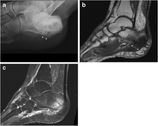
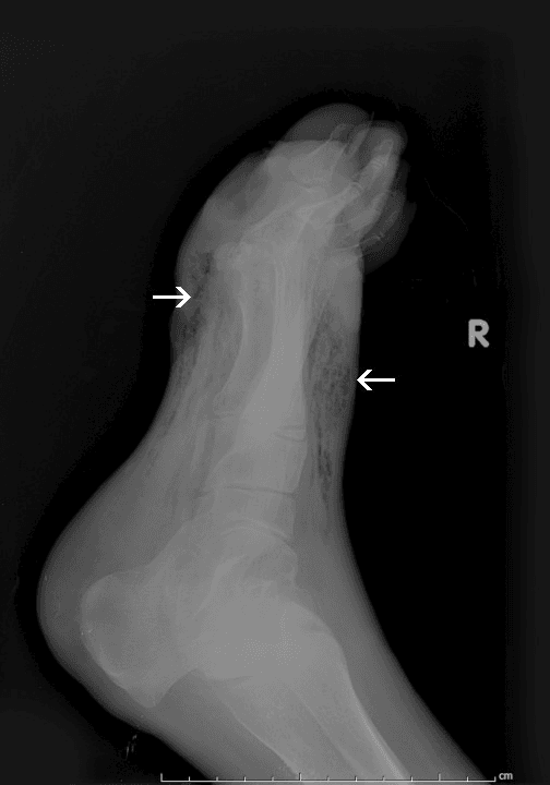
The classic MRI triad is (1) low T1 signal in the marrow, (2) high T2/STIR signal in the marrow, and (3) cortical destruction or periosteal reaction. Confidence rises further when there is adjacent soft-tissue oedema or an ulcer tracking to bone.
The low T1 signal is the one that matters most, because it reflects replacement of normal fatty marrow by infection - high T2/STIR alone is oedema, and oedema is non-specific. That distinction is exactly why Charcot arthropathy is so hard to separate from osteomyelitis: both light up on STIR. What favours infection is a focal abnormality directly beneath an ulcer with a sinus tract and cortical breach, whereas Charcot is typically periarticular, midfoot, multi-bone and follows the joint rather than the ulcer.
Decision points. Four combinations settle most cases:
- Mild infection, negative probe-to-bone and normal radiographs: treat as soft tissue infection only
- Moderate infection with a positive probe-to-bone: MRI to confirm osteomyelitis
- Severe infection: emergency surgery; imaging must not delay debridement
- Any infection with ischaemia (ABI below 0.7): urgent vascular consultation
Probe-to-Bone Test - Original Description (Grayson)
Probe-to-Bone Test - Outpatient Validation (Lavery)
Management
Principles. The severity grade sets the antibiotic and the urgency of surgery, deep cultures direct the antibiotic, and source control, off-loading, perfusion and glycaemic control decide whether the wound heals. A multidisciplinary diabetic foot team reduces amputation rates by 50% compared with standard care.
Antibiotic Therapy
Mild infection. Treat as an outpatient with oral antibiotics against S. aureus and Streptococcus spp, the primary pathogens of acute mild infection, for 1-2 weeks when the infection is confined to soft tissue:
- Cephalexin 500mg four times daily, or
- Amoxicillin-clavulanate 875/125mg twice daily, or
- Clindamycin 300mg three times daily if penicillin-allergic
Moderate infection. Admit for intravenous therapy covering MRSA, Gram-negatives including Pseudomonas, and anaerobes, for 2-3 weeks for soft tissue infection or 4-6 weeks if osteomyelitis has been surgically debrided:
- Vancomycin 15-20mg/kg IV every 8-12 hours (target trough 15-20 for serious infection), plus
- Piperacillin-tazobactam 4.5g IV every 6 hours or 3.375g every 4 hours
- If penicillin-allergic: vancomycin plus ciprofloxacin 400mg IV every 12 hours plus metronidazole 500mg IV every 8 hours
Severe infection. Broad-spectrum cover with ICU support, for a minimum of 4-6 weeks and often longer depending on clinical response:
- Vancomycin 15-20mg/kg IV every 8-12 hours (or linezolid 600mg IV every 12 hours), plus
- Meropenem 1g IV every 8 hours or imipenem-cilastatin 500mg IV every 6 hours, plus
- Consider adding metronidazole 500mg IV every 8 hours if there is extensive necrosis
In moderate and severe infection, start empiric therapy immediately after the cultures are taken; do not wait for the results.
Antibiotic Duration in Nonsurgically Treated Diabetic Foot Osteomyelitis
Surgical Management
- Severe infection (PEDIS 4) with systemic toxicity: emergency debridement within 24 hours.
- Necrotising fasciitis or gas gangrene: immediate debridement, a life-saving intervention.
- Abscess or undrained purulent collection: incision and drainage required.
- Extensive necrotic tissue: debridement necessary for infection control.
- Osteomyelitis: resection of infected bone improves cure rates and shortens antibiotic duration.
Debridement. The operation has five goals: remove all necrotic and infected tissue, obtain deep tissue and bone cultures, reduce the bacterial burden, convert a chronic wound into an acutely healing one, and define the extent of infection. Set up with the patient supine and a thigh tourniquet available (see the alert on its use below), adequate IV access for resuscitation, broad-spectrum antibiotics already given, and preparation for possible amputation if involvement proves extensive. Then work through the wound:
- Excise all necrotic skin and subcutaneous tissue
- Remove the callus around the ulcer edges (keratinocytes impair healing)
- Open all deep spaces and compartments
- Probe for tracking sinuses and abscess pockets
- Resect obviously infected bone (dark, mushy and non-bleeding on inspection)
- Send multiple deep tissue specimens for culture (3-5 samples)
- Irrigate copiously with 3-6 litres of normal saline
How far to go. Debride to bleeding viable tissue, judged by the 4 Cs: colour (pink or red), consistency (firm), contraction with stimulus, and circulation (bleeding). Be aggressive but preserve functional structures where possible, and leave the wound open for delayed closure or healing by secondary intention. In severe infection the first operation is damage control: remove the obviously infected tissue and obtain cultures, reassess tissue viability at 48-72 hours with further debridement if needed (serial debridements every 48-72 hours as required), and perform the definitive reconstruction only once infection is controlled and granulation tissue is forming.
Operations for osteomyelitis. The choice between bone resection and the level of amputation depends on the extent of infection, perfusion, the soft tissue available for cover, the patient's mobility goals and their overall medical state.
Bone resection with preservation of function. For localised bone infection with viable surrounding soft tissue and adequate perfusion: excise the infected segment en bloc with a margin of healthy bleeding bone, as in partial phalangeal resection, metatarsal head resection or calcanectomy. It preserves foot length and function, with success in 80-85% when the resection is complete.
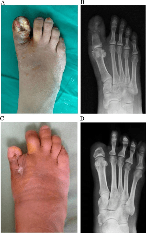
Ray amputation. For osteomyelitis of a toe and its metatarsal with enough soft tissue to close: resect the whole ray, toe and metatarsal, en bloc. The central rays (second, third and fourth) are better tolerated than the border rays. It removes all infected tissue and allows primary closure, and the patient is usually ambulatory in a normal shoe with a filler.
Transmetatarsal amputation. For forefoot infection involving several rays where the heel pad is adequately perfused: a long plantar flap, a short dorsal flap and bevelled metatarsals, preserving at least 50% of first metatarsal length if possible. It maintains functional ambulation in a modified shoe, healing in 70-80% with proper selection (adequate perfusion).
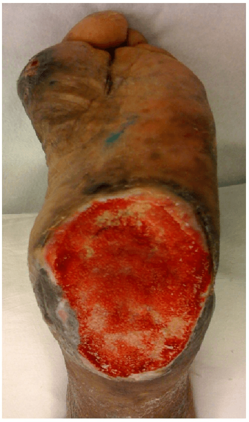
Partial calcanectomy. For calcaneal osteomyelitis, often from a pressure ulcer, with adequate soft tissue cover: excise the infected portion of the calcaneus, preserving the Achilles insertion if possible. 40-50% of the calcaneus can be removed while retaining some weight-bearing, at the cost of prolonged off-loading during healing.
Major amputation. Below-knee or above-knee amputation is for infection uncontrolled by lesser procedures, life-threatening sepsis or a non-salvageable limb, and the decision balances limb salvage against the mortality risk of severe sepsis. Below-knee is preferred for its better functional outcome when gastrocnemius perfusion is adequate; above-knee follows a failed below-knee amputation or severe PAD.
Negative pressure wound therapy. Vacuum-assisted closure is an adjunct to debridement and antibiotics, not a replacement, applied only after infection is controlled and necrotic tissue removed. It reduces oedema and wound size, promotes granulation, removes exudate and bacteria, prepares the bed for closure and may shorten time to healing; several RCTs show faster healing than standard dressings, particularly in post-surgical wounds over 10cm². Use black polyurethane foam for most wounds and white polyvinyl alcohol foam if less granulation is wanted, at continuous -125mmHg, changing the dressing every 48-72 hours.
- Indications: large wounds after debridement (over 10cm²); exposed bone, tendon or joint once infection is controlled; heavily exudating wounds; a bridge to delayed primary closure or skin grafting
- Contraindications: active untreated infection with necrotic tissue; exposed arteries; malignancy in the wound bed; untreated osteomyelitis
Adjunctive Therapies
Off-loading. The total contact cast is the gold standard for plantar ulcers: it reduces plantar pressure by 85-90% and heals 80-90% of ulcers at 12 weeks. A removable cast walker (CAM boot) is an alternative if the patient is reliable, since compliance is critical with removable devices; felted foam off-loads specific pressure points, a heel off-loading boot suits posterior ulcers, and crutches or a wheelchair enforce non-weight bearing, which is required for midfoot or hindfoot involvement. Off-loading continues until the wound has closed completely, typically 6-12 weeks at minimum, and may need permanent footwear modification afterwards.
Glycaemic control. The inpatient target is 140-180 mg/dL, deliberately less stringent during acute illness, and the outpatient goal an HbA1c under 7%, individualised; hypoglycaemia impairs wound healing and is to be avoided. Most patients need insulin during the acute infection, as a basal-bolus regimen or an infusion in the ICU, returning to oral agents once stable. An HbA1c above 8% is associated with delayed healing, each 1% rise in HbA1c increases infection risk by 20%, and hyperglycaemia impairs neutrophil function and collagen synthesis.
Hyperbaric oxygen. Its indications are controversial: a chronic non-healing ulcer despite optimal treatment, a compromised host (advanced age, comorbidities), and ischaemic wounds with PAD after revascularisation. The protocol is 100% oxygen at 2.0-2.5 atmospheres in 90-120 minute sessions, typically 30-40 treatments over 6-8 weeks. The evidence shows a modest reduction in amputation (NNT 10-15) with most benefit in ischaemic wounds, but high-quality evidence is limited and not all insurers cover it.
Growth factors and biologics. Becaplermin gel (Regranex), recombinant PDGF-BB applied daily to a clean ulcer bed, may improve healing rates by 10-15% but carries a black box warning for cancer risk with prolonged use. Bioengineered skin substitutes (Dermagraft, Apligraf, EpiFix) may accelerate healing in chronic ulcers but are expensive, with limited evidence of superiority. Platelet-rich plasma, an autologous platelet concentrate carrying multiple growth factors, has conflicting evidence of efficacy and is not standard of care.
Revascularisation in the Ischaemic Infected Foot
The angiosome concept. Which vessel to target when restoring perfusion to a diabetic foot wound is decided by the angiosome concept. The foot is supplied by three source arteries dividing into six angiosomes, three-dimensional vascular territories each fed by a single source artery, with adjacent territories linked by choke (collateral) vessels.
- Angiosome(s) Supplied
- Medial calcaneal (heel), medial plantar (instep), lateral plantar (lateral sole and forefoot)
- Wounds It Feeds
- Most plantar and heel wounds
- Angiosome(s) Supplied
- Dorsum of the foot
- Wounds It Feeds
- Dorsal foot wounds
- Angiosome(s) Supplied
- Lateral calcaneal (lateral heel) and the anterior perforating branch (lateral ankle)
- Wounds It Feeds
- Lateral heel and ankle wounds; an important collateral source
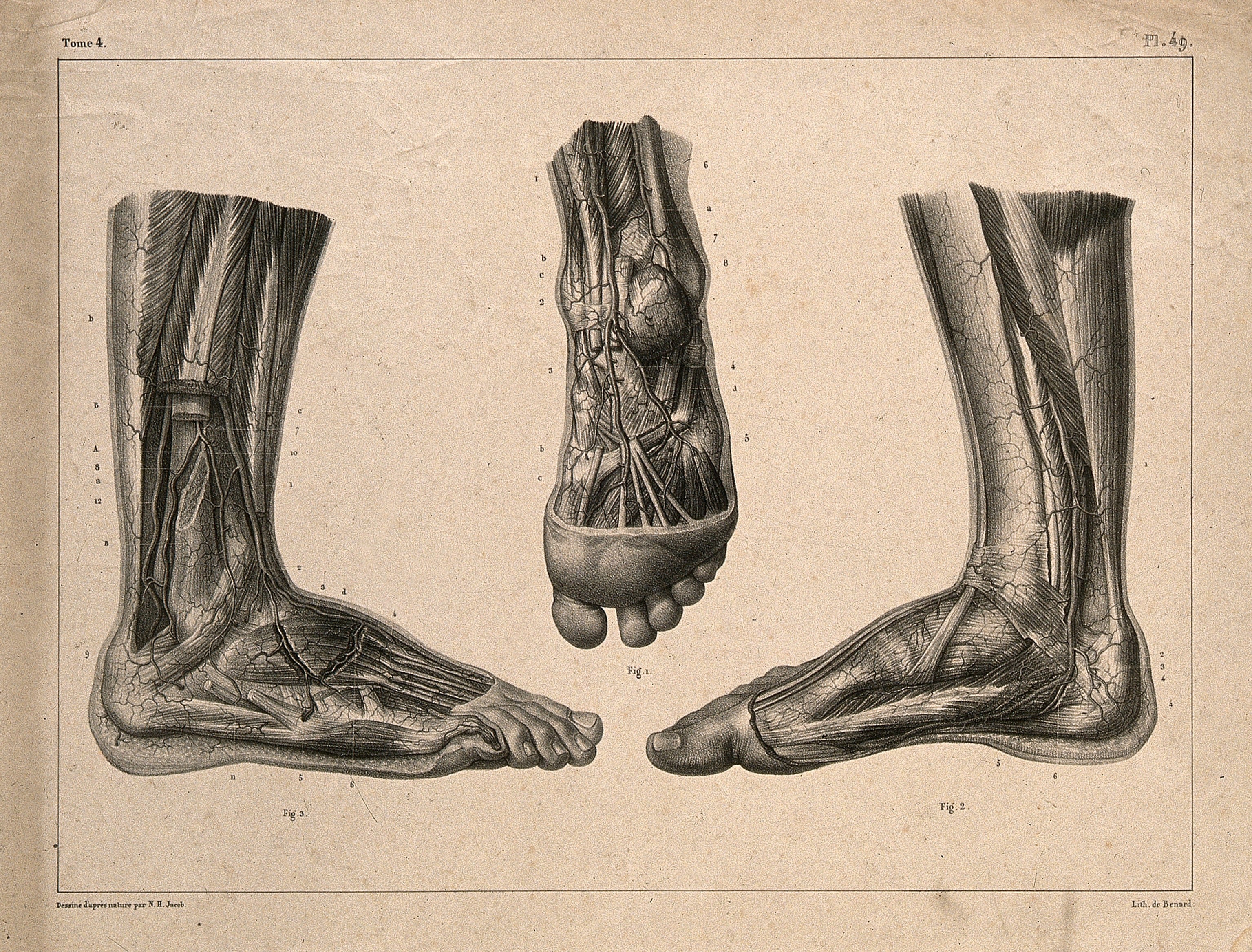
Angiosome-directed (direct) revascularisation restores inline flow along the specific source artery feeding the angiosome that contains the wound, and is associated with better wound healing and limb salvage than indirect (any-patent-vessel) revascularisation.
The practical qualifier matters as much as the principle: when only indirect flow can be achieved, choke vessels and a complete pedal arch can still compensate, so indirect revascularisation remains worthwhile when the direct target is not amenable. In other words, direct is the preference, not a precondition - do not decline to revascularise a limb because the ideal target vessel is unavailable.
Timing and modality. Where chronic limb-threatening ischaemia coexists with infection, revascularisation is part of management, and two questions arise: how it is sequenced with infection control, and whether to revascularise by open bypass or endovascular means.
- Approach
- Source control first - drain pus, debride necrotic tissue and control sepsis (life before limb); revascularise before definitive closure or reconstructive amputation
- Evidence / Caveat
- An actively septic foot needs urgent debridement before or alongside revascularisation; definitive healing and closure await restored perfusion
- Approach
- Yes when chronic limb-threatening ischaemia is present - the wound will not heal without inline perfusion
- Evidence / Caveat
- Assess with pulses, ABI/TBI and toe pressures; absent perfusion predicts non-healing
- Approach
- Surgical vein bypass favoured with a good single-segment great saphenous vein and acceptable surgical risk; endovascular-first favoured for high surgical risk, no suitable vein, or infrapopliteal disease
- Evidence / Caveat
- BEST-CLI: bypass had fewer major adverse limb events when an adequate vein was available; BASIL-2: an endovascular-first strategy gave better amputation-free survival in infrapopliteal disease - individualise
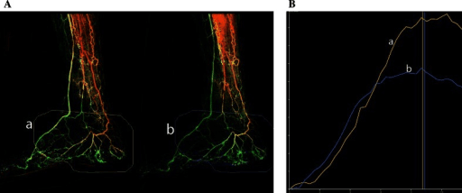
No diabetic foot infection in an ischaemic limb will heal without revascularisation. The order is fixed: control the infection first, then restore perfusion, then close or reconstruct. Debriding sepsis cannot wait for a vascular study, but definitive closure should not be attempted into a limb that cannot perfuse it.
The open-versus-endovascular choice is individualised on conduit (vein) availability, the level and pattern of disease, surgical risk and life expectancy - which is precisely why BEST-CLI and BASIL-2 reached differing conclusions: they enrolled different populations, and the availability of a good single-segment saphenous vein is the variable that separates them.
Management Algorithm
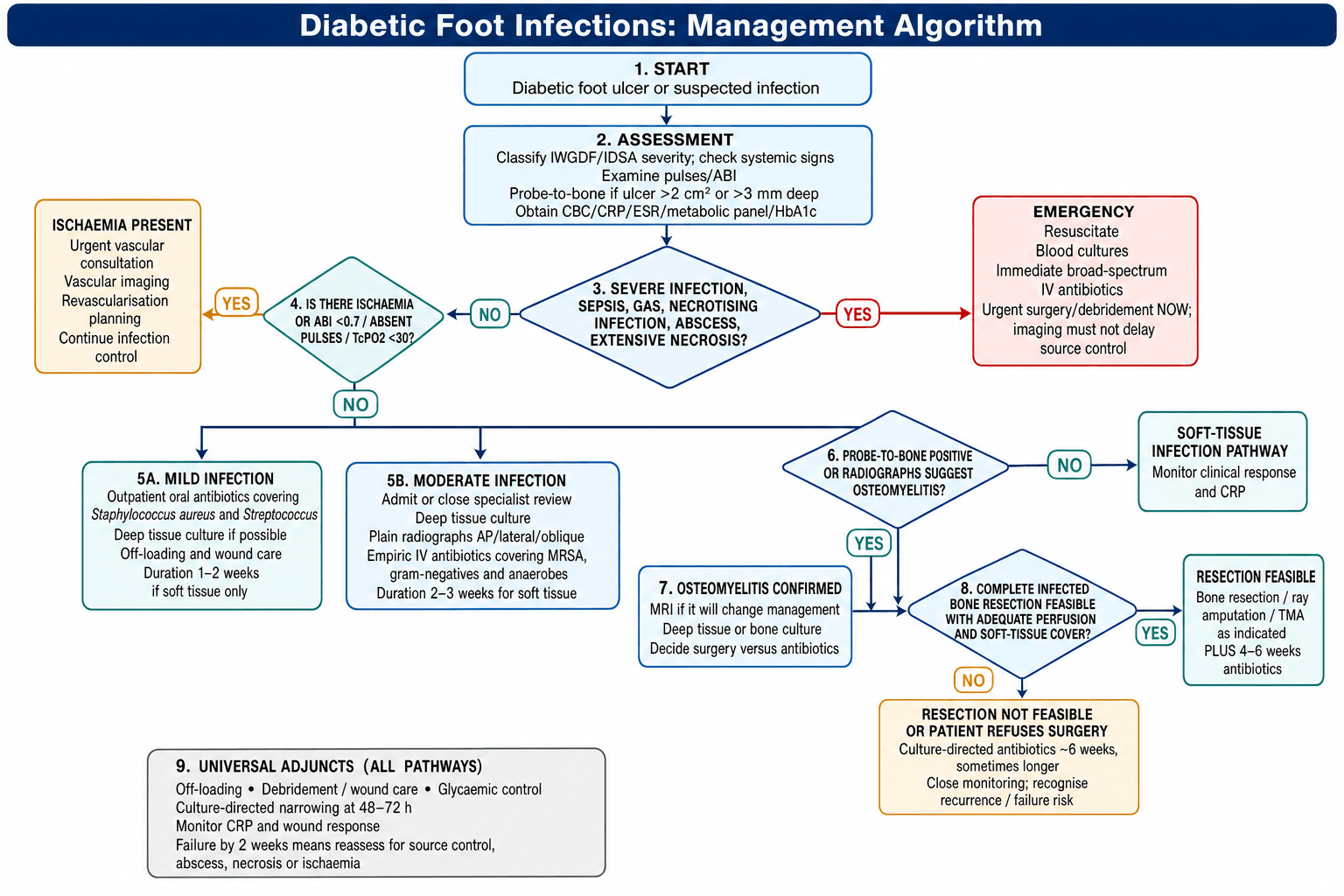
IDSA Clinical Practice Guideline for Diabetic Foot Infections
Complications
The complications belong to the antibiotics, to the surgery and to the disease itself.
Antibiotic complications.
- C. difficile colitis (10-15% with broad-spectrum antibiotics)
- Antibiotic-associated diarrhoea
- Vancomycin: nephrotoxicity, red man syndrome
- Linezolid: bone marrow suppression, peripheral neuropathy (with prolonged use over 6 weeks)
- Fluoroquinolones: tendon rupture, QT prolongation, peripheral neuropathy
- Allergic reactions and drug rashes
Surgical complications.
- Persistent infection after debridement (10-15%)
- Need for a higher-level amputation (15-20%)
- Wound dehiscence (20-30% in ischaemic limbs)
- Phantom limb pain after major amputation
- Falls and fractures from altered biomechanics after partial foot amputation
- Pressure ulcers at new weight-bearing sites
Long-term complications.
- Recurrent ulceration (30-40% at 1 year, 60% at 3 years)
- Charcot arthropathy in the denervated foot
- Contralateral limb ulceration (50% within 3 years)
- Progression of PAD requiring revascularisation
- Worsening renal function from diabetes and sepsis
- Reduced quality of life and mobility
Postoperative Care
Wound care. For the first 48-72 hours inspect the wound daily, dressing it wet-to-dry or with negative pressure therapy and keeping the bed moist but not macerated. After that choose the dressing by the exudate, measure the wound weekly to track progress, and watch for signs of recurrent infection. Off-loading and glycaemic control continue as set out under Adjunctive Therapies.
When healing stalls. If healing is delayed despite adequate treatment, repeat the ABI and TBI, consider CT angiography or MRA, and involve vascular surgery for revascularisation; healing is unlikely without adequate perfusion (TcPO2 above 30mmHg).
Discharge. The patient goes home when all of these are met:
- Afebrile for 48 hours
- Wound improving with controlled drainage
- Tolerating oral intake and antibiotics
- Reliable, with appropriate support
- Follow-up arranged within 1 week
Outcomes and Prognosis
Diabetic foot infections carry significant morbidity and mortality, and the outcome depends on infection severity, vascular status and the timeliness of appropriate treatment.
What to expect.
- Soft tissue infection with adequate treatment: 85-90% resolve without amputation
- Osteomyelitis with surgical resection: 70-80% heal without major amputation
- Severe infection with sepsis: 60-70% limb salvage, 15-20% mortality
- Forefoot amputation (TMA or ray): 70-80% heal, 50% ambulatory at 1 year
- Major amputation: primary healing in 60-70% of below-knee and 85-90% of above-knee amputations
Predictors of a poor outcome. Patient factors are advanced age (over 75), end-stage renal disease on dialysis, severe PAD (ABI below 0.5, TcPO2 below 30), poor glycaemic control (HbA1c above 9%), severe immunosuppression, malnutrition (albumin below 3.0 g/dL) and active smoking. Wound factors are size over 4cm², depth to bone or joint, duration over 6 months, a heel or midfoot location (worse than the forefoot), wet gangrene, polymicrobial infection with resistant organisms (MRSA, Pseudomonas), and a failed previous amputation or debridement. Infection factors are PEDIS grade 4, necrotising soft tissue infection, systemic sepsis needing ICU, osteomyelitis with extensive bone involvement, SIRS or septic shock, delayed presentation (more than 1 week of symptoms) and gas in the soft tissues on imaging.
Five years on. After a diabetic foot infection:
- Recurrent ulceration: 60-65%
- Minor amputation (toe, ray, TMA): 30-35%
- Major amputation (below- or above-knee): 15-20%
- Contralateral limb ulceration or amputation: 40-50%
- Cardiovascular event (myocardial infarction, stroke): 25-30%
- Mortality: 40-50%, primarily from cardiovascular disease and sepsis
Multidisciplinary Care and Amputation in Diabetic Foot Ulcers
Diabetic Foot Ulcer Recurrence and Long-Term Risk
Predictors of Amputation in Infected Diabetic Foot Ulcers (Eurodiale cohort)
Guidelines, Registries & Global Practice
Global Epidemiology
The global pooled prevalence of diabetic foot ulceration is 6.3% (Zhang 2017, Ann Med; PMID 27585063), and approximately 18.6 million people worldwide develop a diabetic foot ulcer each year (Armstrong 2023, JAMA; PMID 37395769). Around 50-60% of ulcers become infected, and roughly 20% of moderate-to-severe infections lead to lower-extremity amputation.
- Pooled Prevalence
- 13.0% (95% CI 10.0-15.9)
- Notes
- Highest regional prevalence; USA 13.0%, Canada 14.8%
- Pooled Prevalence
- 7.2% (95% CI 5.1-9.3)
- Notes
- High prevalence with limited access to multidisciplinary care
- Pooled Prevalence
- 5.5% (95% CI 4.6-6.4)
- Notes
- Large absolute burden given diabetes prevalence
- Pooled Prevalence
- 5.1% (95% CI 4.1-6.0)
- Notes
- Belgium notably high at 16.6%
- Pooled Prevalence
- 3.0% (95% CI 0.9-5.0)
- Notes
- Lowest regional prevalence; Australia 1.5%
Ulcers are more prevalent in men than women and in type 2 than type 1 diabetes; affected patients tend to be older with longer diabetes duration, hypertension, retinopathy and a smoking history. Indigenous and socially disadvantaged populations (including Indigenous Australians, and Black, Hispanic and Native American populations in the USA) carry substantially higher rates of ulceration and amputation.
Major Guidelines Side by Side
- Classification
- Mild / Moderate / Severe (Lipsky 2012, PMID 22619242)
- Key Recommendation
- Antibiotics only for clinically infected wounds; deep tissue (not swab) culture; MRI when osteomyelitis suspected
- Evidence Basis
- GRADE-rated consensus guideline
- Classification
- IWGDF/IDSA severity grades, aligned with PEDIS
- Key Recommendation
- Severity-stratified empiric therapy; bone biopsy as reference standard for osteomyelitis; multidisciplinary team
- Evidence Basis
- Systematic-review based consensus
- Classification
- Uses IWGDF/IDSA severity
- Key Recommendation
- Refer suspected infection to the multidisciplinary foot care service within 24 hours; do not use systemic antibiotics for uninfected ulcers
- Evidence Basis
- Guideline appraisal of evidence
- Classification
- Severity-based
- Key Recommendation
- Empiric anti-staphylococcal/streptococcal cover for mild infection; broaden for moderate-severe and add anaerobic cover for ischaemic/necrotic wounds
- Evidence Basis
- Antimicrobial stewardship consensus
There is broad international agreement on the core principles: grade severity, reserve antibiotics for infected wounds, culture deep tissue rather than superficial swabs, image with plain films first then MRI, and refer urgently to a multidisciplinary foot service. Genuine variation lies mainly in empiric antibiotic choice (driven by local resistance patterns and drug availability) and in resource-dependent access to MRI, revascularisation and specialist teams.
Practice Variation and Registry Context
- High-resource centres: rapid-access foot clinics, MRI, vascular intervention, prompt deep-tissue culture
- Limited-resource settings: later presentation, less imaging and revascularisation, higher amputation rates
- Telehealth and structured screening programmes help bridge access gaps in remote regions
- Multidisciplinary care associated with lower major amputation (3.2% vs 4.4%; OR 0.40, 95% CI 0.32-0.51; Armstrong 2023, PMID 37395769)
- Core team: podiatry, infectious diseases, vascular surgery, endocrinology, orthopaedics, wound-care nursing
- National diabetic foot programmes and audits track amputation rates as a quality metric across health systems
Exam Viva Point - Global Framing: Frame management around the world standard of care rather than one country's system: (1) grade severity with the IWGDF/IDSA classification, (2) culture deep tissue not superficial swabs, (3) plain radiographs then MRI for suspected osteomyelitis, (4) severity-stratified empiric antibiotics adjusted for local resistance and drug availability, and (5) urgent referral to a multidisciplinary foot service, which reduces major amputation. Be ready to discuss regional differences in epidemiology, antibiotic choice and access to imaging and revascularisation.
MCQ Practice Points
Q: What clinical finding differentiates osteomyelitis from soft tissue infection in a diabetic foot ulcer?
A: Probe-to-bone test (positive predictive value 89%). Using a sterile blunt probe, ability to touch bone through the ulcer indicates osteomyelitis. Other indicators: ulcer size greater than 2cm², duration greater than 2 weeks, ESR greater than 70mm/hr. MRI has highest sensitivity/specificity (90%/80%) for diagnosis. X-ray changes lag 2-3 weeks behind infection.
Q: What organisms should empiric antibiotic therapy cover in a limb-threatening diabetic foot infection?
A: Broad-spectrum coverage: Gram-positives (including MRSA), Gram-negatives (including Pseudomonas), AND anaerobes. Typical regimen: piperacillin-tazobactam + vancomycin OR meropenem + vancomycin. Tailor therapy based on deep wound cultures (NOT superficial swabs). Duration: 2-4 weeks for soft tissue, 4-6 weeks for osteomyelitis (or until amputation margin healed).
Q: What is the IDSA/IWGDF classification system for diabetic foot infections and its implications?
A: Grade 1: Uninfected. Grade 2: Mild (superficial, less than 2cm cellulitis). Grade 3: Moderate (cellulitis greater than 2cm, lymphangitis, deep abscess). Grade 4: Severe (systemic toxicity, SIRS, metabolic instability). Grades 3-4 require hospitalization, IV antibiotics, and urgent surgical evaluation. Grade 4 infections have 50% amputation rate.
Q: What is the role of vascular assessment in diabetic foot infections?
A: Essential for all diabetic foot infections. Measure: Ankle-brachial index (ABI) - but may be falsely elevated due to arterial calcification. Toe-brachial index (TBI) greater than 0.7 or toe pressure greater than 30mmHg suggests adequate perfusion for healing. Absent pedal pulses require vascular surgery referral. Revascularization before major amputation may allow limb salvage.
Q: What is the recommended surgical approach for diabetic foot osteomyelitis of the first metatarsal head?
A: Options: (1) Conservative debridement with 4-6 weeks antibiotics, (2) Partial first ray amputation (metatarsal head resection), (3) Full first ray amputation. Decision based on: extent of infection, bone viability, soft tissue coverage, vascular status, patient function. First ray amputation causes significant gait disturbance. Spare as much length as possible while achieving clear margins.
Viva Scenarios
Practise clinical reasoning and management decisions out loud
“A 62-year-old man with poorly controlled type 2 diabetes presents to emergency with 2-day history of rapidly progressive right foot swelling, erythema extending to ankle, and purulent drainage from plantar ulcer. He is febrile to 38.7°C, HR 110, BP 95/60. Examination shows extensive cellulitis, crepitus in forefoot, and probe-to-bone positive at ulcer. WBC 18,000, lactate 3.2. How would you manage this patient?”
“You are seeing a 58-year-old woman in clinic with 4-month history of painless plantar ulcer under 2nd metatarsal head. She has type 2 diabetes for 15 years, HbA1c 8.5%. Examination shows 3cm diameter ulcer, probe-to-bone positive, mild surrounding erythema 1cm from wound edge, no systemic signs. How would you classify and manage this infection?”
Pathophysiology Triad
- Neuropathy (sensory, motor, autonomic)
- PAD (macro and microvascular)
- Immunologic dysfunction (impaired neutrophils, hyperglycemia)
- Together create perfect storm for infection
IWGDF/IDSA Classification
- Uninfected (no signs)
- Mild (PEDIS 2): cellulitis less than 2cm, outpatient oral antibiotics
- Moderate (PEDIS 3): erythema greater than 2cm or deep tissue, IV antibiotics, admission
- Severe (PEDIS 4): SIRS, systemic toxicity, emergency surgery
Microbiology
- Acute/Mild: S. aureus, Streptococcus (monomicrobial 70%)
- Chronic/Moderate-Severe: polymicrobial with MRSA 20-30%, gram-negatives, anaerobes
- Always get deep tissue culture not superficial swab
Probe-to-Bone Test
- PPV 89% (Grayson, inpatients); sensitivity 87%, specificity 91% (Lavery, outpatients)
- Use in ulcers greater than 2cm² or depth greater than 3mm
- Positive test = hard gritty bone felt at ulcer base with sterile metal probe
Imaging
- Plain radiographs first (3 views)
- MRI gold standard for osteomyelitis (sens 90%, spec 79%)
- Classic MRI triad: low T1, high T2/STIR, cortical destruction
- Obtain ABI for all patients
Antibiotics
- Mild: oral cephalexin or amoxicillin-clavulanate 1-2 weeks
- Moderate: IV vancomycin PLUS piperacillin-tazobactam 2-3 weeks
- Severe: vancomycin PLUS carbapenem
- Osteomyelitis: 4-6 weeks if resected, 12 weeks if conservative
Surgery
- Severe infection (PEDIS 4): emergency debridement less than 24 hours
- Necrotizing infection: immediate surgery
- Abscess: I&D required
- Osteomyelitis: resection (ray amputation, TMA, partial calcanectomy) shortens antibiotic duration and improves cure
Outcomes
- Soft tissue only: 85-90% heal
- Osteomyelitis with surgery: 70-80% heal
- Severe with sepsis: 60-70% salvage, 15-20% mortality
- 5-year: 60% recurrent ulcer, 30% minor amputation, 15% major amputation, 50% mortality
- MDT reduces amputation 50%
Evidence Base
Key Evidence Summary
- Level I evidence supports surgical resection for forefoot osteomyelitis with 80-90% remission rates vs 60-70% with antibiotics alone
- Antibiotics-only acceptable in patients with high surgical risk or minimal bone involvement
- IWGDF 2019 guidelines recommend 6 weeks antibiotics if surgery leaves residual infection, 3-5 days if complete bone resection achieved
- Grayson 1995 landmark study: PPV 89% in high-prevalence population
- Lavery 2007 validation: Sensitivity 87%, Specificity 91%
- Test most useful in ulcers greater than 2cm² with visible bone or depth greater than 3mm
- Cochrane review 2020: MDT care reduces amputation rates by 40-50%
- Teams should include: vascular surgery, infectious diseases, podiatry, endocrinology, wound care, orthotist
- Weekly case conferences improve outcomes