Protecting the Critical Physis
- High Risk: Even Type II injuries have 30-50% growth disturbance rate.
- Anatomical Reduction: Essential for all types.
- Long-Term Follow-Up: Mandatory for at least 2 years, and annually to skeletal maturity.
- Vascular Risk: Popliteal artery is close to the physis.
- Growth Arrest Management: Bar excision or epiphysiodesis depending on bar size.
- “This is NOT a benign fracture
- “Reduction must be anatomical
- “Follow for growth disturbance annually
- “Know bar excision indications
Overview and Epidemiology
Distal femoral physeal injuries are relatively uncommon, accounting for 1-5% of all physeal injuries, but they are high-stakes injuries. The physis they damage is the largest and fastest-growing in the body, so a fracture that looks benign can still cost the child length or alignment.
Who. The mean age is 11-13 years, around the adolescent growth spurt. Boys are affected more often than girls, reflecting their higher-energy mechanisms.
Mechanism. The injury follows one of three mechanisms:
- Hyperextension - puts the popliteal artery at risk
- Varus or valgus stress - sports injuries and motor vehicle accidents
- Direct trauma
Anatomy and Pathomechanics
The physis. The distal femoral physis is the largest physis in the body, and it is not a flat disc. Prominent mamillary processes interdigitate with the metaphysis in all planes, which gives some inherent stability but makes the growth plate a complex three-dimensional surface rather than a clean shear plane, and predisposes it to irregular arrest patterns. The physis is completely intracapsular, with no perichondral ring protection medially and laterally near the collaterals.
Its growth. It contributes 70% of femoral length and 35% of total lower-limb length, approximately 1 cm a year near peak growth. Losing even 1-2 years of growth therefore equates to 1-2 cm of leg-length discrepancy.
Why it is the highest-risk physis. In a relatively flat physis, such as the distal radius or proximal humerus, a shear injury propagates through the mechanically weak hypertrophic zone and zone of provisional calcification, sparing the germinal (reserve and proliferative) cells. Across the undulating distal femur the fracture line is forced to cross the germinal and proliferative zones irregularly, directly injuring the cells that produce growth. Even a "benign" Salter-Harris I or II can therefore leave a permanent germinal-layer injury and a bar.
The consequence. Because this physis grows so fast, any germinal-cell injury turns rapidly into leg-length discrepancy or, if the arrest is partial, angular deformity. That is why the arrest rate stays high whatever the Salter-Harris type, and why surgery does not abolish it: the damage is intrinsic to the injury. It is also the mechanistic basis for the core messages of management:
- Gentle, single-attempt anatomical reduction, to avoid adding iatrogenic germinal injury
- Physeal-respecting fixation
- Mandatory surveillance regardless of fracture type
The popliteal artery. The artery is tethered by its genicular branches as it passes through the popliteal fossa. In a hyperextension injury it can be stretched over the posterior metaphysis.
Classification Systems
Salter-Harris applied to the distal femur:
- Type I - through the physis only; approximately 6-8% of distal femoral physeal fractures. Rare in isolation, and usually seen in infants (birth injuries) or pathological bone
- Type II - through the physis and metaphysis, and the most common pattern at approximately 60% (59% of Arkader's 73 and 70% of Adams' 70 injuries). The metaphyseal (Thurston-Holland) fragment is usually posterolateral
- Type III - through the physis and epiphysis, so intra-articular; usually the medial condyle
- Type IV - crosses all layers; high arrest risk
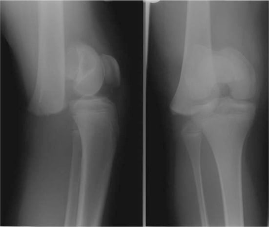
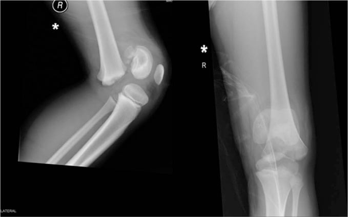
Displacement. Each mechanism has its own displacement pattern:
- Hyperextension - anterior displacement of the distal fragment
- Varus stress - lateral displacement of the distal fragment
- Valgus stress - medial displacement of the distal fragment
Clinical Assessment
History. Establish the mechanism, whether twisting, hyperextension or a direct blow, and ask about neurovascular symptoms such as numbness or a cold foot.
Examination. Inspect for swelling, deformity and skin tenting from a posterior spike, and palpate for tenderness around the distal femur. The neurovascular examination is the critical part:
- Popliteal pulse (may need Doppler), dorsalis pedis and posterior tibial pulses
- Capillary refill and foot warmth
- Peroneal nerve function (foot drop)
- Compartments, for compartment syndrome, especially with a vascular injury
The Neonatal Distal Femoral Physeal Separation
The birth injury is worth knowing separately, because it is radiographically occult and classically misdiagnosed.
Why it is occult. At birth the distal femoral epiphysis is only just ossifying, a small ossific nucleus, and the separation runs largely through cartilage. The plain radiograph therefore shows no obvious fracture line, only subtle displacement of the metaphysis relative to the barely visible epiphysis.
The mimics. A swollen, pseudoparalysed, irritable knee in a neonate is mistaken for septic arthritis of the knee, congenital knee dislocation, or a fracture elsewhere. The physeal separation is missed if the film is read as normal.
Confirming it. Ultrasound is the key test. It shows the cartilaginous epiphysis displaced relative to the metaphysis, with an effusion, and needs no radiation. MRI or a gentle arthrogram are alternatives, and comparison with the other knee helps.
Context and management. It follows a difficult, often breech, delivery. As with any unexplained infant fracture, non-accidental injury must be considered if the history does not fit. Management is gentle splinting in flexion; prognosis is excellent, with rapid remodelling and low arrest risk unlike the older child, so aggressive reduction is avoided.
Investigations
Radiographs. AP and lateral views are standard, and include the knee and proximal tibia. Stress views may help identify an occult Type I, under anaesthesia if needed, but MRI is safer.
CT. For Type III and IV injuries CT maps the intra-articular fracture, defining the articular fragment geometry and guiding screw trajectory. Plain radiographs can underestimate displacement in intra-articular physeal injuries, so cross-sectional imaging is required for operative planning.
MRI. MRI is useful for occult injuries or to assess physeal damage after reduction. It complements CT by showing the cartilaginous physis and the associated soft-tissue injury.
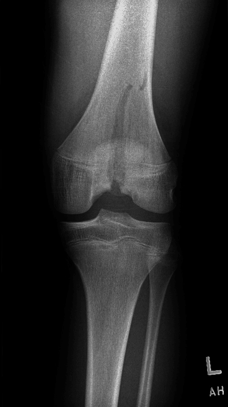
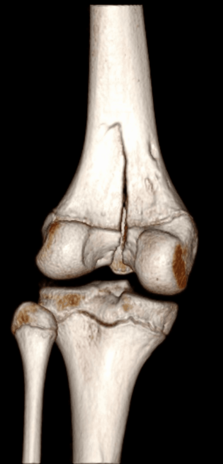
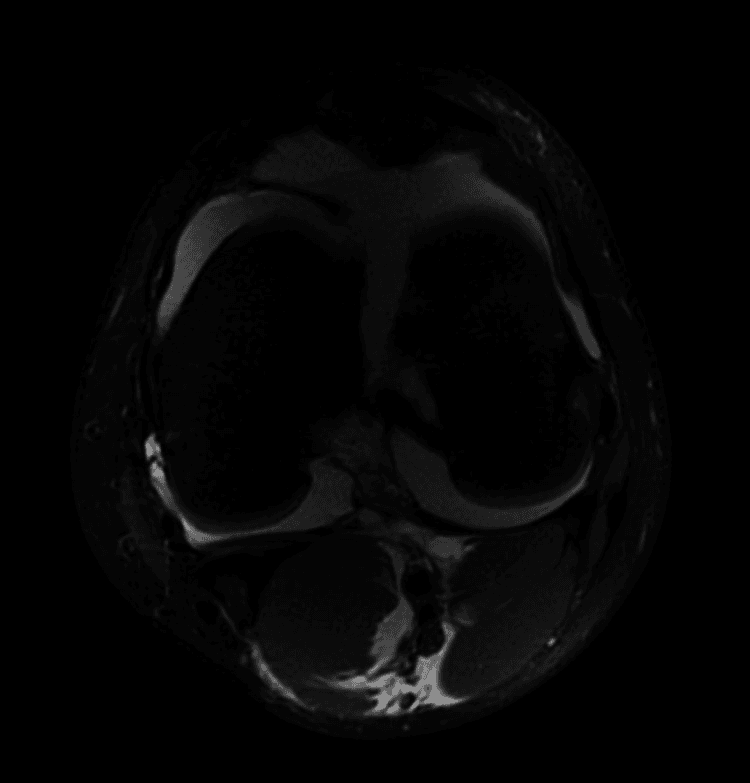
Vascular assessment. Measure the ankle-brachial index if pulses are diminished, and obtain CT angiography if a vascular injury is suspected on hard or soft signs.
Differential Diagnosis
- Distal Femoral Physeal Fracture: Point tenderness over the distal femoral physis.
- Patellar Dislocation: Apprehension sign positive. Patella may be back in place.
- ACL Tear: Hemarthrosis. Lachman positive. Usually post-skeletal maturity.
- Tibial Eminence Fracture: Extension block. X-ray shows avulsed tibial spine.
- Meniscal Injury: Locking, clicking. Usually after twisting.
- Proximal Tibial Physeal Fracture: Tenderness over the proximal tibial physis.
- Pathological Fracture: Through tumor. Night pain, constitutional symptoms.
- Point tenderness over the physis indicates physeal injury.
- Hemarthrosis is common in ligament tears and intra-articular fractures.
- Always get a good lateral X-ray to assess the physis.
Management
Non-displaced or minimally displaced fractures. A long leg cast, watched closely for displacement over the first weeks (protocol under Postoperative Care). Prophylactic pinning should be considered even for non-displaced fractures, because the stakes are high.
Displaced Type I and II. Gentle closed reduction, a single attempt preferred, then percutaneous pinning with smooth K-wires, or with cannulated screws placed in the metaphysis or epiphysis and not crossing the intact physis. Avoid crossing the physis; if you must, use a small diameter and remove it early. A long leg cast follows reduction.
Type III and IV. These are intra-articular, and anatomical reduction is mandatory. Open reduction goes through a medial or lateral approach depending on where the fragment lies, and fixation is with epiphyseal screws parallel to the joint, avoiding the physis, or smooth K-wires as an alternative. A long leg cast follows.
Surgical Technique
Closed reduction and percutaneous pinning. Indicated for displaced Type I and II fractures with an acceptable closed reduction.
- Reduce under fluoroscopy with traction and reversal of the deforming force. For hyperextension injuries, flex the knee and apply posterior force to the distal fragment
- Pass 2-3 smooth K-wires (2.0-2.4 mm) from the metaphysis into the epiphysis, crossing the fracture
- Place the pins divergently for stability, and avoid the intercondylar notch (ACL)
- Cut flush or bury
Open reduction and internal fixation. Indicated for Type III and IV fractures, or a Type II with intra-articular extension.
- Medial parapatellar or lateral approach, depending on the fragment
- Visualise the articular surface, reduce, and fix provisionally with K-wires
- Definitive fixation with 4.0 or 4.5 mm cannulated screws placed within the epiphysis, parallel to the joint surface; threaded hardware does not cross the intact physis
- Bone graft if there is significant comminution or physeal damage
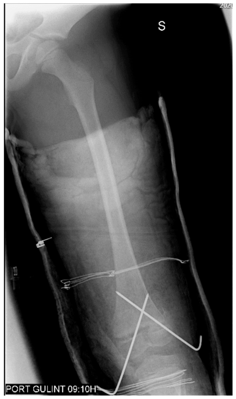
Protecting the physis and the limb. Avoid excessive periosteal stripping, to preserve the blood supply. Document neurovascular status before and after reduction.
Complications
- Rate
- 30-50%
- Prevention/Management
- Anatomical reduction. Avoid iatrogenic damage.
- Rate
- Common (Varus/Valgus), 1° a year
- Prevention/Management
- Bar excision if less than 50%, Osteotomy if more.
- Rate
- Common
- Prevention/Management
- Monitor. Epiphysiodesis or limb lengthening.
- Rate
- 1-2% (approximately 2% for displaced fractures)
- Prevention/Management
- Vascular assessment. Urgent repair if injured.
- Rate
- Rare
- Prevention/Management
- Document pre-op. Avoid traction.
- Rate
- Variable
- Prevention/Management
- Early ROM after healing.
Where the numbers come from. The arrest and vascular figures are conventional teaching ranges. The measured anchors are the cited series: Arkader 2007 found a 40% overall complication rate, growth arrest the commonest, with Salter-Harris grade and displacement predictive; Adams 2020 found 36%, unchanged despite a lower surgical threshold. Yamamura 2023 found 5.0% clinically significant growth disturbance across all lower-limb physes, with the distal femur and proximal tibia the highest-risk.
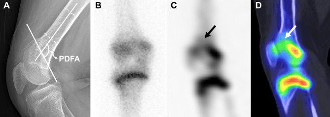
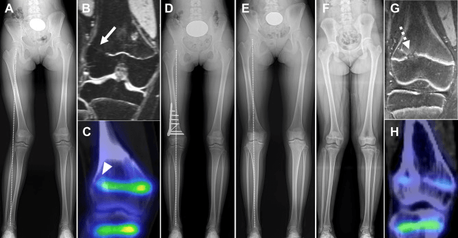
Watching for arrest. Growth-plate asymmetry may appear before clinically obvious deformity, which is why surveillance starts early. The discrepancy that follows is judged on measurement over time, not on a single scanogram, and that series determines the projected discrepancy and the timing of reconstruction.
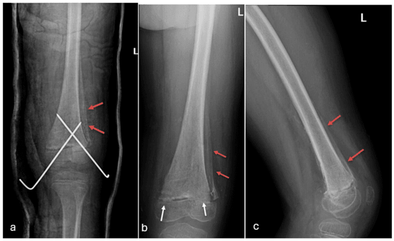
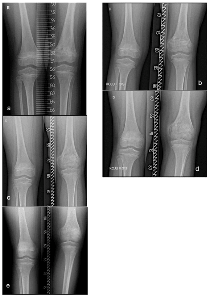
Mapping an arrest. Metabolic imaging can clarify an arrest pattern when the affected physeal segment is difficult to define on radiographs. Bone SPECT/CT maps absent uptake at a bridge against residual activity in the rest of the physis, informing whether bar resection and guided growth remain feasible.
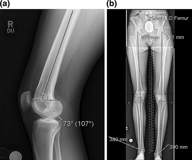
Angle and length together. Deformity analysis must define both the angular and the length components before osteotomy or lengthening, as in this flexion deformity with femoral shortening.
Postoperative Care
Wire removal. Screws can stay; smooth wires come out. Murgai found pin-tract infection rose once pins stayed 30 days or more (11.2% vs 1.4%), so within the 4-6 week window below, removal by about 4 weeks limits pin-tract infection.
Immobilisation and Follow-up Protocol
- Long leg cast (knee 20-30° flexion) for 6 weeks; non-weight bearing initially, then protected weight bearing
- Weekly X-rays for the first 2-3 weeks to watch for displacement
- Smooth-wire removal at 4-6 weeks
- X-ray to assess healing; cast removal if united
- Begin range-of-motion rehabilitation (see Rehabilitation Protocol)
- 6-month and 12-month X-rays with scanograms to assess growth
- Watch for Harris growth-line asymmetry, angular drift and leg-length discrepancy
- Annual follow-up with scanograms until skeletal maturity - Yamamura found every clinically significant growth disturbance declared within 2 years, but late bars are described, so surveillance continues
Rehabilitation Protocol
Rehabilitation
- Long leg cast, knee in 20-30° of flexion; non-weight bearing
- Toe wiggling and calf pumps; hip and ankle ROM within cast constraints
- Cast removal at 6 weeks if healing confirmed; hinged knee brace initially
- Partial weight bearing progressing to full weight bearing
- Active and passive knee ROM; quadriceps and hamstring isometrics
- Progressive resistance and closed kinetic chain exercises (squats, leg press)
- Proprioception and balance training; gait normalisation
- Sport-specific training
- Full ROM and greater than 90% strength
- Clearance by surgeon (confirm no growth disturbance)
Outcomes and Prognosis
Growth arrest. Arrest occurs in 30-50% of cases, regardless of Salter-Harris type. The by-type figures are conventional estimates rather than measurements, since neither cited series publishes arrest rates by Salter-Harris type:
- Type I and II - still a significant risk, conventionally quoted at 25-35%
- Type III and IV - higher, conventionally quoted at 40-60%; Salter-Harris grade did predict complications in Arkader 2007 (P=0.031)
What decides the result. Prognosis depends on the quality of reduction, the amount of growth remaining and the size of any physeal bar. Functional outcomes are generally good if the leg-length discrepancy and angular deformity are managed.
Always warn families about growth arrest, angular deformity and leg-length discrepancy. A 2-year minimum follow-up is mandatory.
Guidelines, Registries & Global Practice
Global epidemiology
- Distal femoral physeal fractures represent roughly 1-5% of all physeal injuries and under 1% of paediatric fractures, but carry disproportionate morbidity.
- Peak incidence is in adolescence (mean age 10-13 across published series), male predominant, typically from sport, road traffic or high-energy mechanisms.
- Clinically significant growth disturbance requiring secondary surgery occurs in around 5% across all lower-limb physeal fractures, but is concentrated in the distal femur — older single-centre distal-femur series report overall complication rates of 36-40%.
Side-by-side guidance (principles converge globally)
- Reduction
- Anatomical, gentle, single attempt
- Fixation
- Smooth K-wires across physis; screws confined to epiphysis/metaphysis
- Follow-up
- To skeletal maturity given arrest risk
- Reduction
- Anatomical for displaced/intra-articular
- Fixation
- CRPP for SH I/II; ORIF for SH III/IV
- Follow-up
- Serial radiographs, scanograms to maturity
- Reduction
- Urgent NV assessment; anatomical reduction
- Fixation
- Stable fixation; document NV status pre/post
- Follow-up
- Structured paediatric trauma follow-up
- Reduction
- Restore physeal + articular anatomy
- Fixation
- Implants avoiding transphyseal threads
- Follow-up
- Counsel + surveil for growth arrest
There is no major guideline disagreement: all converge on anatomical reduction, physeal-respecting fixation, urgent neurovascular assessment for displaced (hyperextension) patterns, and surveillance to maturity. Debate is confined to nuances (prophylactic pinning of non-displaced fractures, transphyseal screw use).
Registry and resource notes
- Paediatric physeal fractures are not captured by arthroplasty registries (NJR, AJRR, AOANJRR); evidence rests on single- and multi-centre cohorts rather than implant registries.
- High-resource settings: ready access to fluoroscopy, CT/MRI for bar mapping, EOS/low-dose scanograms for growth monitoring, and on-site vascular surgery for hyperextension injuries.
- Limited-resource settings: reliance on plain radiographs and clinical leg-length monitoring; physeal bar resection, guided growth and limb-reconstruction services may be unavailable, shifting practice toward contralateral epiphysiodesis or osteotomy for established deformity. Delayed presentation and missed neurovascular compromise are greater risks.
Controversies and Areas of Uncertainty
Prophylactic pinning of non-displaced fractures. A high late-displacement risk argues for stabilisation, but many non-displaced fractures heal well in a cast. Practice varies and no randomised data exist.
Does surgery reduce growth arrest? Adams/Arkader (2020) showed that a lower surgical threshold did not lower the complication rate (36% vs 40%), suggesting that arrest is largely intrinsic to the injury rather than modifiable by treatment.
Transphyseal screw fixation. Smooth wires are preferred, but stable Salter-Harris II fixation sometimes requires metaphyseal screws. Whether brief transphyseal smooth-wire passage independently worsens arrest is debated; Arkader noted a trend, not significance.
Bar resection versus accept-and-reconstruct. For bars under ~25-50% with growth remaining, resection plus guided growth can restore alignment. Rebound deformity and unpredictable physeal recovery (Masquijo 2020) lead some to favour epiphysiodesis or osteotomy.
Optimal imaging for arrest detection. MRI maps bars accurately, but its timing, cost and the need for sedation in young children remain unresolved against serial scanograms.
Deep Dive: Managing Growth Arrest
Types of Arrest
- Complete Arrest: Entire physis stops growing. Results in shortening only (no angular deformity).
- Partial (Central) Arrest: Bar in the center. Causes shortening and may cause "tenting" of the physis.
- Partial (Peripheral) Arrest: Bar on one side. Causes angular deformity as the unaffected side keeps growing.
Evaluation
- Scanogram: Leg length measurement.
- MRI: Maps the physeal bar (location, size).
- Bone Age: Estimate remaining growth.
Management Options
- Bar less than 50%, Greater than 2 years growth remaining: Bar excision + fat/PMMA interposition.
- Bar greater than 50%, or Less than 2 years growth remaining: Bar excision will fail. Consider:
- Contralateral epiphysiodesis (for LLD).
- Limb lengthening (for significant LLD).
- Corrective osteotomy (for angular deformity).
- Guided growth (hemi-epiphysiodesis for angular correction if some growth remains).
Parent's Guide: Understanding Distal Femoral Injuries
What is the distal femoral growth plate? The distal femur (thighbone near the knee) has a growth plate that is responsible for 70% of the thighbone's growth. Injury to this growth plate is serious because it can affect how your child's leg grows.
Why is this injury different? Unlike most other growth plate injuries, the distal femoral growth plate has a HIGH chance (30-50%) of developing problems with growth, even if the treatment is perfect. This may lead to:
- One leg being shorter than the other.
- One leg growing crooked.
What are the treatment options if growth problems occur? If growth problems develop, your doctor may recommend:
- Surgery to remove the damaged area and allow growth to resume.
- Surgery on the opposite leg to slow its growth (so the legs end up the same length).
- Surgery to straighten a crooked leg.
What follow-up is needed? Your child will need regular X-rays for at least 2 years to monitor the growth plate. Please do not miss these appointments, as early detection of problems allows for better treatment.
Surgical Pearls
Reduction Technique
- Apply longitudinal traction with the knee slightly flexed.
- For hyperextension injuries (anterior displacement of the distal fragment), flex the knee and push the distal fragment posteriorly.
- Avoid excessive force. One smooth reduction attempt is better than multiple aggressive attempts.
Pinning Technique
- Enter from the lateral and medial metaphysis, above the physis.
- Direct the wires across the fracture into the epiphysis.
- Diverge the wires for three-point fixation stability.
- Avoid the intercondylar notch (risk of ACL damage).
- Image in two planes (AP and lateral) to confirm position.
Avoiding Iatrogenic Damage
- Use smooth wires (not threaded).
- If threaded screws are used (for epiphyseal fixation), ensure they are parallel to the physis and do not cross it.
- Limit drill passes through the physis.
- Use small diameter implants.
Post-Reduction Checks
- Confirm neurovascular status immediately.
- Document pulses both before and after reduction.
- Obtain X-rays to confirm anatomical reduction.
Comparison: Distal Femur vs Other Physes
- Distal Femur
- 70% femur
- Proximal Tibia
- 55% tibia
- Distal Radius
- 75% radius
- Distal Femur
- 30-50%
- Proximal Tibia
- 20-30%
- Distal Radius
- Less than 5%
- Distal Femur
- Popliteal artery
- Proximal Tibia
- Popliteal artery
- Distal Radius
- Rare
- Distal Femur
- High
- Proximal Tibia
- High
- Distal Radius
- Low
MCQ Practice Points
Q: What percentage of femoral length does the distal femoral physis contribute? A: 70%. This is the highest of any physis in the body.
Q: What is the approximate growth arrest rate for distal femoral physeal fractures? A: 30-50%, regardless of Salter-Harris type.
Q: What vascular structure is at risk in distal femoral physeal injuries? A: Popliteal Artery. It is tethered by genicular branches and can be injured in hyperextension.
Q: What fixation method is preferred for Type II distal femoral physeal fractures? A: Smooth K-wire percutaneous pinning (avoids crossing intact physis with threaded hardware).
Q: What is the maximum physeal bar size amenable to bar excision? A: Less than 50% of the physis width, with at least 2 years of growth remaining.
Q: How long should patients with distal femoral physeal injuries be followed? A: At least 2 years with annual scanograms to skeletal maturity to detect growth disturbance.
Self-Assessment Quiz
Viva Scenarios
Practise clinical reasoning and management decisions out loud
“12-year-old with a Salter-Harris Type II distal femur fracture with anterior displacement of the distal fragment. Pulses palpable.”
“Same patient as above. After closed reduction, the foot is cold and the DP pulse is not palpable.”
“10-year-old, 1 year post Type II distal femur fracture. Now has 2.5cm LLD and 10 degrees of valgus.”
“You are consenting a family for CRPP of a displaced SH II distal femur. What specific risks do you discuss?”
“A neonate is noted to have decreased movement of the right leg after a difficult delivery. X-ray shows physeal widening at the distal femur.”
“16-year-old footballer with a valgus stress injury to the knee. Tender over the medial femoral condyle physis. X-ray shows a Salter-Harris III pattern.”
“8-year-old polytrauma after MVA. Bilateral distal femoral physeal fractures (Type II). Hemodynamically stable after resuscitation.”
KEY FACTS
- 70% Femoral Growth
- 35% Leg Length
- 30-50% Arrest Rate
- Popliteal Artery Risk
TREATMENT
- Anatomical Reduction
- Smooth Pin Fixation
- Avoid Crossing Physis
- 6+ Week Immobilization
COMPLICATIONS
- Growth Arrest
- LLD
- Angular Deformity
- Vascular Injury
FOLLOW-UP
- Weekly X-rays (2-3 wks)
- 6-Month Scanogram
- Annual to Maturity
- Warn Family
Evidence Base
Arkader et al (Predicting Outcome)
- Two-centre series of 73 children (59 boys, mean age 10); 59% Salter-Harris II
- Overall complication rate 40%, growth arrest the most frequent
- Salter-Harris grade (P=0.031) and displacement (48.8% vs 26.6%, P<0.0001) predicted complications; physeal violation by hardware trended worse (65% vs 30%)
Adams, Arkader et al (Lower Surgical Threshold)
- 70 children (mean age 13); 70% SH-II, 84% displaced, 90% treated surgically
- Complication incidence 36% with growth arrest in 20 patients — unchanged versus the pre-2007 40% cohort (P=0.751)
- Lower threshold for surgery did NOT reduce complications; high-energy mechanism and greater displacement carried higher risk
Yamamura et al (Epidemiology of CSGD)
- 1,585 lower-limb physeal fractures; clinically significant growth disturbance (CSGD) incidence 5.0% overall
- EVERY CSGD occurred within 2 years of injury
- Distal femoral and proximal tibial fractures requiring surgery carried the highest CSGD risk
Riseborough, Barrett, Shapiro
- Landmark long-term series of distal femoral physeal fracture-separations
- Leg-length discrepancy and angular deformity are common late sequelae
- Established the high growth-disturbance profile of this physis
Masquijo et al (Bar Resection + Guided Growth)
- 5 children with distal femoral physeal bars (mean bar 16.8% of physis) treated by resection, fat interposition and tension-band plate
- 4 of 5 corrected deformity and resumed longitudinal growth at ~14 months
- Rebound valgus occurred in 2 patients, requiring observation or repeat guided growth
Murgai et al (Percutaneous Pinning Safety)
- 163 distal-femur retrograde percutaneous pinnings (21 physeal fractures)
- Pin-tract infection 6.7%; NO cases of septic arthritis despite intra-articular pins
- Pin duration of 30 days or more increased pin-tract infection (11.2% vs 1.4%)
AO Foundation / AAOS Paediatric Trauma Principles
- Anatomical reduction and stable fixation of physeal fractures to restore physeal and articular alignment
- Smooth K-wires for transphyseal fixation; threaded implants kept within epiphysis or metaphysis
- Counsel families regarding growth-arrest risk and arrange surveillance to skeletal maturity