Limb Salvage | Skeletally Immature | Growth Preservation | Non-Invasive Expansion
- Predicted limb length discrepancy greater than 4cm is the primary indication
- Distal femur contributes 70% of femoral growth (1cm/year)
- Non-invasive mechanisms reduce infection risk vs modular designs
- Growth arrest after resection of physis occurs - calculate expected discrepancy
- Conversion to adult prosthesis typically at skeletal maturity
- “Use multiplier method for limb length prediction
- “Timing of expansion every 3-4 months to match growth
- “Soft tissue envelope limits total expansion achievable
- “Nerve palsy risk with each lengthening episode
Overview and Epidemiology
Expandable endoprostheses allow limb salvage in skeletally immature patients with malignant bone tumours while accommodating the growth still to come. Without them, a child with significant growth remaining would face a limb-length discrepancy (LLD) greater than 4cm after a tumour resection that includes the growth plate.
Who and where. Primary bone sarcomas are rare, approximately 8-9 per million children and adolescents per year worldwide, and osteosarcoma and Ewing sarcoma account for the majority of those requiring a growing reconstruction. Osteosarcoma peaks at 15-19 years and Ewing sarcoma at 10-15 years. The common sites are:
- Distal femur - 40%
- Proximal tibia - 20%
- Proximal humerus - 15%
History. Scales and Sneath developed the first expandable prosthesis at the Royal National Orthopaedic Hospital (Stanmore) in 1976, and the early designs needed open surgery to exchange modular segments. Non-invasive magnetic technology (REPIPHYSIS) arrived in the early 2000s and significantly reduced the operative burden.
Indications. The indications for an expandable prosthesis are:
- Predicted LLD greater than 4cm at skeletal maturity
- A malignant bone tumour whose resection takes the physis
- A patient and family who accept multiple procedures and prolonged follow-up
- An adequate soft-tissue envelope for reconstruction
A pathological fracture through the tumour is a relative contraindication.
GROWGROW - Indications for Expandable Prosthesis
Hook:Think GROW - these prostheses allow continued skeletal growth in children
Anatomy of Growth and LLD Prediction
Physeal contribution. Accurate prediction of the discrepancy is essential for planning, because resecting a physis arrests the growth it would have supplied, and that contribution varies by site.
- Contribution to Bone Length
- 70% of femur
- Growth Rate
- 10mm/year
- Contribution to Bone Length
- 57% of tibia
- Growth Rate
- 6mm/year
- Contribution to Bone Length
- 30% of femur
- Growth Rate
- 4mm/year
- Contribution to Bone Length
- 43% of tibia
- Growth Rate
- 5mm/year
- Contribution to Bone Length
- 80% of humerus
- Growth Rate
- 8mm/year
- Contribution to Bone Length
- 75% of radius
- Growth Rate
- 5mm/year
- Contribution to Bone Length
- 80% of ulna
- Growth Rate
- 5mm/year
The multiplier method. Paley's multiplier method gives the most accurate prediction of LLD, using skeletal age:
- Bone-age radiograph of the left hand and wrist
- Read the multiplier from the published tables for skeletal age and sex
- Remaining growth = current limb length x (multiplier - 1)
- Predicted LLD = the share of that remaining growth the resected physis would have supplied, since it supplies none once resected
A worked example. A 10-year-old boy (skeletal age 10) with osteosarcoma of the distal femur needs a resection that takes the physis. His femur is 35cm long and the multiplier for a 10-year-old boy is 1.28, so the femur has 35 x (1.28 - 1) = 9.8cm still to grow. The distal femur would have supplied 70% of that, a predicted LLD of 6.9cm, and he needs an expandable prosthesis.
Growth prediction has inherent uncertainty. Chemotherapy and radiotherapy can blunt remaining growth by an uncertain amount. Always counsel families that actual discrepancy may differ from predicted values. Plan for worst-case scenario when selecting prosthesis expansion capacity.
Timing of expansion. Lengthening is typically done every 3-4 months to match physiological growth, more often in younger children who grow faster, aiming for 5-10mm per episode.
The soft tissues set the limits. Nerve stretch, muscle contracture and skin tension limit a single lengthening to 10-15mm at most, and the soft-tissue envelope limits the total a prosthesis can deliver, typically 80-100mm. If growth outruns that capacity, the prosthesis may need to be exchanged.
Acute lengthening greater than 15mm risks nerve palsy, particularly the peroneal nerve in lower limb prostheses. Symptoms include foot drop and numbness. If detected, immediate shortening may be required. Pre-operative counselling must include this risk.
Classification Systems - the Henderson Failure Modes
The five modes. Henderson and colleagues reviewed 2,174 tumour endoprostheses across five institutions (534 failures) and defined five primary modes of failure, each stratified by amputation risk and treatment urgency. It is the shared vocabulary examiners expect for endoprosthetic complications, and the one registries use to standardise reporting.
- Failure Mode
- Soft-tissue failure
- What It Means
- Instability, dislocation, tendon/extensor-mechanism failure, wound breakdown
- Typical Management
- Soft-tissue or flap reconstruction, bracing
- Failure Mode
- Aseptic loosening
- What It Means
- Loss of fixation at the bone-implant interface
- Typical Management
- Revision with longer or uncemented stem
- Failure Mode
- Structural failure
- What It Means
- Fracture of the implant, stem, or expansion mechanism
- Typical Management
- Component exchange or revision
- Failure Mode
- Infection
- What It Means
- Deep periprosthetic infection
- Typical Management
- DAIR (early), two-stage revision or amputation (late)
- Failure Mode
- Tumour progression
- What It Means
- Local recurrence involving the reconstruction
- Typical Management
- Re-resection or amputation
Mapping the complications. The expansion-mechanism breakage shown under Complications is a Type 3 structural failure, and the elbow dislocation after distal humeral reconstruction is a Type 1 soft-tissue failure. The aseptic loosening (20-30%) and deep infection (10-15%) figures are Type 2 and Type 4.
What is commonest depends on where you look. Infection was the most common mode in Henderson's own series, whereas aseptic loosening dominated in the pooled literature. Failure mode depends on anatomic location and on time to failure, so cumulative reporting that lumps all sites together masks site-specific trends: report failures per anatomic location, not in aggregate.
In a viva on endoprosthetic complications, classify each problem by Henderson type as you go: it signals a structured, literature-anchored approach. In the growing child the same taxonomy applies, but expansion-mechanism (Type 3) and repeat-surgery-driven infection (Type 4) carry extra weight because the implant is lengthened many times over years.
Henderson Failure-Mode Classification for Tumour Endoprostheses
- Multicentre review of 2,174 tumour endoprostheses across five institutions; 534 failures analysed
- Five failure modes defined: soft-tissue (1), aseptic loosening (2), structural (3), infection (4), tumour progression (5)
- Infection was the commonest mode in this series; aseptic loosening dominated the pooled literature
- Failure mode depends on anatomic location and time-to-failure - avoid cumulative aggregate reporting
Antibiotic Prophylaxis Duration: the PARITY Trial
The trial. PARITY was a blinded, multicentre randomised superiority trial across 48 sites in 12 countries. It enrolled patients with a primary bone tumour, a sarcoma invading the femur or tibia, or oligometastatic femoral or tibial disease who required excision and endoprosthetic reconstruction, the population that includes paediatric and adolescent limb-salvage patients. They were randomised to a 1-day or a 5-day postoperative regimen of intravenous cephalosporin (cefazolin or cefuroxime).
The result. Prolonging prophylaxis to 5 days did not reduce surgical site infection: SSI within 1 year occurred in 15.0% of the 5-day group and 16.7% of the 1-day group (hazard ratio 0.93, 95% CI 0.62 to 1.40, P equals 0.73). Antibiotic-related complications were significantly more common with the longer course, 5.1% against 1.6% (hazard ratio 3.24, P equals 0.02).
What to take from it. A short, approximately 24-hour, prophylactic regimen is appropriate; extending antibiotics does not lower infection and causes more harm. An SSI rate of roughly 15-17% in a controlled trial also shows how infection-prone tumour endoprostheses are, consistent with the 10-15% deep-infection figure. PARITY settles the five-days-versus-one question for oncologic endoprostheses; general perioperative infection-prevention bundles (skin preparation, normothermia, timing of the first dose) are covered in the surgical-site-infection topic.
PARITY is Level I evidence you can quote directly. In the growing child each lengthening episode is a fresh potential inoculation event, so infection vigilance is lifelong - but the answer is meticulous technique and surveillance, not longer prophylaxis.
PARITY - Duration of Antibiotic Prophylaxis for Tumour Endoprostheses
- Blinded RCT, 48 sites in 12 countries, 604 analysed; lower-limb bone tumour requiring endoprosthetic reconstruction
- 1-day vs 5-day postoperative IV cephalosporin; primary outcome surgical site infection within 1 year
- SSI 15.0% (5-day) vs 16.7% (1-day), HR 0.93 (95% CI 0.62-1.40, P=0.73) - no benefit from a longer course
- Antibiotic-related complications higher with 5 days (5.1% vs 1.6%, HR 3.24, P=0.02)
Clinical Assessment
Oncological assessment. The staging imaging is set out under Investigations. Biopsy is by core needle, with the tract planned so that it can be excised with the tumour. The response to neoadjuvant chemotherapy is assessed clinically and radiologically, and the resection is planned for wide margins, a minimum of 2cm from the tumour.
Growth and function. The bone age, limb-length measurement and multiplier calculation predict the discrepancy at skeletal maturity. Document the baseline joint range of motion for later comparison.
The patient and family. Five domains are assessed:
- Assessment
- Chronological and skeletal
- Implication
- Expansion capacity needed
- Assessment
- Ability to attend follow-up
- Implication
- Critical for outcomes
- Assessment
- Expectations of multiple procedures
- Implication
- Counselling required
- Assessment
- Family circumstances
- Implication
- Impacts rehabilitation
- Assessment
- Coping mechanisms
- Implication
- Mental health support
Expandable or not. Five factors decide it.
- Favouring Expandable
- Greater than 4cm
- Favouring Non-Expandable
- Less than 2cm
- Favouring Expandable
- Younger (greater than 4 years growth remaining)
- Favouring Non-Expandable
- Adolescent near skeletal maturity
- Favouring Expandable
- Distal femur, proximal tibia
- Favouring Non-Expandable
- Upper limb (discrepancy better tolerated)
- Favouring Expandable
- Adequate for reconstruction
- Favouring Non-Expandable
- Compromised by tumour/radiation
- Favouring Expandable
- Expected survival greater than 2 years
- Favouring Non-Expandable
- Poor prognosis - prioritise palliation
Investigations
Radiographs. Full-length views of the affected limb, and a chest radiograph as a baseline.
MRI of the entire bone, so that skip lesions are not missed. It also shows joint involvement, soft-tissue extension and the proximity of the neurovascular structures.
CT of the chest stages pulmonary metastases, and CT of the limb provides 3D planning for a custom prosthesis. Systemic staging is completed with a bone scan or PET-CT per protocol.
Growth assessment. A bone-age radiograph of the left hand and wrist, a scanogram or CT for accurate limb-length measurement, and then the multiplier calculation.
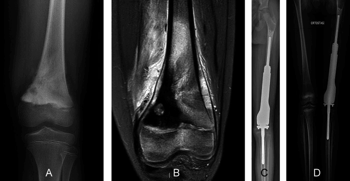
Expansion Mechanisms
Four families of mechanism lengthen the implant, and they trade the number of operations against mechanical reliability.
- Procedure Required
- Outpatient, external magnetic field
- Advantages
- No surgical procedures, lower infection risk
- Disadvantages
- Mechanical failure 15-20%, MRI incompatible
- Procedure Required
- Small incision to access port
- Advantages
- Lower infection than modular, reliable
- Disadvantages
- Requires anaesthesia, incision each time
- Procedure Required
- Open surgery to exchange segments
- Advantages
- Technically reliable, proven track record
- Disadvantages
- Highest infection risk, multiple surgeries
- Procedure Required
- Spring-loaded electromagnetic expansion, almost always outpatient
- Advantages
- Controlled expansion with spring
- Disadvantages
- Limited total expansion, spring less precise than active expansion
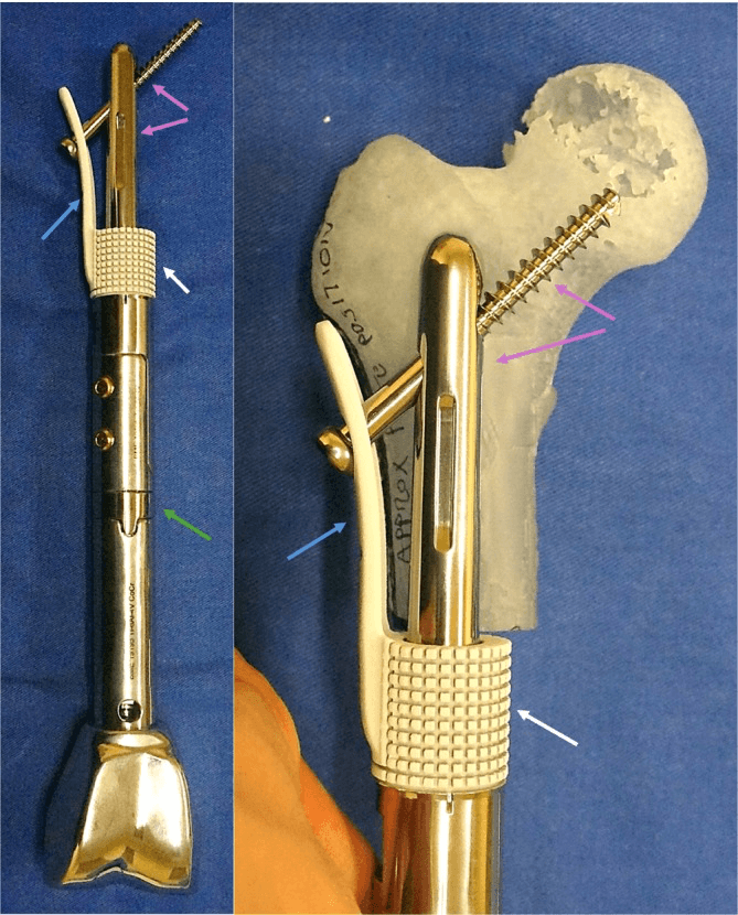
Why they are preferred. Non-invasive prostheses are the current preferred technology where available, because lengthening becomes an outpatient procedure with no incision. That removes the repeated operations, reduces infection risk and carries a psychological benefit for the child.
What they cost. Magnetic systems have higher mechanical failure rates than modular designs, and traditional designs are MRI incompatible. They are expensive and not available in all centres.
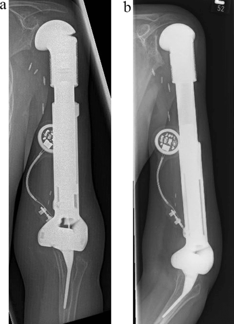
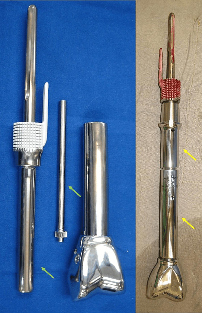
REPIPHYSIS (Wright Medical/Stryker). An external rotating magnetic field (ERC) activates an internal gearbox. Expansion is done as an outpatient without anaesthesia, takes 15-30 minutes and typically gains 4-8mm per session, with up to 100mm of total expansion.
Juvenile Tumour System (JTS, Stanmore Implants). An external electromagnetic field drives an internal motor. The design is modular for customisation, and MRI-conditional designs are now available.
Non-invasive mechanisms have 15-20% mechanical failure rates. Revision to modular prosthesis or exchange may be required. Regular radiographic monitoring for expansion function is essential.
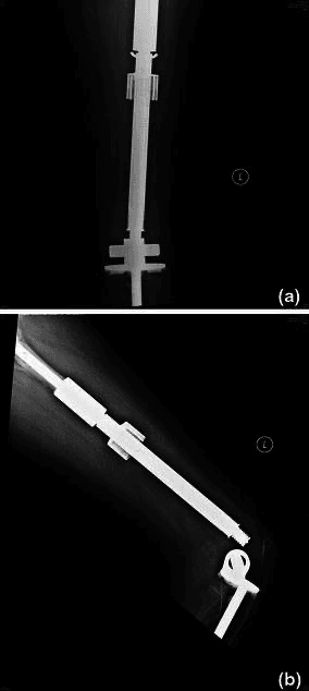
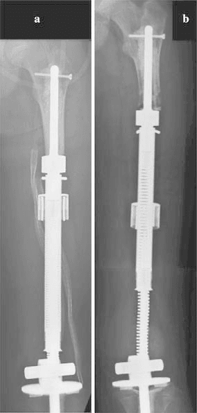
Differential of Reconstruction Options
The expandable endoprosthesis is one of several ways to reconstruct a skeletally immature limb, and examiners expect you to compare them and justify your choice.
- Growth Mechanism
- Internal lengthening (magnetic/modular/spring)
- Best Suited To
- Large predicted LLD, intra-articular distal femur/proximal tibia
- Key Drawback
- High infection and mechanical-failure burden over lifetime
- Growth Mechanism
- Ankle becomes knee; foot grows with child
- Best Suited To
- Very young child, large soft-tissue resection, infection-prone settings
- Key Drawback
- Cosmetic acceptance; requires prosthetic fitting
- Growth Mechanism
- No active growth (biological reconstruction)
- Best Suited To
- Diaphyseal or selected metaphyseal defects, joint-sparing
- Key Drawback
- Nonunion, fracture, resorption, disease transmission risk
- Growth Mechanism
- Living graft can hypertrophy and remodel
- Best Suited To
- Diaphyseal intercalary defects, distal radius
- Key Drawback
- Stress fracture, donor-site morbidity, prolonged protection
- Growth Mechanism
- None (definitive)
- Best Suited To
- Neurovascular involvement, recurrent infection, poor prognosis
- Key Drawback
- Limb loss; durable and low maintenance
For a very young child or where repeated lengthening/surveillance is impractical, rotationplasty offers durable, high-function, low-maintenance limb salvage with growth potential. It is the most important "differential" to raise against an expandable prosthesis in viva - showing you weigh durability and resource needs, not just technology.
Management Algorithm
- Confirm the oncology first. Staging complete, biopsy tract excisable, wide (R0) margins achievable: the reconstruction is chosen only after oncological clearance is planned (Morrison 2026).
- Calculate the predicted LLD from the bone age, the multiplier method and the expected growth of the resected physis.
- Branch on the discrepancy. A predicted LLD of 4cm or less: consider a non-expandable or short modular reconstruction, with or without contralateral epiphysiodesis. Greater than 4cm: an expandable endoprosthesis is indicated.
- Choose the mechanism. Non-invasive magnetic where available, for fewer procedures and lower infection exposure; modular where reliability and availability dominate, remembering the Ruggieri survival caveat under Controversies.
- Weigh the alternatives honestly. In the very young child, with poor soft tissues, an irradiated field (Jeys 2007 - postoperative radiotherapy raised infection from 9.8% to 35.3%) or limited follow-up resources, rotationplasty, biological reconstruction or amputation may serve better.
- Plan the whole pathway at the outset - the expansion schedule to maturity, surveillance and conversion to an adult prosthesis - and document it with the family before the first incision.
Surgical Technique
The team. The multidisciplinary meeting brings together the orthopaedic oncologist, paediatric oncologist, radiologist, pathologist and rehabilitation specialist.
Imaging review. Confirm the tumour extent and planned margins, identify the neurovascular proximity, take the measurements for custom prosthesis sizing and plan the soft-tissue reconstruction.
Selecting the prosthesis. The total expansion capacity must cover the predicted LLD with a margin to spare:
- Decision
- Based on centre expertise and availability
- Decision
- Cemented vs cementless (age-dependent)
- Decision
- Adequate fixation in remaining bone
- Decision
- Based on soft tissue sacrifice
- Decision
- Predicted LLD plus margin
Consent. The discussion covers:
- Limb salvage compared with amputation
- Deep infection risk of 10-15%
- Mechanical failure and revision
- The need for conversion at skeletal maturity
- Functional expectations, using the MSTS range under Outcomes
Complications
Failure of the lengthening mechanism is specific to these implants. The Henderson classification in the next section gives all of them a common vocabulary.
- Incidence
- 10-15%
- Risk Factors
- Multiple surgeries, chemotherapy immunosuppression
- Management
- Debridement and antibiotics, often requires amputation
- Incidence
- 20-30%
- Risk Factors
- Young active patients, cemented stems
- Management
- Revision surgery with longer stem
- Incidence
- 15-20%
- Risk Factors
- Non-invasive mechanisms, patient weight
- Management
- Prosthesis exchange or conversion
- Incidence
- 10-15%
- Risk Factors
- Poor initial coverage, radiation
- Management
- Flap coverage, revision
- Incidence
- 5-10%
- Risk Factors
- Rapid expansion, cumulative lengthening
- Management
- Shortening, observation, rarely permanent
Infection. Management follows the timing:
- Timing
- Less than 4 weeks
- Management
- Antibiotics, wound care
- Timing
- Less than 6 weeks
- Management
- DAIR (debridement, antibiotics, implant retention)
- Timing
- Greater than 6 weeks
- Management
- Two-stage revision or amputation
- Timing
- Recurrent
- Management
- Amputation often required
Single-stage revision is rarely successful.
Deep periprosthetic infection is the leading cause of amputation after limb salvage. Success rates for infection eradication in expandable prostheses are lower than in primary arthroplasty, driven by chemotherapy immunosuppression, a large implant surface and often irradiated soft tissue. No paediatric series cited here reports a two-stage reimplantation success rate - counsel that eradication is uncertain and amputation remains a real endpoint.
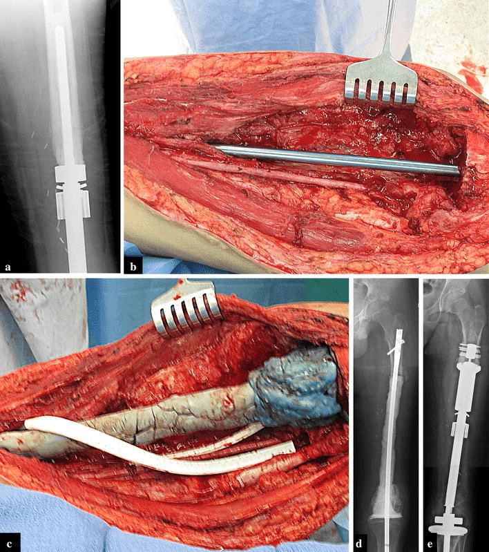
Mechanical failure. The failure modes differ by design:
- Non-invasive systems - motor burnout (most common), gear mechanism slippage, telescoping section jamming, electromagnetic coil malfunction
- Modular systems - locking mechanism failure, taper corrosion, component dissociation
Management is revision to an alternative expansion mechanism, conversion to a non-expandable prosthesis if near maturity, or a custom prosthesis for complex failures.
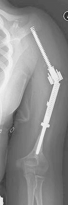
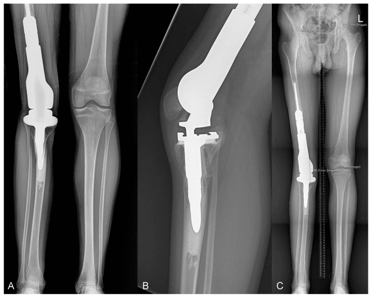
Loosening. Aseptic loosening at the stem is why surveillance is lifelong and extends beyond the completion of growth.
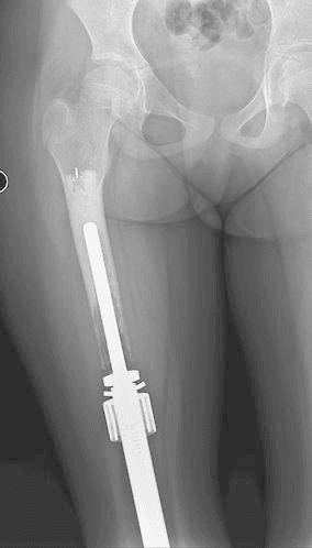
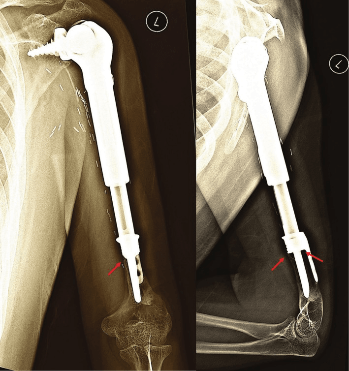
Soft-tissue failure includes instability and dislocation (Henderson Type 1).
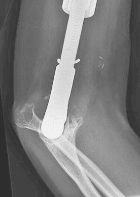
Growth-related problems. Asymmetric growth produces angular deformity, joint contracture limits function, and the lengthening can over- or under-correct the limb.
The implant is not permanent. Expect conversion to an adult prosthesis at skeletal maturity and multiple revisions over a lifetime.
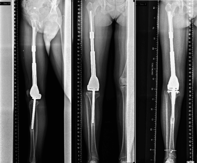
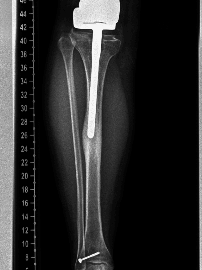
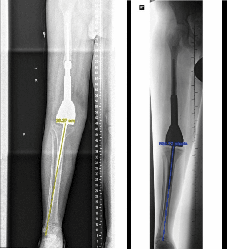
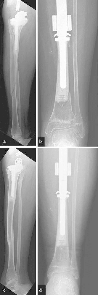
Amputation after initial limb salvage is a real and countable outcome. The best paediatric figure comes from the systematic review in the Evidence section: 5.2% of children underwent subsequent amputation, against 9.5% reported in adults. Families must be counselled at the outset that amputation remains a possible endpoint of limb salvage, not a failure of it.
Postoperative Care and Rehabilitation
The first days. An inpatient stay of days 0-3 for wound monitoring, with:
- DVT prophylaxis, mechanical and pharmacological
- Drain management
- Pain control, PCA then oral
- Non-weight bearing initially
Rehabilitation. Physiotherapy referral starts a four-phase programme that ends in lifelong surveillance.
Aims: wound healing, preventing contracture, protected mobility.
- Protected (touch or partial) weight bearing with a walking frame
- Active-assisted range of motion
- Quadriceps/hamstring isometrics
- Wound surveillance for infection
Aims: increase weight bearing, strengthen muscles.
- Progress to full weight bearing as tolerated
- Strengthening exercises
- Gait training
- Hydrotherapy if wound healed
Aims: return to school and activities, independent mobility.
- Sport-specific rehabilitation (non-contact)
- Stair climbing, community ambulation
- Psychological support
- Ongoing expansion procedures
Aims: monitor for oncological recurrence and prosthesis function.
- Regular expansion to match growth
- Oncological surveillance (chest imaging, local imaging)
- Prosthesis function assessment
- Conversion planning at skeletal maturity
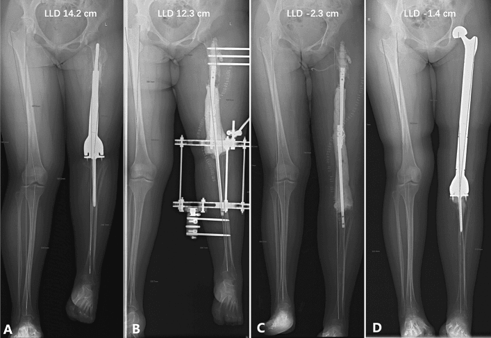
Conversion to an adult prosthesis. The expandable implant is exchanged at or near skeletal maturity, or earlier when its expansion capacity is exhausted or mechanical failure precludes further expansion. The conversion is an elective revision to a non-expandable prosthesis; it often needs a longer stem for fixation, may need bone grafting of expansion gaps, and is the moment to reassess joint constraint.
Outcomes and Prognosis
Oncological outcomes. Local recurrence is 5-10% with wide margins and higher with inadequate ones, and surveillance MRI is recommended. Five-year survival is 65-70% for osteosarcoma and 70-75% for Ewing sarcoma, and the response to chemotherapy is the key prognostic factor.
Implant survival at 5, 10 and 15 years:
- Implant Survival
- 70-80%
- Notes
- Reported higher with non-invasive; contested (see Controversies)
- Implant Survival
- 50-65%
- Notes
- Revision or conversion expected
- Implant Survival
- 30-40%
- Notes
- Multiple revisions likely
Function. Mean Musculoskeletal Tumor Society scores in the paediatric systematic review ranged from 71.0% to 86.8%, depending on reconstruction type and anatomical site. MSTS is surgeon-rated, not a patient-reported outcome. Scores are lower than for non-expandable prostheses in adults, and are affected by the multiple procedures. Limb salvage succeeds in 85-90%, with some activity restriction recommended and high-impact sports avoided.
Quality of life. Generally good when limb salvage succeeds, though repeated procedures have a psychological impact and support services are important.
Limb salvage with expandable prosthesis provides equivalent oncological outcomes to amputation when wide margins are achieved. Functional outcomes favour limb salvage for lower limb tumours. The choice should be individualised based on tumour location, patient factors, and family preferences.
Guidelines, Registries & Global Practice
Global Epidemiology
Primary bone sarcomas of childhood and adolescence are rare worldwide (approximately 8-9 per million children/adolescents annually). Osteosarcoma and Ewing sarcoma dominate, with peak incidence during the pubertal growth spurt. The proportion needing a growing reconstruction is driven by age at presentation and tumour site - distal femoral and proximal tibial lesions most often require growth-accommodating implants because they straddle the most active lower-limb physes.
Side-by-Side Guidance
- Position on Pediatric Limb Salvage
- Centralised sarcoma-unit care; wide (R0) margin is non-negotiable, reconstruction follows; expandable endoprosthesis is a standard growth-accommodating option
- Position on Pediatric Limb Salvage
- Suspected bone sarcoma referred to a specialist supra-network centre; MDT decides limb salvage vs amputation
- Position on Pediatric Limb Salvage
- Wide excision with limb-sparing reconstruction where margins and function allow; endoprosthesis among accepted reconstructions
- Position on Pediatric Limb Salvage
- Chemotherapy backbone with surgery timed to neoadjuvant response; surgery centralised in trial centres
Across all major bodies the principles converge: referral to a specialist sarcoma centre, MDT decision-making, R0 resection before reconstruction, and shared decision-making with family. Genuine differences are organisational (network structure) rather than oncological.
Registry and Outcome Notes
There is no dedicated international registry for paediatric expandable prostheses; evidence is dominated by single-centre and multi-institutional case series (Level IV). Adult tumour-endoprosthesis registries and the Henderson failure-mode classification (soft tissue, aseptic loosening, structural, infection, tumour progression) are widely applied to standardise reporting. Collaborative groups (e.g. PARITY trial network for infection prophylaxis) are improving the evidence base.
High- vs Limited-Resource Practice Variation
- Well-resourced centres: Non-invasive magnetic/electromagnetic systems (REPIPHYSIS, JTS, Stanmore non-invasive) with outpatient lengthening; 3D-printed custom implants; routine MDT and long-term surveillance.
- Limited-resource settings: Higher reliance on modular/minimally invasive expansion or non-expandable reconstruction; rotationplasty and amputation remain important, durable, cost-effective options where growing implants or repeated lengthening logistics are not feasible.
- Universal principle: Implant choice should match the local capacity for repeated follow-up, expansion, and revision - a sophisticated implant without reliable surveillance can underperform a simpler, durable reconstruction.
Controversies and Areas of Uncertainty
Non-invasive versus modular survival. Non-invasive systems reduce surgical episodes and infection exposure, yet some series (e.g. Ruggieri, where the open Kotz outperformed the Repiphysis) report lower implant survival for early magnetic devices. The optimal mechanism remains unsettled and is centre- and era-dependent.
Growth prediction. The multiplier method assumes normal growth, which treatment can blunt (Growth Prediction Uncertainty, above). Over- or under-estimation of the final LLD is common, and how aggressively to oversize expansion capacity is debated.
Cemented or cementless. Cementless or compress fixation may improve long-term survival in young active patients, but the evidence is limited and stem fixation in the immature skeleton is not standardised.
When to stop expanding. There is no consensus on the LLD to accept against the risks of repeated lengthening (nerve palsy, contracture, mechanical failure). Some advocate accepting a minor LLD with a shoe raise over pushing for every millimetre.
What the literature cannot yet answer.
- No randomised data compare expandable endoprosthesis with rotationplasty or biological reconstruction; all comparisons are Level IV.
- Patient-reported and long-term quality-of-life outcomes are inconsistently reported, limiting true device comparison.
- PARITY answers five days against one, but it was a largely adult trial that tested no shorter regimen, so the optimal prophylaxis duration for these high-risk implants in children is still being defined.
MCQ Practice Points
Q: What is the primary indication for an expandable prosthesis?
A: Predicted limb-length discrepancy greater than 4 cm at skeletal maturity after physeal resection.
Q: What is the distal femoral physeal contribution to growth?
A: About 70% of femoral growth, roughly 10 mm per year.
Q: What is the commonest reason for amputation after limb salvage?
A: Deep periprosthetic infection.
Q: What did the PARITY trial show?
A: A 5-day IV cephalosporin course did NOT reduce surgical site infection versus 1 day (15.0% vs 16.7%) and caused more antibiotic-related complications (5.1% vs 1.6%).
Q: What did Ruggieri 2013 find comparing the Kotz and Repiphysis prostheses?
A: The open Kotz prosthesis had significantly higher implant survival (p=0.026); only 3 of the 9 children reaching skeletal maturity had limb-length equality.
Q: What is the effect of postoperative radiotherapy on infection (Jeys 2007)?
A: Infection rose from 9.8% to 35.3%; 10-year infection-free survival fell to 44.8%.
Clinical Decision Scenarios
Practise clinical reasoning and management decisions out loud
“An 8-year-old boy presents with a 3-month history of knee pain. Radiographs and MRI confirm a high-grade osteosarcoma of the distal femur with metaphyseal involvement but no joint invasion. Staging shows no metastatic disease. He is receiving neoadjuvant chemotherapy with good response.”
“A 12-year-old girl had an expandable prosthesis inserted for proximal tibial Ewing sarcoma 18 months ago. She now presents with a draining sinus over the prosthesis with purulent discharge. She is afebrile but inflammatory markers are elevated (CRP 85, WCC 14).”
“A 14-year-old boy with a distal femoral non-invasive expandable prosthesis (inserted at age 10 for osteosarcoma) attends for routine expansion. The external magnetic device is applied but no expansion is achieved. Radiographs confirm the prosthesis has not lengthened despite multiple attempts.”
Key Indications
- Predicted LLD greater than 4cm at skeletal maturity
- Malignant bone tumour requiring physeal resection
- Distal femur contributes 70% of femoral growth (1cm/year)
- Proximal tibia contributes 57% of tibial growth (0.6cm/year)
Expansion Mechanisms
- Non-invasive (REPIPHYSIS, JTS) - lower infection, higher mechanical failure
- Modular (Stanmore) - higher infection from repeat surgery, reliable
- Self-expanding (Phenix) - spring mechanism, limited capacity
- Expansion every 3-4 months, 10-15mm per episode maximum
Critical Complications
- Deep infection 10-15% - leading cause of amputation
- Aseptic loosening 20-30%
- Mechanical failure 15-20% (non-invasive)
- Nerve palsy with over-expansion
Surgical Pearls
- Gastrocnemius flap for proximal tibia coverage
- Calculate LLD using multiplier method pre-op
- Select prosthesis with capacity exceeding predicted LLD
- Plan for conversion to adult prosthesis at maturity
Exam Triggers
- Child with bone sarcoma requiring physeal resection
- Calculating expected limb length discrepancy
- Comparing expansion mechanism options
- Managing infected expandable prosthesis
Evidence and Guidelines
Pediatric Limb-Salvage Outcomes - Systematic Review
- 60 studies (all Level IV) of endoprosthesis, allograft and APC reconstruction in children
- Infection was the primary mode of failure across all reconstruction types and locations
- Subsequent amputation lower in children (5.2%) than reported in adults (9.5%) (p=0.013)
- MSTS scores ranged 71.0% to 86.8%; residual leg-length discrepancy 13.4% at latest follow-up
The headline figures on this page are conventional teaching ranges anchored in the cited series: deep infection of 10-15% and its link to amputation (Groundland 2016; Jeys 2007; the 15-17% one-year SSI rate in the controlled PARITY trial), implant survival and the Kotz-versus-Repiphysis trade-off (Ruggieri 2013), the early Phenix experience (Neel 2003), non-invasive expansion results (Torner 2016), and the failure-mode taxonomy (Henderson 2011). The growth-contribution percentages (distal femur 70%, proximal tibia 57%) and the per-year growth rates are standard growth-plate teaching, and the multiplier method is Paley's. No randomised trial compares an expandable prosthesis with rotationplasty or biological reconstruction, and no paediatric series cited here reports a two-stage reimplantation success rate for infection - so none is quoted.