Small Round Cell Tumor | Second Most Common Pediatric Bone Malignancy | Multimodal Treatment Required
- Second most common primary bone malignancy in children (after osteosarcoma)
- EWS-FLI1 translocation t(11;22) present in 85% - diagnostic hallmark
- Onion-skin periosteal reaction classic radiographic finding
- Multimodal treatment mandatory: chemotherapy + surgery ± radiation
- Poor response to chemotherapy (under 90% necrosis) is worst prognostic factor
- “Fever and elevated inflammatory markers mimic infection - biopsy essential
- “VDC-IE protocol: Vincristine, Doxorubicin, Cyclophosphamide alternating with Ifosfamide, Etoposide
- “Surgical margins more important than degree of necrosis for local control
- “Metastases to lung, bone, bone marrow - whole-body staging required
Overview and Epidemiology
Ewing sarcoma is the second most common primary bone malignancy in children and adolescents, after osteosarcoma, and accounts for 10-15% of all primary bone tumours. Before chemotherapy arrived in the 1970s five-year survival was under 10%; multimodal treatment has taken it to 70% for localised disease. It is a highly aggressive small round cell tumour and it is treated urgently.
Who. Three quarters of patients are aged 10-20, a further 15% are 20-30, and only 10% are over 30. Males outnumber females 1.5:1. About 85% of patients are white, and the tumour is rare in African and Asian populations.
Where. The femur and the pelvis are the commonest sites:
- Femur - 25%, the commonest single site
- Pelvis - 20%, the worst prognosis of any site because of the size these tumours reach
- Tibia and fibula - 15%
- Humerus - 10%
- Ribs - 10%
- Axial skeleton - 20% (vertebrae, scapula)
The diaphysis. In 90% of extremity cases the tumour sits in the diaphysis of a long bone, which distinguishes Ewing from osteosarcoma, a metaphyseal tumour; lymphoma of bone may occupy either the metaphysis or the diaphysis. A pelvic tumour lies deep and its symptoms are delayed, so it is often large by the time anyone finds it; axial disease can present late, with neurological or visceral symptoms.
It is not only a bone tumour. True Ewing sarcoma is one member of the Ewing sarcoma family of tumours (ESFT), a group unified by the same EWSR1-ETS fusion, and the same CD99/NKX2-2 phenotype, regardless of where it arises. The members are skeletal Ewing sarcoma, extraskeletal (extraosseous) Ewing sarcoma arising primarily in soft tissue with no bone of origin, the Askin tumour (Ewing of the chest wall and thoracopulmonary region), and peripheral primitive neuroectodermal tumour (pPNET), the more neurally differentiated end of the same spectrum.
Extraskeletal Ewing tends to occur in slightly older patients and in axial, paraspinal or chest-wall sites. It is treated on the same multi-agent chemotherapy backbone as bone Ewing, but local control follows soft-tissue sarcoma surgical principles: wide excision of the soft-tissue mass, with radiotherapy where margins are constrained. The unifying concept for a viva is that the Ewing family is defined by its molecular fusion, not by whether it started in bone.
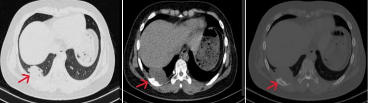
Pathophysiology and Molecular Biology
The fusion. The t(11;22)(q24;q12) translocation joins EWS to FLI1 and creates the EWS-FLI1 fusion oncogene in 85% of cases; the remaining 15% carry variant translocations such as EWS-ERG. The fusion product behaves as an aberrant transcription factor, driving uncontrolled proliferation of primitive neuroectodermal cells. FISH or RT-PCR for an EWS rearrangement is mandatory for diagnosis: it confirms Ewing sarcoma and separates it from the other small round cell tumours.
The cells. Sheets of small, round, blue cells with a high nuclear-to-cytoplasmic ratio, arising from primitive neuroectodermal cells (mesenchymal stem cells). The cytoplasm holds PAS-positive glycogen, and the cells express CD99 (MIC2) strongly, in a diffuse membrane pattern, with nuclear NKX2-2 and FLI1 supporting the diagnosis. None of the three is specific on its own.
How it grows. Permeative growth through the marrow spaces, lytic destruction of the cortex, periosteal reaction in an onion-skin or sunburst pattern, and extensive soft-tissue extension are all characteristic. Spread is early and haematogenous, to lung, bone and bone marrow.
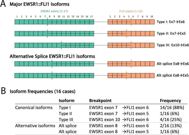
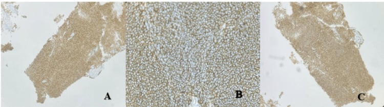
A small round blue cell tumour that looks like Ewing, and may even be CD99-positive, is not necessarily Ewing. The current WHO classification separates true Ewing sarcoma, defined by an EWSR1-ETS fusion, classically EWS-FLI1, from a group of "Ewing-like" undifferentiated round cell sarcomas that are EWSR1-FISH-negative and driven by different genetics — principally CIC-rearranged sarcomas (usually CIC-DUX4) and BCOR-rearranged sarcomas. These mimics have variable, patchy CD99 and distinct immunoprofiles (CIC sarcomas are often WT1/ETV4-positive; BCOR sarcomas BCOR/SATB2-positive), tend to be more soft-tissue and axial, and carry a worse prognosis with a poorer response to standard Ewing chemotherapy. The exam point answers the classic distractor "small round blue cells but EWSR1-negative (or CD99-negative)": that tumour is a Ewing-LIKE sarcoma, not Ewing, which is exactly why molecular confirmation of an EWSR1 rearrangement, not CD99 alone, is required before committing a patient to a Ewing protocol.
Classification and Staging
Ewing sarcoma is always high-grade, so of the Enneking stages only IB, IIB and III are ever used, and most tumours are IIB at presentation: high-grade, extracompartmental, no metastases. The system asks three questions — grade, compartment, metastases — and the compartment is the one worth getting right. Intracompartmental means the tumour is confined within the bone cortex or within a single anatomic compartment such as the anterior thigh; extracompartmental means it has broken through the cortex into soft tissue, or has crossed an anatomic barrier such as a joint or a neurovascular bundle.
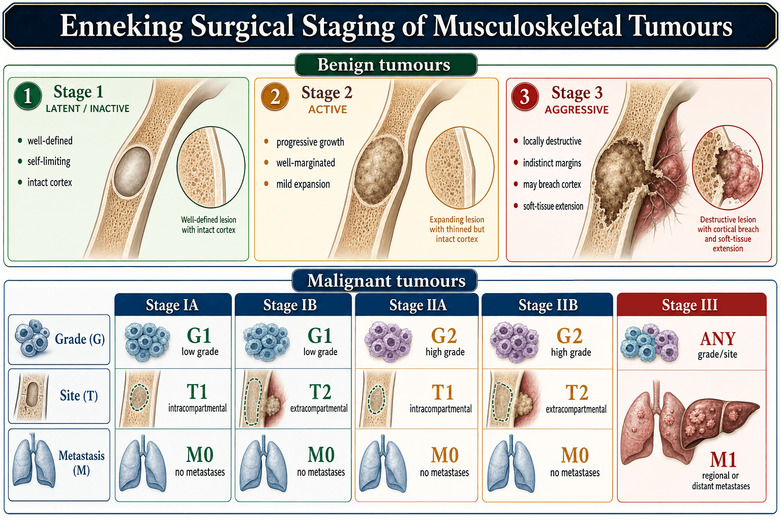
The Enneking system is the gold standard for bone sarcomas, built on grade, local extent and metastases.
- Grade
- Low-grade
- Extent
- Intracompartmental
- Metastases
- None
- 5-Year Survival
- Not applicable (Ewing always high-grade)
- Grade
- High-grade
- Extent
- Intracompartmental
- Metastases
- None
- 5-Year Survival
- 70-75%
- Grade
- Low-grade
- Extent
- Extracompartmental
- Metastases
- None
- 5-Year Survival
- Not applicable (Ewing always high-grade)
- Grade
- High-grade
- Extent
- Extracompartmental
- Metastases
- None
- 5-Year Survival
- 65-70%
- Grade
- Any grade
- Extent
- Any extent
- Metastases
- Present (regional or distant)
- 5-Year Survival
- 25-30%
Clinical Presentation
The history. Localised bone pain in 90%, progressive and worse at night, with a palpable soft-tissue mass in 60%. Systemic features are common: fever in 30%, weight loss, malaise. Symptoms have usually run for 2-6 months before the diagnosis is made, an average of 3, and 30% report an injury beforehand, which is coincidental rather than causative.
The examination. Visible swelling, skin erythema and venous distension over a warm, tender mass, with soft-tissue extension common. A lower limb tumour produces a limp and a restricted range of movement. The neurovascular structures are usually intact, because invasion is rare, and regional nodes are rarely involved, unlike soft tissue Ewing.
The trap. Around 30% of patients arrive with fever, a raised white cell count and a raised ESR, and are treated for osteomyelitis. The features that separate them are the duration, months rather than weeks, the diaphyseal location, and the large soft-tissue mass.
- Ewing Sarcoma
- Months (average 3)
- Osteomyelitis
- Days to weeks
- Ewing Sarcoma
- Low-grade, intermittent
- Osteomyelitis
- High-grade, persistent
- Ewing Sarcoma
- Diaphysis
- Osteomyelitis
- Metaphysis
- Ewing Sarcoma
- Large, firm
- Osteomyelitis
- Small or absent
- Ewing Sarcoma
- None
- Osteomyelitis
- Improves within 48-72 hours
Always biopsy a suspected infection that does not respond to antibiotics within 48-72 hours, particularly a lytic diaphyseal lesion with a soft-tissue mass.
- Typical age
- 10-20 years (peak 15)
- Distinguishing histology / IHC
- Uniform small round cells, PAS-positive glycogen, strong diffuse membranous CD99, NKX2-2 positive
- Key genetics
- EWSR1-FLI1 t(11;22) (85%); EWSR1-ERG variant
- Typical age
- 10-20 years (peak 16)
- Distinguishing histology / IHC
- Pleomorphic cells producing malignant osteoid; SATB2 positive
- Key genetics
- Complex karyotype, TP53/RB1 alterations
- Typical age
- Bimodal (10-20 and over 60)
- Distinguishing histology / IHC
- Larger cells, LCA/CD45 positive, CD20 (B) or CD3 (T) positive; CD99 negative
- Key genetics
- Various B/T-cell rearrangements
- Typical age
- Under 5 years
- Distinguishing histology / IHC
- Rosettes, neuropil, PHOX2B / synaptophysin positive; raised urinary catecholamines
- Key genetics
- MYCN amplification
- Typical age
- Children
- Distinguishing histology / IHC
- Rhabdomyoblasts; desmin, myogenin, MyoD1 positive
- Key genetics
- PAX3/7-FOXO1 (alveolar)
- Typical age
- Any
- Distinguishing histology / IHC
- Neutrophilic inflammation, organisms on culture; no clonal tumour cells
- Key genetics
- Not applicable
Investigations
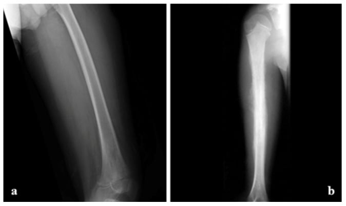
Radiographs first. AP and lateral views of the affected bone plus the joint above and below. The classic picture is a permeative lytic lesion in the diaphysis with moth-eaten bone destruction, a wide zone of transition, onion-skin (lamellated) periosteal reaction, a Codman triangle of elevated periosteum, and a large soft-tissue mass. A sunburst pattern occurs but is less common than in osteosarcoma. The periosteal reaction is suggestive rather than pathognomonic: infection and other aggressive tumours stay in the differential until the biopsy returns.
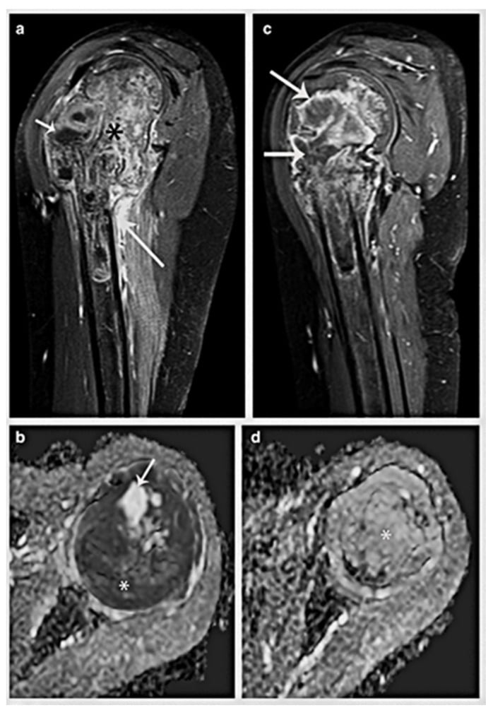
MRI with contrast is the gold standard for local extent. It defines intramedullary involvement, soft-tissue extension, the neurovascular relationship and skip lesions, and it is what the surgical plan is built on. Marrow signal is low on T1 and high on T2 along with the surrounding oedema, and viable tumour enhances heterogeneously. Map the entire involved bone, including marrow length, cortical breach, the soft-tissue component and the joint: the soft-tissue mass may be disproportionately large compared with the radiographic osseous change.
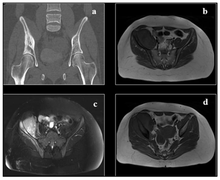
CT answers a different question. It shows cortical destruction and any mineralisation, while MRI shows the marrow and the soft tissues. Stage the whole compartment on both, and never choose a biopsy trajectory before the MRI has been reviewed.
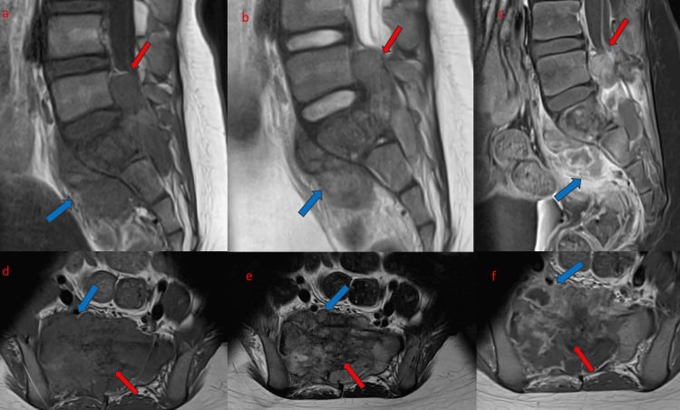
Staging is whole-body, and it is completed before treatment starts.
- High-resolution CT chest for pulmonary metastases: present in 15% at diagnosis, and the nodules may be under 5mm. Repeat every 3 months during treatment
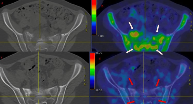
- PET-CT or bone scan for skeletal metastases: 10% at diagnosis. PET-CT also tracks chemotherapy response, a falling SUV indicating a good response
- Bilateral iliac crest marrow aspirate and biopsy for marrow disease: 5% at diagnosis, and it carries a prognosis similar to metastatic disease
Perform an incisional or core needle biopsy along the longitudinal axis of the limb, in line with the planned surgical incision, because the contaminated tract must be excised en bloc with the tumour. Avoid transverse incisions. Send fresh tissue for cytogenetics (FISH for an EWS rearrangement), flow cytometry, and microbiology if infection is in the differential, and coordinate with oncology and pathology before the biopsy rather than after it. The resection specimen also goes fresh to pathology, so that necrosis can be assessed.
The bloods support a suspicion rather than settle it. Anaemia in 30% and leucocytosis in 25%; ESR and CRP raised in 50%, non-specifically; LDH raised in 60%, where it carries prognostic weight. Alkaline phosphatase is normal or mildly raised. Renal and hepatic function are the baselines chemotherapy will be judged against.
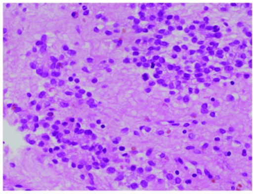
Histology decides. Sheets of uniform small round blue cells; PAS-positive glycogen, diastase-sensitive; CD99 (MIC2) strong and diffuse on the membrane in 95%; and the EWS-FLI1 fusion on FISH or RT-PCR in 85%. The negative stains earn their place too: LCA negative argues against lymphoma, desmin negative against rhabdomyosarcoma.
Management: Multimodal Treatment Paradigm
Chemotherapy comes before surgery, always. Ewing sarcoma is a systemic disease from the day it is found, and local control waits 10-14 weeks while induction chemotherapy runs. Treating first does three things an operation cannot: it treats micrometastases early, it shrinks the tumour so that resection is easier and a borderline case may convert to limb salvage, and it measures chemosensitivity in a resected specimen, where over 90% necrosis marks the good responder.
Operating before chemotherapy is a critical error that worsens survival and increases morbidity.
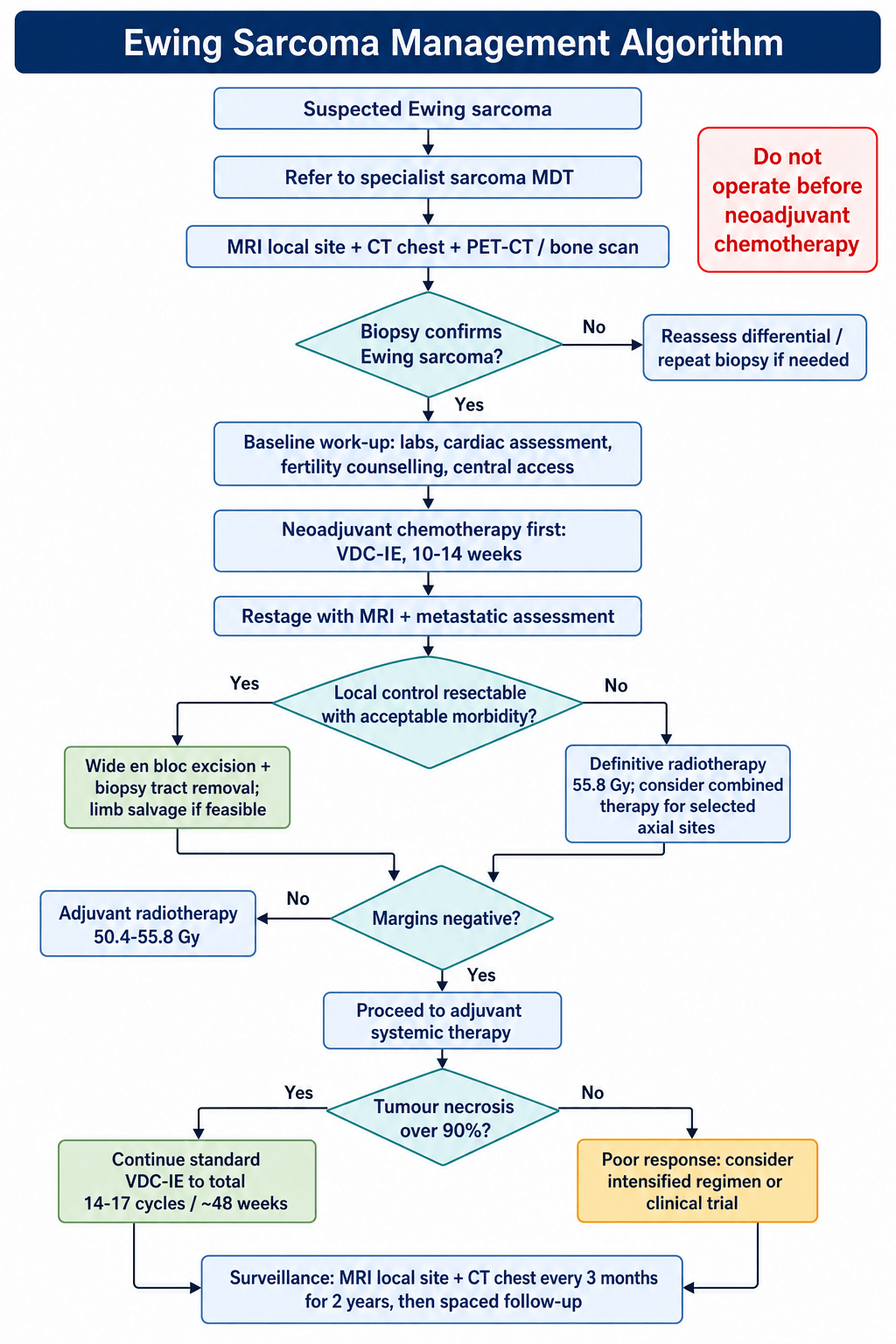
Choosing local control. Surgery is preferred wherever it is feasible. Local recurrence runs 5-10% after surgery against 10-20% after radiotherapy, surgery avoids radiation-induced growth arrest in a skeletally immature patient and the 1-2% at 10 years risk of radiation-induced sarcoma, and only a resected specimen yields a necrosis figure. Ewing is radiosensitive, though, and definitive radiotherapy achieves excellent local control at the sites — pelvis, spine, skull base — where resection would carry unacceptable morbidity.
Multimodal Treatment Timeline
Biopsy confirmation and complete staging (MRI, CT chest, PET-CT, bone marrow biopsy). Specialist sarcoma multidisciplinary tumour board review. Counsel patient and family, including fertility counselling. Baseline bloods and cardiac assessment. Central line placement.
VDC-IE protocol, typically 5-6 cycles over 10-14 weeks. Monitor for cardiotoxicity, haemorrhagic cystitis and neutropenia. Restaging MRI at week 10 to assess response.
Wide surgical excision with negative margins where feasible; radiotherapy (55.8 Gy) for unresectable tumours of the pelvis or spine. Pathological assessment of percent necrosis.
Continue VDC-IE to a total of 14-17 cycles, approximately 48 weeks of treatment. Good responders continue the standard protocol; poor responders are considered for an intensified regimen or a clinical trial.
CT chest and MRI of the local site every 3 months for 2 years, every 6 months to year 5, then annually. Monitor for cardiac, pulmonary and second-malignancy late effects.
- Staging
- IB or IIB, over 90% necrosis
- Treatment
- Wide excision + reconstruction
- Key Pearl
- Limb salvage in 85-90% - margins matter more than necrosis
- Staging
- IIB extracompartmental
- Treatment
- Chemotherapy + radiation (surgery if resectable)
- Key Pearl
- Radiation for unresectable sites - 55.8 Gy standard dose
- Staging
- Stage III with lung mets
- Treatment
- Intensive chemotherapy + local control + lung surgery
- Key Pearl
- Whole lung radiation if unresectable - survival drops to 30%
Alternating VDC and IE cycles are the backbone, given over 48 weeks in total, approximately 14-17 cycles depending on the protocol.
VDC cycles run at weeks 1, 7 and 13 onwards:
- Vincristine 2 mg/m² IV (maximum 2 mg) on day 1
- Doxorubicin 75 mg/m² IV over 48 hours on days 1-2
- Cyclophosphamide 1200 mg/m² IV on day 1
IE cycles run at weeks 4, 10 and 16 onwards:
- Ifosfamide 1800 mg/m² IV daily for 5 days
- Etoposide 100 mg/m² IV daily for 5 days
- Mesna 1800 mg/m² IV daily, for uroprotection against ifosfamide
VDC-IEVDC-IE Chemotherapy Protocol
Hook:VDC-IE cycles: VDC for cycles 1,3,5 then IE for cycles 2,4,6 - alternating reduces resistance!
Doxorubicin cardiotoxicity: ECHO or MUGA at baseline, 6 months and the end of treatment; cumulative dose limit 450-550 mg/m². Ifosfamide haemorrhagic cystitis: prevented by mesna, which binds the acrolein metabolite. Neutropenic fever: G-CSF support. Secondary malignancy: etoposide raises the risk of acute myeloid leukaemia at 2-5 years.
Prognostic Factors
Necrosis is the factor treatment can change. Over 90% tumour necrosis after neoadjuvant chemotherapy carries 75% five-year survival against 50% below that threshold, and it sets the intensity of what follows: good responders continue the standard protocol, poor responders are considered for an intensified regimen or a trial. For local control, though, surgical margins matter more than necrosis.
Size and margins. A tumour over 8cm does worse than one under 8cm, and it is positive or marginal margins, not the necrosis figure, that drive local recurrence.
Age. Younger patients do better than older ones. The quantified comparison below draws the line at 15 years, and the adverse group has also been defined as those over 18.
- Favorable
- Localized disease
- Unfavorable
- Metastatic (lung, bone, marrow)
- Impact on 5-Year Survival
- 70% vs 25-30%
- Favorable
- Over 90% necrosis
- Unfavorable
- Under 90% necrosis
- Impact on 5-Year Survival
- 75% vs 50%
- Favorable
- Under 200 ml
- Unfavorable
- Over 200 ml
- Impact on 5-Year Survival
- 70% vs 50%
- Favorable
- Extremity (distal)
- Unfavorable
- Pelvis, axial skeleton
- Impact on 5-Year Survival
- 75% vs 50%
- Favorable
- Under 15 years
- Unfavorable
- Over 15 years
- Impact on 5-Year Survival
- 70% vs 55%
- Favorable
- Normal
- Unfavorable
- Elevated
- Impact on 5-Year Survival
- 70% vs 55%
Surgical Technique
The goal. An en bloc resection through normal tissue with the pseudocapsule intact, reconstructed so that the limb works and so that adjuvant chemotherapy can restart on time. Wound healing is part of the oncological plan, because the adjuvant chemotherapy waits for it.
Enneking's margins name what was actually achieved, and the words matter in a viva:
- Intralesional - through tumour, inadequate
- Marginal - through the reactive zone, high recurrence
- Wide - through normal tissue, the goal for cure
- Radical - the entire compartment, rarely needed
Timing. Surgery follows 10-14 weeks of chemotherapy, once the patient has recovered from its toxicity and the counts allow — platelets over 100 and white cells over 3 — which is usually 2-3 weeks after the last cycle. Coordinate the date with the oncology team rather than announcing it.
The biopsy tract goes with the specimen. Whether the diagnosis came from an incisional or a needle biopsy, the tract is contaminated tissue and is excised en bloc; nothing contaminated may be left behind.
Margins. Aim for 2-3cm of soft tissue where the anatomy allows, 2cm being the usual working figure, with a cuff of normal muscle around the tumour. Structures that have been contaminated are sacrificed with the specimen, and frozen section of the margins intraoperatively confirms what has been achieved. The specimen itself goes to pathology fresh, for assessment of necrosis. Neurovascular structures are preserved if they are not encased, which is usually the case.
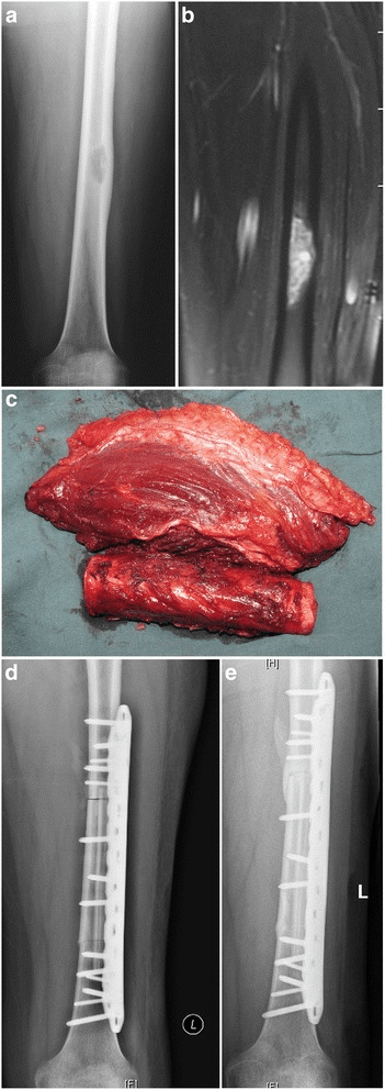
Neurovascular judgement is made on the imaging, before the incision. MRI shows the relationship, angiography is added if encasement is suspected, and a vascular surgeon is involved early if a bypass is likely.
- A vessel displaced but not encased is preserved
- An encased vessel is sacrificed and reconstructed
- A critical nerve is sacrificed only if it is encased
Soft-tissue cover is planned with the resection. Anticipate the defect, and choose between local muscle flaps — gastrocnemius, soleus, latissimus — and a free flap for a large defect, with plastic surgical input where needed. Chemotherapy delays healing, so close without tension, keep drains away from the prosthesis, and allow for postoperative radiotherapy if the margins are close.
Reconstruction follows the defect:
- Endoprosthetic replacement with a modular tumour prosthesis, the commonest choice for long bones
- Structural allograft, or an allograft-prosthetic composite
- Vascularised fibula for an intercalary defect
- Rotationplasty as a specialised limb-sparing option
- Amputation where limb salvage is not safe
Tumour prostheses, custom or modular, allow the length to be set on the table, are cemented or press-fit, and come as growing designs for the skeletally immature. The sequence is fixed: resect en bloc with wide margins, measure the resection length precisely, prepare the bone, assemble the modular components, fix them, reconstruct the soft tissues around the prosthesis, and close over drains.
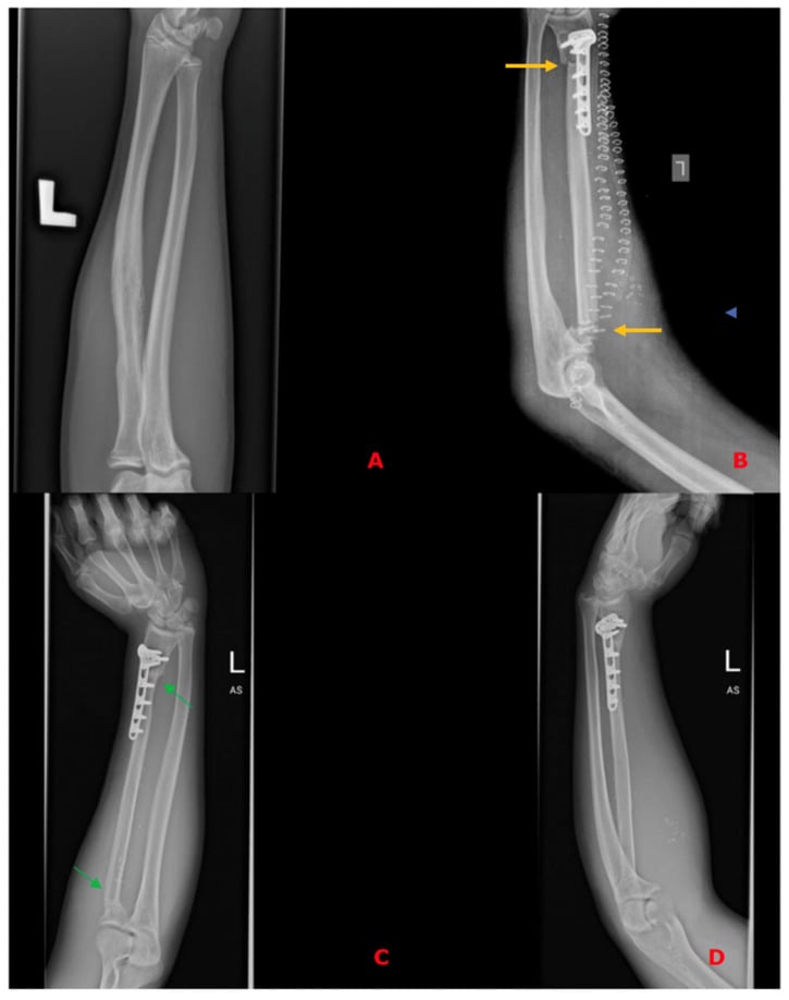
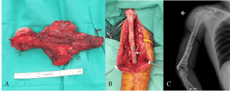
- Surgical Options
- Distal femoral resection
- Reconstruction
- Modular endoprosthesis, allograft-prosthetic composite
- Special Considerations
- Most common site, excellent function possible
- Surgical Options
- Proximal tibial resection
- Reconstruction
- Endoprosthesis, allograft composite
- Special Considerations
- Extensor mechanism reconstruction critical
- Surgical Options
- Proximal humeral resection
- Reconstruction
- Endoprosthesis, allograft, spacer
- Special Considerations
- Shoulder function limited but hand function preserved
- Surgical Options
- Pelvic resection (Type I-IV)
- Reconstruction
- Reconstruction varies, may be 'defect left'
- Special Considerations
- High morbidity, radiation often preferred
- Surgical Options
- Vertebrectomy if feasible
- Reconstruction
- Instrumented fusion, cage reconstruction
- Special Considerations
- Often radiation primary; surgery for instability
- Surgical Options
- Chest wall resection
- Reconstruction
- Mesh, flap reconstruction
- Special Considerations
- May need thoracic surgery involvement
Rotationplasty (the Van Nes procedure) is for the young patient with a distal femoral or proximal tibial tumour in whom conventional limb salvage would give poor function. The tumour-bearing segment is resected and the distal limb rotated 180 degrees, so that the knee becomes the new hip and the ankle the new knee: plantarflexion now does the work of knee flexion, and the patient is fitted with a below-knee type prosthesis. Function is better than after an above-knee amputation, and the cosmetic price is the reason it needs discussing honestly.
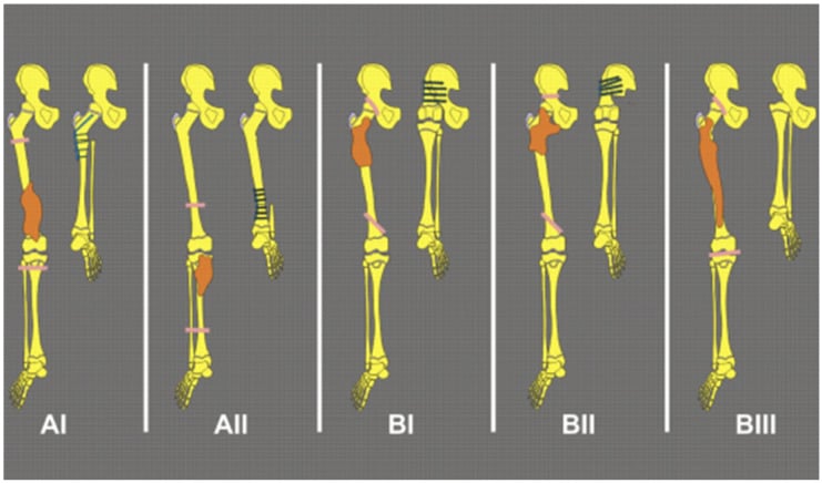
With modern technique and multimodal therapy, limb salvage achieves oncological outcomes equivalent to amputation, which is exactly why the margin can never be traded for the reconstruction.
A wide margin is mandatory for cure, and a positive margin dramatically increases local recurrence. Amputation remains an option if limb salvage is unsafe. Any proposal to compromise a margin goes back to the multidisciplinary team first.
Complications
Treatment is long, and the chemotherapy complications are predictable from the agents, which is why the regimen is worth knowing drug by drug.
- Incidence
- 5-10% (clinical), 30% (subclinical)
- Causative Agent
- Doxorubicin (cumulative dose)
- Management
- Baseline and serial ECHO; dexrazoxane cardioprotection; limit cumulative dose
- Incidence
- 10-30% without mesna
- Causative Agent
- Ifosfamide
- Management
- Mesna uroprotection; aggressive hydration; monitor urinalysis
- Incidence
- 40-60%
- Causative Agent
- Myelosuppression (all agents)
- Management
- G-CSF support; prophylactic antibiotics; urgent admission if fever
- Incidence
- 1-2% at 10 years
- Causative Agent
- Etoposide, alkylating agents
- Management
- Long-term surveillance; no prevention available
- Incidence
- 5-15%
- Risk Factors
- Chemotherapy-induced neutropenia, endoprosthesis
- Management
- Antibiotics; irrigation and debridement; prosthesis retention vs removal
- Incidence
- 5-10%
- Risk Factors
- Positive margins, poor chemo response
- Management
- Re-excision or radiation; metastatic workup
- Incidence
- Variable
- Risk Factors
- Skeletally immature patients
- Management
- Expandable prosthesis; contralateral epiphysiodesis
Radiotherapy's complications follow the field and the age of the patient: growth arrest where the field crosses a physis in a skeletally immature patient, radiation-induced sarcoma in 1-2% at 10 years with a latency of 5-20 years, pathological fracture through weakened bone in 5-10%, and soft-tissue fibrosis with chronic pain and reduced movement. IMRT and proton therapy reduce all of these significantly compared with older techniques.
Late effects outlive the treatment. Infertility follows alkylating agents and pelvic radiotherapy in 20-40%, which is why fertility preservation is discussed before the first cycle rather than after it. Ifosfamide leaves nephrotoxicity in 10-20%, and second malignancies reach 5-10% at 20 years after radiotherapy and alkylating agents, so surveillance is lifelong. Growth disturbance in a child irradiated across a physis needs limb-length monitoring.
Metastatic progression is the disease's own complication, and the lung is where it happens.
- Lung: metastases in 15% at diagnosis, and a further 30% develop them during treatment. Detected on three-monthly CT chest. Treated with intensified chemotherapy, surgical resection if oligometastatic (under 5 nodules), or whole lung radiation if unresectable. Five-year survival 30%
- Bone and bone marrow: 10% bone and 5% marrow at diagnosis, found on PET-CT or bone scan and bilateral marrow biopsy. Treated with systemic chemotherapy and radiotherapy to symptomatic sites. Five-year survival 25%, similar to lung metastases
- Local progression: pathological fracture in 10%, neurovascular compression, and rarely compartment syndrome
Metastatic disease at presentation is the worst prognostic factor, reducing five-year survival from 70% to 25-30%.
Postoperative Care
The wound sets the pace. Closed suction drainage comes out when the output falls under 30 mL/24h, and the wound is inspected daily for haematoma and infection. Analgesia and mobilisation are the other tasks of the first few days. Perioperative antibiotics cover 24-48 hours, dressings are changed cleanly, and the threshold for suspecting infection stays low, because these patients are immunosuppressed and an endoprosthesis is unforgiving.
Rehabilitation is dictated by the reconstruction. After an endoprosthesis, partial weight-bearing with crutches starts immediately and progresses to full over 4-6 weeks, earlier than any biological reconstruction allows; high-impact activity is avoided for life, and the implant is watched for the rest of the patient's life for loosening, infection and implant failure, with revision for wear or loosening in a proportion of patients. An allograft is protected for 3-6 months until serial radiographs show incorporation, and it carries its own risks: nonunion 10-20%, allograft fracture 15-25%, infection 10-15%, and resorption over time. Range of motion begins when the wound is stable, under physiotherapy, with occupational therapy for an upper limb reconstruction and walking aids for as long as they are needed.
Chemotherapy restarts within 2-4 weeks, once the wound has healed and the counts have recovered, taking the total treatment duration to approximately 12 months, and the resected specimen is reported before then: percent necrosis, which grades the response, and margin status, which decides whether radiotherapy is added. Both change what happens next, so chase the report.
Surveillance is intensive early because the lung is the site that fails. CT chest and MRI of the local site run every 3 months for 2 years, every 6 months to year 5, then annually thereafter, or as the institutional protocol dictates, with the first reassessment at about three months. Alongside it, monitor cardiac function for doxorubicin, renal function for ifosfamide, fertility, and second malignancy, particularly after radiotherapy.
Seek immediate evaluation for new pain at the surgical site, a palpable mass or swelling, new pulmonary symptoms, any new bone pain, or constitutional symptoms such as fever and weight loss.
Outcomes
Survival tracks two things: whether there were metastases at diagnosis, and how the tumour answered the chemotherapy.
- 5-Year EFS
- 75-80%
- 5-Year OS
- 80-85%
- Key Factors
- Best outcomes, continue standard therapy
- 5-Year EFS
- 45-55%
- 5-Year OS
- 55-65%
- Key Factors
- Consider intensified/experimental therapy
- 5-Year EFS
- 30-35%
- 5-Year OS
- 35-40%
- Key Factors
- Whole lung irradiation may improve outcomes
- 5-Year EFS
- 15-20%
- 5-Year OS
- 20-25%
- Key Factors
- Worst prognosis, often palliative
- 5-Year EFS
- 10-20%
- 5-Year OS
- 15-25%
- Key Factors
- Prognosis poor; depends on site, timing
Function after limb salvage is good rather than normal: most patients reach 70-85% of the MSTS functional score (Enneking) (Enneking), an upper limb reconstruction preserves hand function, and a lower limb reconstruction leaves the patient walking with or without aids, with quality of life comparable to amputation. The long-term costs are an endoprosthesis needing revision in 10-20% at 10 years, leg length discrepancy in a growing child, a permanent ban on high-impact activity, and chronic pain in some patients.
After amputation survival is oncologically equivalent, prosthetic function is generally good, and some studies report higher rates of activity participation; there are no implant-related complications and the solution is more durable. What it costs is body image, and that deserves the same honest conversation as the functional gain.
Recurrence declares itself locally or distantly, and the distant pattern dominates. Local recurrence runs 10-15% where the margins were adequate and radiation was given, and is higher with inadequate margins; it is treated with re-resection where possible, radiotherapy and chemotherapy, with 20-30% long-term survival afterwards. Distant recurrence goes to the lungs in 50-60% of cases, and also to bone, bone marrow and other soft tissues; treatment is systemic chemotherapy with or without local control of the metastases, and five-year survival is 15-25%.
With modern multimodal therapy most patients with localised Ewing sarcoma are cured, and metastatic disease remains a significant challenge. Completing the chemotherapy is what controls the disease systemically.
Guidelines, Registries & Global Practice
Global Epidemiology
Ewing sarcoma is the second most common primary bone malignancy in children and adolescents, with an annual incidence of approximately 1 to 3 per million in populations of European ancestry. It is markedly less common in populations of African and East Asian ancestry, a disparity now attributed in part to germline variation at the EGR2 locus and ancestry-related differences in regulatory microsatellites. Population-based registry data (SEER) confirm a male predominance and a peak in the second decade, and identify pelvic/axial site, tumour size over 8cm, older age and metastatic extent as the principal adverse-risk factors (Shi 2020).
- Local control
- Wide surgical excision preferred; definitive RT for unresectable axial sites; post-op RT for close/positive margins or poor response
- Systemic therapy
- Neoadjuvant VDC-IE, interval-compressed (q2-weekly) for localised disease
- Evidence level
- Category 1 / 2A (RCT-backed)
- Local control
- Surgery where adequate margins achievable; RT for inoperable sites or as adjuvant; combined surgery+RT for marginal margins/poor response
- Systemic therapy
- VIDE / VDC-IE backbone; busulfan-melphalan high-dose consolidation for selected high-risk (EE2008/R2 evidence)
- Evidence level
- Grade A/B
- Local control
- Management exclusively in designated supraregional bone-sarcoma centres; MDT decision on surgery vs RT
- Systemic therapy
- Euro Ewing protocol chemotherapy within national trials
- Evidence level
- Expert consensus + RCT
- Local control
- Treatment at tertiary paediatric/sarcoma centres; surgery preferred, RT for axial sites
- Systemic therapy
- Standard VDC-IE agents within international protocols
- Evidence level
- Consensus / adopted RCT
- Contribution
- Established VDC-IE backbone for localised disease
- Key result
- 5-yr EFS 69% vs 54% with addition of ifosfamide/etoposide
- Contribution
- Established interval compression (q2-weekly)
- Key result
- 5-yr EFS 73% vs 65%; durable at 10 years
- Contribution
- Defined local-control hierarchy
- Key result
- Local failure 7.5% surgery vs 26.3% definitive RT
- Contribution
- Population-level risk stratification
- Key result
- Pelvic site OR 2.49 and size over 8cm OR 1.91 for metastasis
Practice Variation
The principal axis of international variation is high-dose chemotherapy with autologous stem-cell rescue: European protocols (EE2008/R2) incorporate busulfan-melphalan consolidation for selected high-risk localised patients, whereas North American practice has not adopted routine high-dose consolidation. Interval-compressed chemotherapy is standard in paediatric North American practice but is applied more selectively in adults. Relapsed/refractory disease is increasingly managed within the international rEECur platform, which is establishing the most active salvage regimen. Across all systems, care is mandated within designated bone-sarcoma reference centres with a specialist multidisciplinary team (orthopaedic oncologist, paediatric/medical oncologist, radiation oncologist, radiologist, pathologist).
- Histological + molecular confirmation (EWSR1 rearrangement) before starting therapy
- Neoadjuvant chemotherapy before any local treatment
- Biopsy planned by the resecting surgeon (tract excised en bloc)
- Fertility preservation discussed before alkylating chemotherapy
- Informed consent for chemotherapy (cardiotoxicity, hemorrhagic cystitis, infertility, secondary malignancy risks)
- Informed consent for surgery (infection, local recurrence, limb length discrepancy, neurovascular injury)
- Tumor board documentation of treatment plan
- Pathology confirmation of EWS-FLI1 fusion before starting chemotherapy
- Discussion of fertility preservation (sperm banking for males, oocyte preservation for females) before starting chemotherapy - alkylating agents cause infertility
- Long-term surveillance plan documented (cardiac monitoring, secondary malignancy screening)
- Delayed diagnosis (misdiagnosed as osteomyelitis or sports injury)
- Operating before chemotherapy (incorrect sequencing)
- Inadequate surgical margins leading to local recurrence
- Failure to discuss fertility preservation before chemotherapy
MCQ Practice Points
Q: What is the most common chromosomal translocation in Ewing sarcoma? A: t(11;22)(q24;q12) creating the EWS-FLI1 fusion oncogene, present in 85% of cases. The remaining 15% have variant translocations (e.g., EWS-ERG). This fusion is detected by FISH or RT-PCR and is diagnostic for Ewing sarcoma, distinguishing it from other small round cell tumors.
Q: What immunohistochemical marker is most sensitive for Ewing sarcoma? A: CD99 (MIC2) with strong diffuse membrane staining, positive in 95% of Ewing sarcomas. However, CD99 is not entirely specific (can be positive in lymphoblastic lymphoma, synovial sarcoma). The combination of CD99 positivity, PAS-positive glycogen, and EWS-FLI1 fusion confirms the diagnosis.
Q: What is the correct sequence of treatment for localized Ewing sarcoma? A: Neoadjuvant chemotherapy → Local control (surgery or radiation) → Adjuvant chemotherapy. Total treatment duration is approximately 48 weeks. Operating before chemotherapy is a critical error that worsens outcomes by failing to treat micrometastases and assess chemotherapy response.
Q: What is the most important modifiable prognostic factor in Ewing sarcoma? A: Percent tumor necrosis after neoadjuvant chemotherapy. Patients with over 90% necrosis have 75% 5-year survival vs 50% for under 90% necrosis. This finding guides adjuvant therapy intensity. However, surgical margins are more important than necrosis for local control.
Q: How do you differentiate Ewing sarcoma from osteomyelitis clinically? A: Key features: Ewing has longer symptom duration (months vs weeks), diaphyseal location (osteomyelitis favors metaphysis), large soft tissue mass, and no response to antibiotics. Always biopsy a suspected infection that does not improve within 48-72 hours of antibiotics, especially with lytic diaphyseal lesion and soft tissue mass.
Q: What percentage of Ewing sarcoma patients present with metastatic disease, and what are the most common sites? A: 25% present with metastases. Most common sites: Lungs (15%), bone (10%), and bone marrow (5%). Metastatic disease at presentation reduces 5-year survival from 70% to 25-30%. Complete staging includes CT chest, PET-CT or bone scan, and bilateral bone marrow biopsy.
Exam Viva Scenarios
Practise clinical reasoning and management decisions out loud
“A 14-year-old boy presents with a 3-month history of progressive right thigh pain and swelling. He has had intermittent fevers. X-ray shows a permeative lytic lesion in the mid-femoral diaphysis with an onion-skin periosteal reaction and soft tissue mass. ESR is elevated. His GP started him on antibiotics 2 weeks ago with no improvement. What is your assessment and management?”
“A 16-year-old girl with biopsy-proven Ewing sarcoma of the distal femur has completed 14 weeks of VDC-IE chemotherapy. Restaging MRI shows excellent response with 60% reduction in soft tissue mass. The tumor remains 2cm from the distal femoral physis. She is skeletally immature (Risser 2). Discuss your approach to local control.”
“A 17-year-old presents with Ewing sarcoma of the proximal humerus. Staging workup reveals 4 pulmonary nodules (5-12mm) on CT chest and bone marrow involvement (5% tumor cells on bilateral iliac crest biopsy). How would you manage this patient?”
Key Pathology
- EWS-FLI1 fusion t(11;22) in 85% - diagnostic hallmark
- Small round blue cells, PAS-positive glycogen
- CD99 strong diffuse membrane staining (95%)
- High-grade malignancy, early hematogenous spread
Clinical Presentation
- Age 10-20 years, diaphysis of long bones (90%)
- Painful swelling, fever (30%), elevated ESR - mimics infection
- Femur and pelvis most common (60% of cases)
- 25% present with metastases (lung, bone, marrow)
Staging Workup
- MRI local site - assess extent, soft tissue involvement
- CT chest - detect lung metastases (15% at diagnosis)
- PET-CT or bone scan - skeletal metastases (10% at diagnosis)
- Bilateral bone marrow biopsy - marrow involvement (5%)
Treatment Algorithm
- Neoadjuvant chemo FIRST (VDC-IE protocol, 10-14 weeks)
- Local control: Wide excision (preferred) or radiation (55.8 Gy)
- Adjuvant chemo to complete 48 weeks total (14-17 cycles)
- Assess percent necrosis: over 90% = good prognosis
Surgical Pearls
- Limb salvage in 85-90% of extremity tumors
- Wide margins (2cm healthy tissue) more important than necrosis
- Excise biopsy tract en bloc with tumor
- Expandable prosthesis for skeletally immature patients
Prognostic Factors
- Metastases at diagnosis: 70% vs 25-30% survival (localized vs metastatic)
- Over 90% necrosis post-chemo: 75% vs 50% survival
- Tumor volume under 200ml: Better prognosis
- Pelvic tumors: Worse prognosis (large, difficult resection)
Evidence Base and Key Trials
INT-0091 (Grier): Addition of Ifosfamide and Etoposide
- Multi-centre RCT: 518 eligible patients with Ewing sarcoma / PNET of bone
- Standard VACD (vincristine, doxorubicin, cyclophosphamide, dactinomycin) alternating with ifosfamide-etoposide (IE) vs VACD alone
- Non-metastatic disease (n=398): 5-year EFS 69% (IE arm) vs 54% (standard), P=0.005; OS 72% vs 61%, P=0.01
- No benefit from IE in the 120 patients with metastatic disease at diagnosis
CESS/EICESS: Local Therapy in Localised Ewing Tumours (Schuck)
- Pooled analysis of 1058 patients from CESS 81, CESS 86 and EICESS 92 trials
- Local failure: 7.5% after surgery (with or without post-operative RT) vs 26.3% after definitive radiotherapy (P=0.001)
- Pre-operative RT achieved 5.3% local failure (comparable to surgery)
- Irradiated patients were a negatively selected group with unfavourable (axial) sites
AEWS0031 (Womer): Interval-Compressed Chemotherapy
- RCT (Children's Oncology Group): 568 eligible patients with localised Ewing sarcoma randomised to standard (every 21 days) vs interval-compressed (every 14 days) VDC-IE
- 5-year EFS: 73% (compressed) vs 65% (standard), P=0.048
- Toxicity similar between the two arms with filgrastim support
- 10-year follow-up (Cash 2023) confirmed durable benefit: 10-year EFS 70% vs 61%, OS 76% vs 69%, no excess second malignancies
Picci: Chemotherapy-Induced Necrosis as Prognostic Factor
- 118 patients with localised extremity Ewing sarcoma treated with pre-operative chemotherapy then surgery
- Histological grading of response: grade III (no viable tumour) 5-year DFS 95% vs grade II 68% vs grade I (macroscopic viable tumour) 34% (P less than 0.0001)
- Histological response had the strongest correlation with outcome of all parameters tested
- Method identifies poor responders who may benefit from intensified adjuvant therapy
May: EWS-FLI1 is a Transforming Chimeric Transcription Factor
- The t(11;22) translocation fuses the EWS transactivation domain to the FLI1 DNA-binding (ETS) domain
- EWS-FLI1 transformed NIH 3T3 cells (focus formation, anchorage-independent growth) - demonstrating oncogenic activity
- Deletion of either the EWS domain or the FLI1 DNA-binding domain abolished transformation
- Established the molecular mechanism: a chimeric aberrant transcription factor drives tumourigenesis
Shi: Population-Based Risk Factors for Metastasis and Prognosis (SEER)
- Large SEER population-based cohort of Ewing sarcoma patients
- Pelvic primary (OR 2.49) and tumour over 8cm (OR 1.91) independently predicted metastasis at diagnosis
- Worse overall survival with older age, larger size, pelvic site, black race (HR 2.10) and metastatic extent (HR 3.26)
- Chemotherapy was strongly associated with improved survival (HR 0.47)