Progressive Collapse | PTT Dysfunction | Staged Reconstruction
- Johnson and Strom classification - guides surgical reconstruction strategy
- Rigid vs flexible - determines need for osteotomy vs arthrodesis
- Too many toes sign - pathognomonic for hindfoot valgus on clinical exam
- Single heel rise test - inability indicates PTT incompetence
- Stage II divide - IIA (flexible hindfoot) vs IIB (forefoot driven, fixed forefoot varus)
- “Stage IIB requires cotton test and first ray plantar flexion osteotomy
- “FDL is preferred tendon transfer over FHL (better excursion, less donor morbidity)
- “Lateral column lengthening (Evans) corrects forefoot abduction
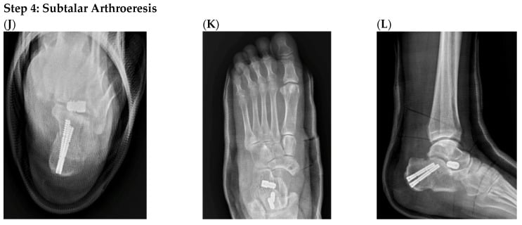
- “Stage III requires subtalar or triple arthrodesis for rigid deformity
Overview and Epidemiology
Adult acquired flatfoot deformity (AAFD) is progressive collapse of the medial longitudinal arch caused by posterior tibial tendon (PTT) dysfunction. It is the most common cause of acquired flatfoot in adults, and it is a spectrum rather than a single lesion: isolated tenosynovitis at one end, a severe fixed deformity with ankle involvement at the other. Treatment is chosen by where on that spectrum the foot sits, so the staging is the first thing to get right.
Who. The peak is at 40-60 years, with a 3:1 female predominance. Obesity is the major risk factor, through increased mechanical load. The other recognised associations are:
- Diabetes - associated with tendon degeneration
- Hypertension - vascular compromise to the tendon
- Previous trauma to the medial ankle
- Inflammatory arthropathy - rheumatoid and seronegative arthritis
- Steroid use - tendon weakening
Natural history. Left alone the deformity progresses from stage I to stage IV over years, and the talonavicular, subtalar and ankle joints become arthritic. What it costs the patient:
- Pain with prolonged walking or standing
- A compensatory externally rotated, antalgic gait
- A significant impact on mobility and on footwear
Pathophysiology and Mechanisms
The Posterior Tibial Tendon
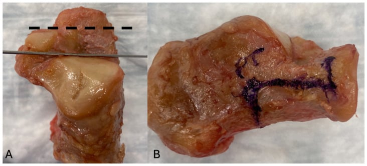
The muscle and its tendon. Tibialis posterior arises from the posterior tibia, fibula and interosseous membrane and is the strongest invertor of the foot. Its tendon runs posterior and inferior to the medial malleolus in a fibro-osseous tunnel and then fans out across the midfoot. The primary insertion is the navicular tuberosity; secondary slips reach all three cuneiforms and the bases of the second to fourth metatarsals.
What each insertion does. The multiple slips distribute force across the midfoot and support the transverse arch. The navicular insertion holds the keystone of the medial arch, and detachment there causes immediate arch collapse. Once the tendon fails, eversion is unopposed because peroneus brevis dominates.
Where it fails. There is a hypovascular zone 2-4 cm proximal to the navicular insertion, and that is where degenerative tendinopathy develops and where the tendon eventually ruptures.
Why the tendon matters so much. The PTT provides over 50% of the dynamic support to the medial longitudinal arch during the stance phase. The spring ligament and plantar fascia are static restraints; the PTT actively resists collapse during gait, and nothing else does that job. Lose it and the foot drifts into the classic triad of arch collapse, hindfoot valgus and forefoot abduction.
Static Stabilisers
The spring ligament. The plantar calcaneonavicular ligament is the primary static stabiliser of the medial arch and has two components. The superomedial calcaneonavicular ligament is the main load-bearing structure and supports the talar head; the inferior calcaneonavicular ligament reinforces the plantar aspect. As the PTT loses function the spring ligament fails by attrition, the talar head progressively uncovers and the arch collapses.
The windlass mechanism. The plantar aponeurosis runs from the calcaneal tuberosity forwards beneath the metatarsal heads to the plantar plates and proximal phalanges. At terminal stance, dorsiflexion of the metatarsophalangeal joints, especially the hallux, winds the aponeurosis around the metatarsal heads like a cable around a drum, shortening the calcaneus-to-forefoot distance. The arch rises and stiffens, and a flexible foot becomes a rigid lever for push-off.
The partnership, and how it fails. Arch height depends on three contributors acting together: the dynamic stabiliser (the PTT), the static ligamentous stabilisers (spring ligament, plantar aponeurosis and plantar ligaments) and the bony architecture. As the PTT fails and the spring ligament attenuates, load is transferred onto the plantar fascia. An intact windlass partly compensates early, but with progressive collapse and forefoot abduction the lever arm and hallux purchase are lost, the windlass becomes mechanically inefficient and the arch can no longer be re-tensioned at push-off. Realigning the medial column and forefoot, by lateral column lengthening, a Cotton osteotomy or medial column fusion, re-establishes an effective windlass, which is part of why these procedures raise the arch and improve push-off rather than simply changing static alignment.
The Deformity in Three Planes
Examiners ask you to describe the deformity, and the answer is systematic: one plane at a time.
- Coronal - hindfoot valgus. Subtalar eversion, lateral talar shift and uncovering of the posterior facet; from behind, the too many toes sign.
- Sagittal - arch collapse (planus). Plantar flexion of the talus, navicular sag, an increased talo-first metatarsal angle and attenuation of the spring ligament.
- Axial - forefoot abduction. Talonavicular uncovering, the lateral border of the forefoot swinging out (the other half of the too many toes sign) and compensatory external rotation of the tibia.
Classification Systems
Johnson and Strom is the most widely used system and the one still examined; the Myerson modification splits Stage II; Bluman grades the tendon on MRI; and the 2020 PCFD consensus is the current international standard. Know all of them, and know which question each answers.
The stages. Each stage carries its own treatment.
- Clinical Findings
- PTT pain, swelling medial ankle. Normal alignment. Single heel rise positive. No too many toes sign.
- Radiographic Features
- Normal alignment. No arch collapse. No hindfoot valgus.
- Treatment
- Conservative: CAM boot 6-12 weeks, UCBL orthotic, NSAIDs, PT
- Clinical Findings
- Hindfoot valgus flexible. Arch collapses weight-bearing. Single heel rise negative. Too many toes positive. Cotton test negative.
- Radiographic Features
- Hindfoot valgus (AP: talar head uncovering). Lateral: sag at talonavicular, increased talo-1st MT angle.
- Treatment
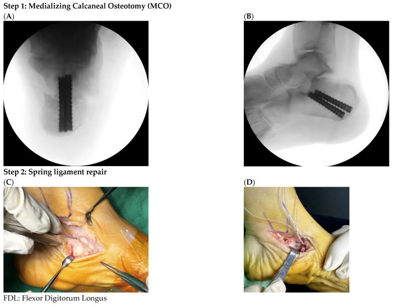
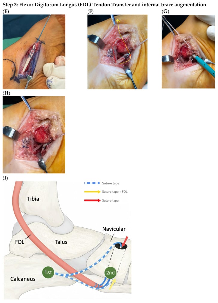
- Medializing calcaneal osteotomy + FDL transfer ± lateral column lengthening
- Clinical Findings
- As IIA PLUS fixed forefoot varus (does not correct). Cotton test positive (forefoot drives hindfoot valgus).
- Radiographic Features
- As IIA PLUS forefoot varus visible on lateral radiograph with hindfoot corrected.
- Treatment
- As IIA PLUS medial cuneiform plantar flexion osteotomy (Cotton osteotomy)
- Clinical Findings
- Rigid hindfoot valgus (does NOT correct passively). Subtalar stiffness. Talonavicular pain/arthritis.
- Radiographic Features
- Subtalar arthritis. Talonavicular arthritis. Fixed hindfoot valgus. No passive correction on stress views.
- Treatment
- Subtalar arthrodesis or triple arthrodesis (± tendon transfer if Stage IIIA)
- Clinical Findings
- As Stage III PLUS lateral ankle pain. Deltoid insufficiency. Ankle valgus tilt.
- Radiographic Features
- Ankle valgus tilt on AP ankle (tilted talus). Tibiotalar arthritis. Widening of the medial clear space with lateral tibiotalar joint-space narrowing.
- Treatment
- Tibiotalocalcaneal (TTC) arthrodesis. Consider total ankle replacement in select cases.
The Stage II split (Myerson modification). Both IIA and IIB are flexible at the hindfoot; what separates them is the forefoot. In IIA the hindfoot corrects when the forefoot is unloaded and the Cotton test is negative, so hindfoot procedures alone will do. In IIB there is a fixed forefoot varus with the first ray dorsiflexed: the hindfoot valgus persists when the first ray is elevated (Cotton test positive) because the forefoot varus is driving the hindfoot valgus as a compensation. That foot needs its forefoot corrected as well as its hindfoot, and a reconstruction that leaves the forefoot alone fails.
Clinical Assessment
History. Where it hurts tells you what is failing: medial ankle pain is the tendon, lateral pain is peroneal overload or the sinus tarsi, plantar pain is arch strain. Ask whether the onset was an acute injury or insidious degeneration, how the deformity has progressed over months to years, and what it now costs the patient in walking distance, stairs and uneven ground. Footwear is a useful witness: shoes that are difficult to fit and a medial heel that wears out. The red flags are inflammatory symptoms (rheumatoid arthritis), neuropathy (diabetes) and acute swelling (DVT).
Examination. Standing first, then seated.
- Standing (weight-bearing): too many toes sign from behind, arch height against the other side, hindfoot valgus, forefoot abduction, compensatory external tibial rotation, and the single heel rise on each foot separately
- Seated (non-weight-bearing): palpation of the PTT for swelling, tenderness or a gap; hindfoot flexibility under valgus stress; the Cotton test; the Silfverskiöld test; a neurovascular examination
Too many toes sign. Stand behind the patient, who stands with the feet shoulder-width apart. Normally only the fifth and part of the fourth toe show lateral to the heel; more than two toes visible on the affected side is positive and indicates forefoot abduction with hindfoot valgus. Sensitivity is 90% for Stage II or greater.
Single heel rise. The patient stands on one leg and rises onto the toes while you watch the hindfoot from behind. Normally the heel swings into varus as the PTT contracts and the arch rises. With PTT dysfunction the hindfoot stays in valgus or everts further, or the patient cannot rise at all, which means complete rupture or dysfunction. This is the gold standard for PTT function, and inability to perform it puts the foot at Stage II or greater.
Hindfoot flexibility. With the patient seated and the foot unloaded, grasp the heel and try to bring the valgus hindfoot to neutral or varus. If it corrects, the deformity is flexible (Stage II): a soft-tissue problem of tendon and spring ligament without fixed bony or arthritic change. If it does not, it is rigid (Stage III), which means subtalar or talonavicular arthritis or a fixed bony deformity, and the operation changes from osteotomy to arthrodesis.
Cotton test. With the patient seated, elevate the first ray by plantarflexing the first metatarsal-cuneiform joint and watch the hindfoot. If the valgus corrects, the forefoot is flexible and the test is negative: Stage IIA. If the valgus persists, a fixed forefoot varus is driving it and the test is positive: Stage IIB.
Silfverskiöld test. Dorsiflex the ankle with the knee extended, then again with the knee flexed to 90 degrees. Dorsiflexion that improves with the knee flexed means an isolated gastrocnemius contracture. Equinus drives valgus, so a tight gastrocnemius exacerbates the hindfoot deformity and may need a recession added to the reconstruction; the result changes the operative plan.
Both feet. Approximately 20% of patients have bilateral involvement, often asymmetric, so always examine and compare both feet: the "normal" side may be early, asymptomatic AAFD. Miss that and you compare against an abnormal baseline, lose the chance of early intervention on the quiet side, and inherit a dissatisfied patient when the other foot progresses after surgery.
Differential Diagnosis of the Adult Flatfoot
- Distinguishing features
- Acquired, usually unilateral and progressive; flexible early then rigid; positive too-many-toes and failed single heel rise
- Key test / investigation
- Weight-bearing radiographs + MRI (PTT and spring ligament); flexibility on examination
- Distinguishing features
- Younger or rigid flatfoot, painful peroneal spasm, restricted subtalar motion from the outset
- Key test / investigation
- CT (bony bar) or MRI (fibrous/cartilaginous coalition); often bilateral
- Distinguishing features
- Neuropathy (diabetes), warm swollen foot, rocker-bottom collapse, often painless out of proportion to deformity
- Key test / investigation
- Loss of protective sensation; radiographs/MRI of midfoot; check inflammatory and metabolic status
- Distinguishing features
- Polyarticular symptoms, morning stiffness, synovitis, often bilateral and symmetrical
- Key test / investigation
- Inflammatory markers, autoantibodies (RF, anti-CCP), erosive changes on radiographs
- Distinguishing features
- History of injury, deformity localised to midfoot, tarsometatarsal malalignment
- Key test / investigation
- Weight-bearing radiographs (Lisfranc alignment), CT for articular detail
- Distinguishing features
- Lifelong, flexible, painless, bilateral and symmetrical; normal single heel rise
- Key test / investigation
- Clinical - normal PTT function and no progression; no imaging needed if asymptomatic
- Distinguishing features
- Talar head sag with relatively preserved PTT strength; isolated medial sag
- Key test / investigation
- MRI shows spring ligament tear; PTT may be intact - changes reconstruction emphasis
Investigations
Weight-bearing radiographs, always. Non-weight-bearing films underestimate the deformity, so every film used to judge its magnitude is taken standing.
- AP foot - talonavicular uncovering (lateral subluxation of the navicular on the talus), forefoot abduction, the talo-first metatarsal angle
- Lateral foot - calcaneal pitch, the lateral talo-first metatarsal (Meary) angle, plantar flexion and sag of the talus
- AP ankle, if Stage III or IV is suspected - valgus tilt of the talus, tibiotalar arthritis, widening of the medial clear space from deltoid insufficiency
- Hindfoot alignment (Saltzman) view - taken standing with the beam angled 20 degrees cephalad from behind the ankle; it measures the weight-bearing axis of the hindfoot and quantifies the valgus
- Normal Value
- 20-30 degrees
- AAFD Finding
- Less than 18 degrees (arch collapse)
- Clinical Significance
- Decreased angle indicates loss of arch height
- Normal Value
- 0-5 degrees
- AAFD Finding
- Greater than 10 degrees (arch sag)
- Clinical Significance
- Measures alignment of talus and first metatarsal - should be collinear
- Normal Value
- Less than 7 degrees
- AAFD Finding
- Greater than 20 degrees (uncovering)
- Clinical Significance
- Lateral subluxation of navicular off talar head - forefoot abduction
- Normal Value
- 0 ± 3mm from midline
- AAFD Finding
- Greater than 5mm lateral deviation (valgus)
- Clinical Significance
- Quantifies hindfoot valgus deformity for surgical planning
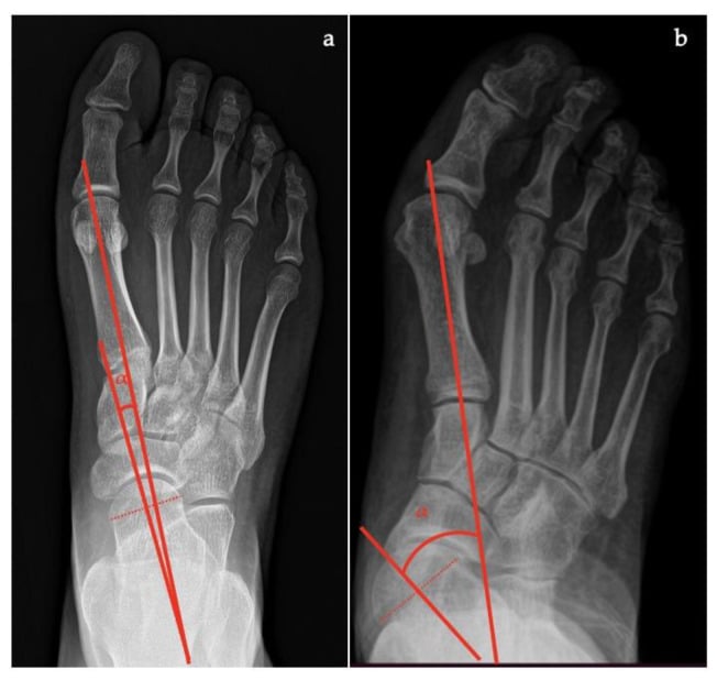
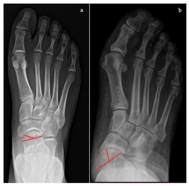
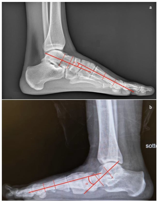
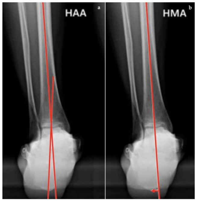
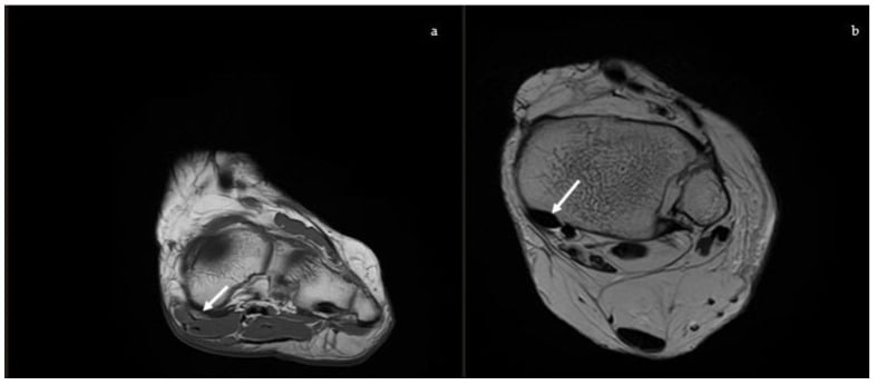
MRI. The second-line study, for an uncertain diagnosis, for assessing PTT integrity when planning surgery, for evaluating the spring ligament, and for ruling out occult fracture or other pathology. It shows both the dynamic and the static medial restraints, which matters when pain or deformity is disproportionate to the radiographs and when tendon or ligament reconstruction is being planned. The tendon is graded by the Bluman system above. Look for:
- PTT - increased T2 signal (tenosynovitis), thinning, discontinuity (tear), fluid in the sheath
- Spring ligament - attenuation, tear, increased signal
- Bone marrow oedema - talar head (overload), navicular stress
- Subtalar and talonavicular arthritis - cartilage loss, subchondral change
CT. For assessing subtalar or talonavicular arthritis before an arthrodesis, for finding a tarsal coalition in the rigid flatfoot, and for planning complex osteotomies. It gives better bony detail than MRI, and 3D reconstructions are especially useful for planning lateral column lengthening or complex deformity correction.
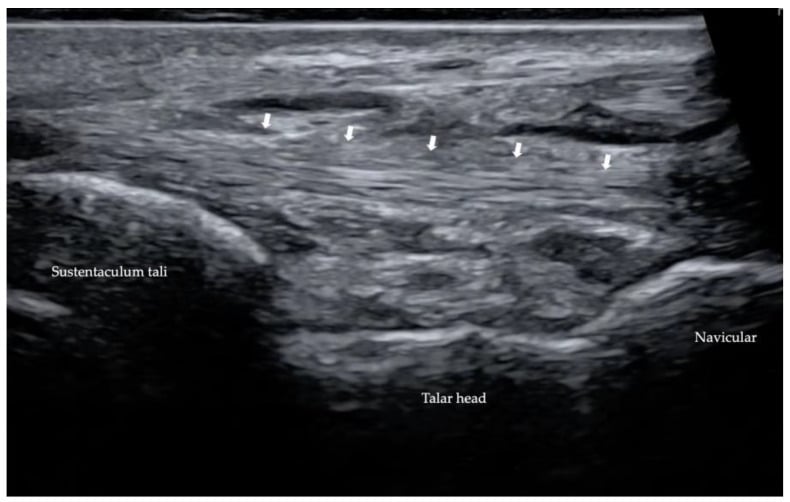
Ultrasound. A limited role: dynamic assessment of the tendon in real time, and telling a complete rupture from a partial tear. It is operator-dependent and not routinely used for staging in most centres, though availability and reliance vary by region and local expertise.
Management Algorithm
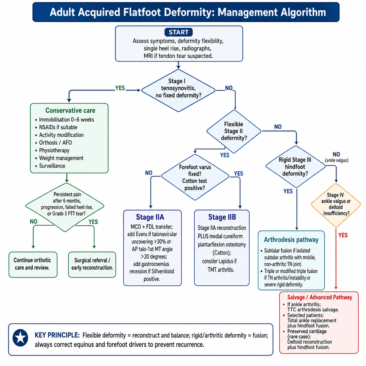
The decision turns on whether there is a deformity at all, whether it is flexible or rigid, and whether the ankle is involved. Stage I is treated conservatively, flexible Stage II is reconstructed around the Cotton test, rigid Stage III is fused, and Stage IV with ankle valgus follows a salvage pathway.
Who. Stage I (tenosynovitis without deformity); mild Stage IIA with minimal symptoms; patients unfit for surgery because of comorbidity; and the patient who, having understood the options, prefers not to have an operation.
Conservative Treatment Protocol
The goals are to reduce inflammation, offload the tendon and control pain.
- Immobilisation in a CAM walker boot or short leg cast, non-weight-bearing or protected weight-bearing
- Oral NSAIDs if there is no contraindication
- Ice for 15-20 minutes three times a day over the medial ankle
- Activity modification: avoid prolonged standing, stairs and uneven ground
- Rest, because the tendon needs unloading for the inflammation to settle
The goals are to restore function, prevent recurrence and hand over to an orthotic.
- Progressive weight-bearing, moving gradually from the boot into supportive footwear
- Physiotherapy: PTT strengthening (resisted inversion), intrinsic strengthening, proprioception
- An orthotic: a UCBL (University of California Berkeley Laboratory) insert or an ankle-foot orthosis
- Footwear with a firm heel counter and medial arch support; no flat flexible shoes
The goals are to prevent progression, maintain function and keep watch.
- Orthotic worn daily in all footwear
- Weight management to reduce the load on the tendon
- Avoid high-impact activity; walking aids if needed
- Review every 6-12 months for progression to Stage II
- Increasing deformity, a too many toes sign or a failed single heel rise means surgical referral
When conservative treatment fails. Surgery is indicated in a Stage I foot for persistent pain despite 6 months of appropriate conservative treatment, for progressive deformity (transition to Stage II), for a failed single heel rise, and for a Grade 3 tear on MRI, because a complete rupture will not heal. Not every Stage I patient progresses: approximately 50% respond to conservative treatment with orthotics. Those with a Grade 3 tear should be offered early FDL transfer, before the deformity develops.
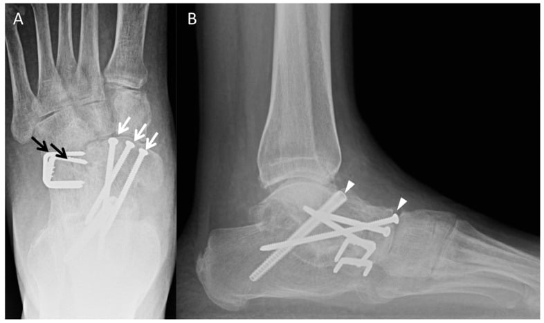
Surgical Technique: Stage II Reconstruction
Pre-operative Planning
Consent. The risks to name, with the figures in the complications table further down:
- Undercorrection and recurrence, which may need revision
- Overcorrection: hindfoot varus (a painful rigid foot) and lateral column overload
- Nerve injury: sural nerve on the lateral approaches, saphenous nerve and tibial nerve branches on the medial
- Infection, superficial and deep, higher in diabetics
- Nonunion of the calcaneal osteotomy or of the Evans graft
- Donor site morbidity: FDL harvest (toe clawing is rare; flexion weakness), iliac crest pain if autograft is used
- A prolonged recovery: strict non-weight-bearing for the first weeks, graded loading over the first 3 months, and 6-12 months to full recovery
- Adjacent joint arthritis in the long term: ankle, subtalar, talonavicular
Equipment.
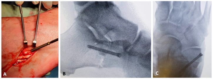
- Implants: cannulated screws (6.5 mm or 7.0 mm for the calcaneus), an interference screw or suture anchor for the FDL, an Evans plate if lateral column lengthening is planned
- Power: oscillating saw for the osteotomies, drill, reamer
- Imaging: C-arm with foot and ankle capability, positioned so a hindfoot alignment view can be obtained
- Bone graft: allograft tricortical iliac crest or femoral head for the Evans; an autograft harvest set if preferred
- Tendon instruments: nerve hooks, right-angle clamps, drill for the navicular tunnel
- Retractors: self-retaining (Weitlaner), Army-Navy, Hohmann
Position. Supine on a radiolucent table (a Jackson table or a standard table with a radiolucent foot extension). Head neutral with airway access; arms on boards or across the chest according to the anaesthetist; pelvis centred and neutral. The operative hip is in neutral or slight external rotation with the knee extended or slightly flexed over a bump; the other leg is abducted and supported on a leg holder or pillow, out of the C-arm's path.
Padding. Sacrum, the non-operative heel and the elbows are padded; the ulnar nerve at the elbow and the peroneal nerve at the fibular head are at risk, the latter if the leg is externally rotated or in a leg holder. The operative heel must be off the table, the ankle hanging free or on a bolster.
Tourniquet. A thigh tourniquet over cast padding, the limb exsanguinated by Esmarch or elevation, inflated to 250-300 mmHg adjusted for the patient's size and blood pressure. Expect 90-120 minutes for multiple osteotomies and a transfer; if the time exceeds 2 hours, deflate and re-exsanguinate. A bloodless field helps the dissection, the tendon identification and the precision of the osteotomies; the costs are tourniquet pain, limited by the anaesthetic, and the time limit.
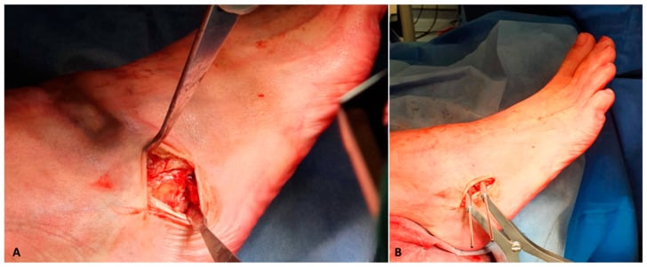
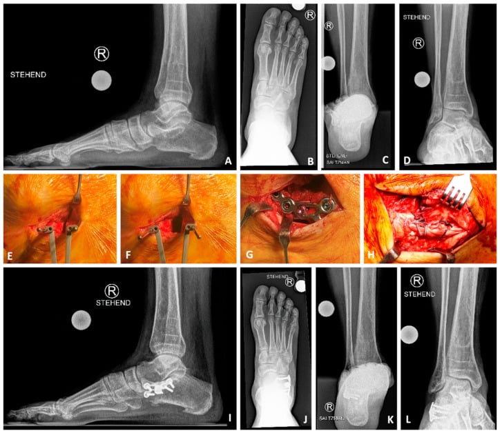
Prep and drape. Circumferential prep of the foot and ankle to mid-calf, and higher if a gastrocnemius recession is anticipated. Stockinette over the foot, then a U-drape or split sheet isolating the foot and ankle, exposing the medial malleolus to the toes (for the FDL and the medial cuneiform) and the lateral calcaneus (for the calcaneal osteotomy through a lateral or oblique approach, and the Evans if needed).
Test the C-arm before you prep. Confirm that AP, lateral and oblique views of the foot and a hindfoot alignment view are all obtainable without repositioning the patient. The Saltzman view is how intraoperative correction is judged after the osteotomy; if you cannot obtain it, you are operating blind on alignment.
Complications
Undercorrection with recurrent deformity is the most common complication, and the one the whole reconstruction is designed to avoid.
- Incidence
- 20-30% (varies by study and definition)
- Risk Factors
- Stage IIB missed (no forefoot correction), inadequate MCO translation, FDL transfer insufficient tension, patient non-compliance (early weight-bearing)
- Management
- Mild: Orthotics, activity modification. Moderate-Severe: Revision surgery (repeat MCO, revision transfer, consider arthrodesis if progression to Stage III)
- Incidence
- 5-10%
- Risk Factors
- Excessive MCO translation (greater than 12mm), over-lengthening Evans (greater than 12mm), FDL transfer overtightened
- Management
- Painful rigid foot, lateral column overload. Mild: Orthotics (lateral heel wedge), shoe modifications. Severe: Revision osteotomy (lateralizing calcaneus) or takedown and redo
- Incidence
- 5-10% (higher if smokers, diabetics)
- Risk Factors
- Smoking, diabetes, inadequate fixation, early weight-bearing, infection
- Management
- If asymptomatic: Observe. If painful: Revision fixation (ORIF with bone graft, consider bone stimulator)
- Incidence
- 10-15% (if bone graft used)
- Risk Factors
- Allograft (higher than autograft), smoking, inadequate fixation
- Management
- Often asymptomatic (graft provides structural support even without union). If painful: Revision with autograft and rigid fixation
- Incidence
- 5-15% (numbness, neuroma)
- Risk Factors
- Lateral approaches (MCO, Evans), inadequate identification and protection
- Management
- Numbness lateral foot/ankle (variable distribution). Usually improves over 6-12 months. Painful neuroma: Nerve blocks, desensitization, rarely neuroma excision
- Incidence
- 5-10%
- Risk Factors
- Diabetes, obesity, smoking, prolonged surgery, inadequate wound care
- Management
- Antibiotics (oral if cellulitis), wound care, dressing changes. Rarely needs debridement.
- Incidence
- 2-5% (higher in diabetics)
- Risk Factors
- Diabetes, immunosuppression, contamination, hardware
- Management
- Debridement, hardware removal (once union achieved), IV antibiotics (6 weeks minimum), consider VAC therapy. May require staged reconstruction or amputation if severe.
- Incidence
- Less than 5% (rare)
- Risk Factors
- Tight closure of FDL harvest site, loss of FDL to lesser toes
- Management
- Usually minimal functional impact (FHL compensates). Clawing: Toe taping, silicone sleeves. Rarely needs surgical correction (IP fusion if fixed deformity)
- Incidence
- 10-20% radiographic, 5-10% symptomatic
- Risk Factors
- Over-distraction (greater than 12mm), intra-articular osteotomy, graft malposition
- Management
- Mild: NSAIDs, activity modification, orthotics. Moderate-Severe: Calcaneocuboid fusion (salvage)
- Incidence
- 10-15% long-term (10+ years)
- Risk Factors
- Undercorrection, high-demand activities, obesity, age at surgery
- Management
- Surveillance with serial radiographs. Symptomatic arthritis: Arthrodesis (subtalar, triple, or TTC depending on location)
The diabetic patient with AAFD carries a higher infection risk (2-3 times baseline), impaired wound healing from neuropathy and vascular disease, higher nonunion rates from metabolic factors, and a risk of Charcot neuroarthropathy progression, especially if neuropathy is present.
Optimise before operating: HbA1c under 7.5% and ideally under 7%, a vascular assessment (ABI, with angiography if abnormal) and a neuropathy assessment. Consider staging the procedures, for example the FDL transfer first and the osteotomies later, to reduce the complexity of any single operation, and plan a longer protected period (12-16 weeks rather than 8-12) to make sure everything heals.
Postoperative Care and Rehabilitation
The shape of it. The osteotomies and the transfer are protected for the first 6 weeks without any load, weight is introduced in a boot only once the 6-week radiographs show healing, and the foot is fully loaded in the boot by 10-12 weeks. Some surgeons keep the foot non-weight-bearing for the full 12 weeks after a complex reconstruction (Stage IIB with multiple osteotomies) or in high-risk patients (diabetic, osteoporotic). Early loading risks nonunion or malunion of the osteotomies, especially the translated calcaneus, hardware failure (screw pull-out, plate breakage), failure of the transfer (anchor pull-out, or elongation with loss of tension) and a hindfoot that settles back into valgus.
Rehabilitation Timeline After Stage II Reconstruction
Protect the osteotomies and the transfer; keep the swelling down.
- Below-knee backslab, converted to a cast at 2 weeks after suture removal
- Strict non-weight-bearing on crutches or a walker
- Leg elevated above heart level as much as possible
- Ice 20 minutes every 2-3 hours over the cast, not directly on skin
- DVT prophylaxis: aspirin 100 mg daily, or enoxaparin if high risk (obesity, prior DVT, prolonged immobility); mechanical prophylaxis (foot pumps, TED stockings) where possible
- Multimodal analgesia (paracetamol, NSAID, opioid as required), opioids weaned by week 2
- Wound check and suture or staple removal at day 10-14
Continue protection, start gentle ankle motion out of the cast, and watch for healing.
- Below-knee circumferential cast, or a removable CAM walker boot
- Still non-weight-bearing: the osteotomies have not healed
- Gentle ankle range of motion out of the cast or boot, plantarflexion and dorsiflexion only, no inversion or eversion; knee and hip exercises against stiffness
- Radiographs at 6 weeks (AP and lateral foot, hindfoot alignment) for osteotomy healing and hardware position
Move to weight-bearing, advance motion, restore gait.
- CAM walker boot, which allows the load to be graded
- Progressive weight-bearing if the radiographs show healing: touch to partial (25-50% body weight) at weeks 6-8, partial to full in the boot at weeks 8-10, full in the boot at weeks 10-12 then into a supportive shoe with an orthotic
- Active ankle motion in all planes, proprioception on a balance board, gait re-education to a heel-toe pattern, FDL strengthening with resisted inversion
- No running, jumping or other high impact; avoid uneven ground
Restore full function, strengthen the supporting muscles, prevent recurrence.
- Supportive athletic or walking shoes with a custom orthotic (UCBL or similar) supporting the arch
- Full unrestricted weight-bearing out of the boot
- Progressive resistance in all four ankle directions, calf strengthening with heel raises, proprioception, sport-specific training if applicable
- Low-impact activity (walking, cycling, swimming) first, then a gradual return to higher impact if appropriate
- Radiographs at 3 and 6 months to confirm union and alignment
Maintain the correction, watch for recurrence, protect the adjacent joints.
- Orthotic worn daily in all footwear, which is critical to preventing recurrence
- Footwear with a firm heel counter and medial arch support; no flat flexible shoes
- Avoid prolonged barefoot walking and high-impact repetitive loading
- Annual radiographs for the first 2-3 years, then as symptoms dictate
- Weight management to keep the mechanical load down
- Teach the patient the signs of recurrence (increasing arch sag, hindfoot valgus, pain) and to report them early
Outcomes and Prognosis
Pain relief is achievable at every stage, but satisfaction falls as motion is sacrificed: the flexible foot reconstructed keeps its joints, the rigid foot fused does not, and the Stage IV salvage trades all ankle and hindfoot motion for a stable platform.
Judging the correction. Postoperative weight-bearing AP, lateral and hindfoot views document the correction in every plane. Compare the Meary alignment, talonavicular coverage, calcaneal pitch and hindfoot axis with the preoperative study rather than judging success from implant position alone.
- Pain Relief
- 50-60% achieve adequate pain relief at 1 year
- Function / Satisfaction
- Moderate functional improvement, ongoing orthotic dependence, compliance variable
- Durability
- 30-40% progress to Stage II within 5 years if Grade 3 PTT tear on MRI
- Pain Relief
- 70-80% good to excellent pain relief
- Function / Satisfaction
- 75-85% patient satisfaction, improved gait, ability to return to low-impact activities
- Durability
- 20-30% recurrence (residual symptoms or progressive deformity) at 5-10 years. Undercorrection most common cause.
- Pain Relief
- 80-85% significant pain relief (if solid fusion achieved)
- Function / Satisfaction
- 65-75% satisfaction (lower than Stage II due to loss of motion). Stable plantigrade foot. Functional limitations on uneven ground.
- Durability
- 10-15% nonunion. 20-30% develop adjacent joint arthritis (ankle, naviculocuneiform) by 10 years post-fusion.
- Pain Relief
- 70-75% pain relief (if solid fusion achieved)
- Function / Satisfaction
- 50-60% satisfaction (significant functional limitation - complete loss of ankle and hindfoot motion). Salvage procedure.
- Durability
- High nonunion risk (10-15%). Patients need lifelong rocker-bottom shoes, significant gait alteration. Quality of life impact substantial.
What predicts a worse result after Stage II reconstruction.
- Obesity (BMI greater than 35) - more mechanical load, more recurrence
- Diabetes - impaired healing, more infection, and neuropathy that disturbs balance
- Smoking - nonunion and wound complications
- Advanced age (greater than 70) - less functional reserve, slower rehabilitation
- Inflammatory arthropathy (RA, seronegative) - the systemic disease keeps progressing
- Stage IIB without forefoot correction - the Cotton test missed, and recurrence inevitable
- Delayed presentation with a longstanding severe deformity - fixed changes and a worse baseline alignment
Counsel patients beforehand about realistic expectations in the light of their risk factors, and consider moving to arthrodesis earlier, a Stage III procedure for a Stage II foot, in the high-risk patient.
Guidelines, Registries & Global Practice
Adult flatfoot from posterior tibial tendon failure is a worldwide condition with no dedicated joint registry and no high-level (RCT) guideline: management is driven by expert consensus (the 2020 PCFD nomenclature) and retrospective case series, so the world standard is a deformity-specific reconstruction tailored to flexibility and the planes of collapse.
Global Epidemiology
- Figure
- Most present in the 5th-6th decade
- Source / note
- Reflected across surgical series (e.g. mean age 54 in PCFD reliability cohort, PMID 34852647)
- Figure
- Female predominance (≈ 62% female in PCFD cohort)
- Source / note
- PMID 34852647 (single-centre, surgical population)
- Figure
- Multiplanar collapse is the norm; isolated single-plane deformity is uncommon
- Source / note
- Class A (hindfoot valgus) 89.5%, class C (forefoot varus / medial column instability) 86.2% (PMID 34852647)
- Figure
- Reviews estimate several million affected adults and rising prevalence with ageing and obesity
- Source / note
- Narrative estimate (PMID 39036227); precise population prevalence is not robustly established
Nomenclature and Classification - Side by Side
- Year / status
- 1989, still widely examined
- What it adds
- Original 4-stage tendon-centred staging (I tenosynovitis to IV ankle valgus)
- Evidence level
- Level V (historical, unvalidated)
- Year / status
- 1997
- What it adds
- Splits Stage II into IIA / IIB (forefoot-driven), adds Stage IV subtypes
- Evidence level
- Level V expert opinion
- Year / status
- 2007 (PMID 17561198)
- What it adds
- Subcategories for forefoot supination, abduction, medial column and ankle; MRI grading
- Evidence level
- Level V expert opinion
- Year / status
- 2020 (PMID 32856474) - current international standard
- What it adds
- Renames to Progressive Collapsing Foot Deformity; class + components A-E, flexible/rigid
- Evidence level
- Level V consensus; reliability tested (PMID 34852647)
Guidelines and Peri-operative Standards (Region-Neutral)
- No AAOS, NICE or EFORT disease-specific guideline exists for adult flatfoot reconstruction - this is consensus-driven (PCFD 2020), unlike arthroplasty topics
- Staged escalation is universal: trial non-operative care (orthoses/bracing, activity and weight management) before reconstruction in flexible disease
- Flexible vs rigid divide drives surgery everywhere: osteotomy plus tendon transfer for flexible deformity, arthrodesis for rigid/arthritic deformity
- Surgical safety: WHO Surgical Safety Checklist is the global standard; prophylactic cephalosporin within 60 min of incision and peri-operative normoglycaemia are near-universal SSI-prevention measures
- Imaging: weight-bearing CT is increasingly used in North America and Europe to quantify peritalar subluxation (PMID 33358266) but remains unavailable in many centres, where weight-bearing radiographs ± MRI remain standard
- VTE prophylaxis: foot/ankle surgery with prolonged immobilisation has no global consensus - chemical prophylaxis (LMWH or aspirin) is used more readily in some systems than others; decisions are individualised on risk
- Lateral column lengthening vs medial column procedures for forefoot abduction varies by surgeon and school rather than by guideline
- Resource setting: in limited-resource settings, bracing and arthrodesis predominate over multi-osteotomy reconstruction owing to implant, graft and rehabilitation costs
Unlike hip and knee arthroplasty, adult flatfoot reconstruction is not captured by any national joint registry. The major registries - NJR (England & Wales), AJRR (USA), AOANJRR (Australia), SHAR (Sweden), the Norwegian and New Zealand registries - track arthroplasty implants, not foot/ankle deformity surgery. Any "flatfoot registry revision rate" should be treated with suspicion. The revision and complication figures available for this reconstruction come from case series and reviews, and quoting them at that level - rather than as registry-grade numbers - is what a viva examiner is listening for.
Key documentation requirements:
-
Informed consent must include:
- Realistic success rate (75-85% good outcomes, but 20-30% recurrence risk)
- Prolonged recovery (3-6 months to full function, 12+ months to final outcome)
- Complications: Undercorrection (20-30%), overcorrection (5-10%), nerve injury (sural nerve 5-15%), infection (superficial 5-10%, deep 2-5%), nonunion (5-15%), progression to arthrodesis (10-15% long-term)
- Alternative treatments: Conservative (orthotic), arthrodesis (if rigid deformity)
- Expected functional outcomes: Improved pain and gait, but NOT return to high-impact sports
-
Common litigation issues:
- Failure to diagnose Stage IIB (missing Cotton test) leading to recurrence - document Cotton test result in notes
- Nerve injury (sural nerve, tibial nerve branches) without pre-operative counseling - must consent for numbness risk
- Infection in diabetic patients without adequate optimization - document HbA1c, pre-operative counseling, glucose control peri-op
- Undercorrection perceived as surgical failure - set realistic expectations (not all patients achieve perfect correction), document degree of deformity pre-op and plan for multi-stage if severe
-
Documentation best practices:
- Pre-operative: Clinical photos (too many toes sign, arch height), weight-bearing radiographs with measurements (angles documented), Cotton test result, Silfverskiöld test, single heel rise test result
- Intraoperative: Fluoroscopy images saved (hindfoot alignment view showing correction), degree of calcaneal translation measured and documented, FDL transfer tension confirmed
- Post-operative: Serial radiographs to monitor healing and alignment, document orthotic prescription and compliance counseling
Frequently asked questions
What is adult-acquired flatfoot deformity?
Adult-acquired flatfoot is collapse of the medial longitudinal arch in an adult who previously had a normal arch, most commonly from posterior tibial tendon dysfunction (PTTD). The triad is loss of the arch, hindfoot valgus and forefoot abduction - the tendon that normally locks and inverts the hindfoot fails, so the spring ligament and other static restraints stretch under load.
What are the stages of posterior tibial tendon dysfunction?
The Johnson and Strom classification guides reconstruction. Stage I: tenosynovitis only - pain and swelling along the tendon with no deformity and a normal (if sore) single heel rise. Stage II: flexible deformity with an incompetent tendon - subdivided into IIA (flexible hindfoot) and IIB (forefoot-driven with fixed forefoot varus). Stage III: the deformity has become rigid and fixed. Stage IV: the ankle is involved, with talar tilt in the mortise from deltoid ligament failure.
What is the too-many-toes sign?
Viewed from behind the standing patient, more than two or three toes are visible lateral to the heel on the affected side. It is pathognomonic for forefoot abduction driven by hindfoot valgus, and is one of the quickest screening signs for adult flatfoot.
What does an abnormal single heel rise test mean?
Asked to rise onto the forefoot on one leg, a competent posterior tibial tendon locks the hindfoot, which inverts as the heel leaves the ground. Inability to perform the rise, or a rise in which the heel stays in valgus rather than inverting, indicates posterior tibial tendon incompetence - the key functional test separating PTTD from a simple flexible flat arch.
Why does flexible versus rigid flatfoot change the operation?
A flexible (stage II) foot can be corrected by joint-preserving reconstruction - flexor digitorum longus transfer, medialising calcaneal osteotomy, lateral column lengthening and spring ligament repair - because the joints still reduce. A rigid (stage III) foot cannot be corrected passively, so realignment must be achieved by arthrodesis, classically a triple fusion. Stage IV adds the ankle and may need deltoid reconstruction or a tibiotalar-including fusion.
MCQ Practice Points
Q: What is the PRIMARY insertion of the posterior tibial tendon and why is it critical for arch support? A: The primary insertion is the navicular tuberosity. This is critical because the navicular is the keystone of the medial longitudinal arch. The PTT's pull on the navicular (via its insertion) lifts the arch during stance phase. When the PTT fails, the navicular sags inferiorly and the arch collapses. The PTT has secondary insertions to all 3 cuneiforms and metatarsal bases 2-4, providing distributed midfoot stability, but the navicular insertion is the main arch supporter.
Q: What distinguishes Stage IIA from Stage IIB in the Johnson and Strom classification, and why does this matter for surgical planning? A: Both are flexible deformities, but the key difference is the forefoot. Stage IIA has a flexible forefoot - the Cotton test is negative (hindfoot valgus corrects when first ray is elevated). Stage IIB has a fixed forefoot varus - the Cotton test is positive (hindfoot valgus persists despite first ray elevation because the forefoot varus is DRIVING the hindfoot valgus compensatorily). This matters because Stage IIB requires additional surgery: medial cuneiform plantar flexion osteotomy (Cotton osteotomy) to correct the forefoot varus. Without this, the reconstruction WILL fail because the uncorrected forefoot drives recurrent hindfoot valgus.
Q: Describe the single heel rise test and what a positive (abnormal) test indicates. A: The patient stands on one leg (the affected side) and attempts to rise up on tiptoes (heel rise off ground). The examiner observes the hindfoot from behind. Normal: As the patient rises, the hindfoot inverts (shifts from valgus to varus) due to tibialis posterior contraction. Abnormal (positive test): The hindfoot remains in valgus or everts further, OR the patient is completely unable to perform a heel rise. This indicates posterior tibial tendon dysfunction - the PTT is incompetent and cannot generate the inversion moment. A positive test signifies Stage II or greater AAFD (Stage I patients can usually still perform heel rise, though may have pain).
Q: Why is FDL preferred over FHL for tendon transfer in AAFD reconstruction? A: FDL advantages: (1) Better excursion - longer muscle belly and arc of contraction provides greater active inversion force. (2) Lower donor morbidity - FHL is the stronger hallux flexor; transferring FHL causes more noticeable hallux weakness and potential IP joint stiffness. FDL loss to lesser toes is rarely symptomatic (FHL compensates). (3) Easier harvest - medial approach (same as navicular exposure) vs deep posterior approach for FHL. (4) Equivalent strength - biomechanical studies show FDL provides sufficient force for arch support. Both FDL and FHL have similar clinical outcomes in terms of pain relief and deformity correction, but FDL has better risk-benefit profile.
Q: What is the most common cause of failure after Stage II AAFD reconstruction, and how can it be prevented? A: The most common cause is undercorrection / recurrent deformity (occurs in 20-30% of patients). Causes include: (1) Missing Stage IIB - failing to perform medial cuneiform osteotomy when forefoot varus is present (Cotton test positive). (2) Inadequate calcaneal translation - not medializing enough (should aim for 8-12mm, confirmed on hindfoot alignment view). (3) Missing lateral column lengthening - when significant forefoot abduction present (talonavicular uncovering greater than 30 degrees, too many toes sign) but LCL not performed. (4) Patient factors - obesity (high BMI increases mechanical load), diabetes, non-compliance with orthotic use post-op. Prevention: Meticulous pre-operative planning (Cotton test, assess forefoot abduction, measure angles), adequate surgical correction (confirm with intraoperative fluoroscopy), address patient risk factors (weight loss, diabetes control), enforce orthotic compliance post-operatively.
Q: What is the evidence base for the FDL transfer plus medialising calcaneal osteotomy in flexible Stage II disease? A: The most-cited series is Myerson, Badekas and Schon (Foot Ankle Int 2004), who reviewed 129 patients with Stage II posterior tibial tendon deficiency treated with medialising calcaneal osteotomy plus FDL transfer to the navicular at a mean 5.2 years. They reported 97% pain relief, a mean postoperative AOFAS hindfoot score of 79, significant radiographic correction in all parameters, and high satisfaction with few complications. This underpins the modern soft-tissue-plus-osteotomy approach. Limitations to quote: it is a retrospective, single-surgeon series with no control arm, AOFAS is not a validated score, and forefoot-driven (Stage IIB) feet were excluded. Note there is no joint registry capturing flatfoot reconstruction (registries such as AOANJRR, NJR and AJRR track arthroplasty, not foot/ankle deformity surgery), so the evidence base is case series and reviews rather than registry data.
Exam Viva Scenarios
Practise clinical reasoning and management decisions out loud
“A 52-year-old female presents with progressive medial foot pain and flattening of her arch over 2 years. She has type 2 diabetes (HbA1c 7.2%) and BMI 32. On examination, you observe a too many toes sign, and she is unable to perform a single heel rise on the affected side. Her hindfoot corrects to neutral when you passively stress it into varus (seated examination). You perform a Cotton test: when you elevate her first ray (plantarflex the first metatarsal), her hindfoot valgus DOES correct. Weight-bearing radiographs show hindfoot valgus with talonavicular uncovering of 25 degrees and decreased calcaneal pitch. What is your assessment and management plan?”
“Walk me through your technique for performing a medializing calcaneal osteotomy as part of a Stage II AAFD reconstruction. Specifically, describe your approach, osteotomy location and orientation, how much translation you aim for, and your fixation method. What nerves are at risk and how do you protect them?”
“A 58-year-old patient returns 18 months after Stage IIA reconstruction (MCO + FDL transfer, no lateral column lengthening) with recurrent medial foot pain and visible return of arch collapse. Radiographs show hindfoot valgus has recurred (calcaneal osteotomy healed in translated position) and talonavicular uncovering has worsened from 20 degrees pre-op to 15 degrees immediately post-op to now 30 degrees. She is obese (BMI 36) and non-compliant with orthotic use (admits she rarely wears it). How do you approach this patient?”
Key Anatomy
- PTT = primary dynamic arch stabilizer (over 50% of support)
- Navicular tuberosity = primary PTT insertion (arch keystone)
- Hypovascular zone 2-4cm proximal to insertion = site of rupture
- Spring ligament = static stabilizer (fails secondary to PTT loss)
- 3-plane deformity: hindfoot valgus (coronal) + arch collapse (sagittal) + forefoot abduction (axial)
Johnson and Strom Classification
- Stage I = PTT tenosynovitis, no deformity, conservative treatment
- Stage IIA = Flexible hindfoot, negative Cotton test, MCO + FDL ± LCL
- Stage IIB = Flexible hindfoot + fixed forefoot varus (positive Cotton), add medial cuneiform osteotomy
- Stage III = Rigid deformity, subtalar/triple arthrodesis
- Stage IV = Ankle valgus tilt (deltoid insufficiency), TTC fusion
Clinical Assessment Algorithm
- Standing: Too many toes sign (forefoot abduction + hindfoot valgus)
- Functional: Single heel rise test (PTT function - negative = incompetent)
- Seated: Hindfoot flexibility test (Stage II flexible, Stage III rigid)
- Cotton test: Elevate first ray - if valgus persists = Stage IIB (forefoot-driven)
- Silfverskiöld test: Gastrocnemius contracture (add recession if positive)
Surgical Decision Points
- Flexible (Stage II) = osteotomy + tendon transfer, Rigid (Stage III) = arthrodesis
- FDL over FHL: Better excursion, lower donor morbidity, easier harvest
- LCL indications: Forefoot abduction greater than 30 degrees (talonavicular uncovering)
- MCO translation: 8-12mm medially (confirm with hindfoot alignment view)
- Post-op: strict non-weight-bearing to 6 weeks, then graded loading in a boot to 12 weeks (multiple osteotomies to heal)
Complications and Prevention
- Undercorrection 20-30% = most common, prevent with adequate MCO + LCL if needed + Cotton test
- Overcorrection 5-10% = hindfoot varus (painful), avoid greater than 12mm translation/LCL
- Sural nerve injury 5-15% = identify and protect during lateral approach
- Nonunion 5-15% = smoking cessation mandatory, avoid NSAIDs during healing
- Recurrence risk factors: obesity, diabetes, orthotic non-compliance, smoking
Evidence Base and Key Trials
FDL Transfer with Medialising Calcaneal Osteotomy for Stage II PTT Deficiency
- Retrospective series of 129 patients with Stage II posterior tibial tendon deficiency (flexible flatfoot without fixed forefoot supination), operated 1990-1997, mean follow-up 5.2 years
- Procedure: medial translational calcaneal osteotomy PLUS flexor digitorum longus transfer to the navicular
- Mean postoperative AOFAS hindfoot score 79 (range 54-93); significant radiographic correction in all four parameters measured
- 97% (125/129) experienced pain relief, 94% improved function, 84% wore shoes comfortably without modification or orthotic support
- Patient satisfaction: entirely satisfied 118, partially satisfied 7, dissatisfied 4; seven significant complications in six patients
Outcomes of Reconstruction of the Flexible Adult-Acquired Flatfoot Deformity
- Narrative review synthesising contemporary clinical and radiographic outcomes for the major procedures used in flexible AAFD reconstruction
- Confirms that combined procedures - FDL transfer, medialising calcaneal osteotomy (MCO), gastrocnemius recession, lateral column lengthening (LCL), Cotton osteotomy or first tarsometatarsal fusion, and spring ligament reconstruction - are frequently used together
- Reports consistent significant improvement in patient-reported outcomes after operative treatment of flexible deformity
- Emphasises tailoring the combination of osteotomies and soft-tissue work to the individual deformity rather than a single fixed algorithm
- Reviews the relative correction power and complications of MCO, LCL and Cotton osteotomies
PCFD Consensus: Classification and Nomenclature for Progressive Collapsing Foot Deformity
- Expert consensus group renamed adult acquired flatfoot deformity (AAFD) / posterior tibial tendon dysfunction (PTTD) to Progressive Collapsing Foot Deformity (PCFD)
- Frames the condition as a complex 3-dimensional deformity with varying hindfoot valgus, forefoot abduction and midfoot varus, rather than a single tendon disease
- Replaces the older numeric staging (Johnson and Strom 1989; Myerson 1997) with a class (severity) and flexible/rigid descriptor system reporting deformity components A-E
- Unanimous consensus (9/9) that prior classification systems are incomplete or outdated and none had been validated
- Strong consensus (8/9) to adopt the new system to stage deformity and guide treatment
Reliability of the New PCFD Classification System
- Retrospective study of 92 feet (84 patients) from a prospective registry assessing intra- and interobserver reliability of the PCFD classification
- Class A (hindfoot valgus) was the most frequent component (89.5%), followed by class C (forefoot abduction) in 86.2%
- Most patients had combined rather than isolated deformity; the commonest subclass was 1ABC (25.4%)
- Interobserver reliability was moderate (Fleiss kappa 0.561); intraobserver reliability was very good (Cohen kappa 0.851)
- Confirms that isolated single-component deformity is uncommon - most feet collapse in multiple planes simultaneously
Posterior Tibial Tendon Rupture: A Refined (Bluman-Myerson) Classification System
- Refines the Johnson and Strom 1989 staging to capture the wider spectrum of deformity seen with posterior tibial tendon rupture
- Adds subcategories accounting for ankle and hindfoot valgus, forefoot supination, forefoot abduction and medial column instability
- Provides stage-specific treatment recommendations, including for early disease and for ankle (deltoid) involvement
- Underpins the MRI-based grading of tendon integrity widely quoted in viva preparation
- Bridges the gap between the simple 4-stage system and the later PCFD consensus
Weight-bearing CT and MRI Correlation of Soft-Tissue Failure in Flexible PCFD
- Retrospective comparative study of 54 patients with flexible PCFD using weight-bearing CT (WBCT) markers of peritalar subluxation and MRI markers of soft-tissue failure
- Posterior tibial tendon degeneration on MRI correlated significantly with sinus tarsi impingement (p = 0.04)
- Spring ligament degeneration correlated with subtalar joint subluxation (p = 0.04)
- Talocalcaneal interosseous ligament involvement was the only finding significantly correlated with subfibular impingement (p = 0.02)
- Demonstrates that bony collapse (peritalar subluxation) and soft-tissue (PTT, spring and interosseous ligament) failure progress together