BMP Osteoinduction | VEGF Angiogenesis | TGF-β Regulation | Clinical rhBMP Applications
- BMPs are the only growth factors that induce ectopic bone (true osteoinduction)
- BMP-2 and BMP-7 signal via Smad1/5/8 to activate Runx2, the master osteoblast regulator
- VEGF couples angiogenesis to osteogenesis - no vessels means no bone formation
- TGF-β is most abundant in bone matrix, released during resorption for coupling
- rhBMP-2 FDA-approved for ALIF L4-S1 and open tibia, serious complications off-label
- “BMPs induce ectopic bone in muscle (Urist 1965 discovery)
- “VEGF secreted by hypertrophic chondrocytes initiates vascular invasion
- “TGF-β biphasic: promotes proliferation, inhibits terminal differentiation
- “Fracture hematoma platelets release PDGF and TGF-β (initiates cascade)
Clinical Imaging
Imaging Atlas


BMPs are unique - only growth factors that induce ectopic bone when implanted in muscle. BMP-2 and BMP-7 signal via Smad1/5/8 to activate Runx2. rhBMP-2 is FDA-approved for ALIF L4-S1 and open tibia fractures but has serious complications.
Recombinant BMP-2 has serious risks: ectopic bone, osteolysis, heterotopic ossification, inflammatory swelling. Off-label cervical spine use caused life-threatening airway compromise. FDA warning against cervical use.
VEGF is essential for bone healing. Couples angiogenesis to osteogenesis via Type H vessels. Secreted by hypertrophic chondrocytes to initiate vascular invasion. Without VEGF, cartilage callus cannot be replaced by bone.
TGF-β has biphasic effects: promotes MSC proliferation but inhibits terminal differentiation. Most abundant growth factor in bone matrix. Released during resorption to recruit osteoprogenitors for coupling.
At a Glance
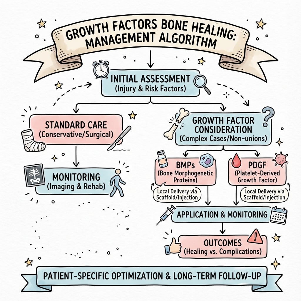
Growth factors orchestrate bone healing through coordinated signaling cascades. BMPs (Bone Morphogenetic Proteins) are the only factors that induce ectopic bone in muscle—true osteoinduction. BMP-2/7 signal via Smad1/5/8 to activate Runx2, the master osteoblast transcription factor. rhBMP-2 is FDA-approved for ALIF L4-S1 and open tibia fractures but has serious complications (ectopic bone, osteolysis, airway swelling) especially off-label in cervical spine. VEGF couples angiogenesis to osteogenesis—secreted by hypertrophic chondrocytes to initiate vascular invasion; no vessels means no bone. TGF-β is most abundant in bone matrix and released during resorption for coupling. PDGF and TGF-β from platelet-rich haematoma initiate the healing cascade.
BMPBMP - Bone Morphogenetic Protein
Hook:BMP makes Bone in Muscle Pouches (ectopic bone formation)
VEGFVEGF - Vascular Coupling
Hook:VEGF = Vessels Enable Growth for Fracture healing
GROWTHGROWTH - Key Growth Factors
Hook:GROWTH factors orchestrate bone healing in coordinated sequence
SMADSMAD - BMP Signaling Pathway
Hook:SMAD pathway: BMP receptor phosphorylates Smad1/5/8 which activates Runx2
Overview and Introduction
Growth factors are signaling proteins that regulate cellular proliferation, differentiation, migration, and matrix synthesis during bone healing. They orchestrate the complex cascade of fracture repair through sequential expression and coordinated cellular responses.
Why growth factors matter clinically:
Understanding growth factor biology is essential for:
- Comprehending fracture healing mechanisms at molecular level
- Using rhBMP-2 and rhBMP-7 safely and effectively
- Developing novel bone healing therapies
- Explaining why certain conditions impair healing (diabetes, smoking, NSAIDs)
- Rational use of biologics (PRP, bone marrow aspirate, growth factor products)
Marshall Urist (1965) discovered bone morphogenetic proteins by demonstrating that demineralized bone matrix (DBM) implanted into muscle induced ectopic bone formation. This proved the existence of osteoinductive factors within bone matrix, leading to decades of work to isolate and clone BMPs. rhBMP-2 was FDA-approved in 2002 after extensive development.
Concepts and Principles of Bone Healing
Three concepts underpin every growth-factor question and frame the rational use of biologics.
| Property | Definition | Driven by | Example material |
|---|---|---|---|
| Osteoinduction | Stimulation of primitive/mesenchymal cells to differentiate into bone-forming cells (can form ectopic bone) | BMPs (and demineralised bone matrix) | rhBMP-2/7, DBM |
| Osteoconduction | Provision of a passive scaffold permitting bone ingrowth along its surface | Surface architecture, not a signal | Hydroxyapatite, β-TCP, allograft |
| Osteogenesis | Direct formation of new bone by transplanted living osteoblasts/progenitors | Viable cells | Fresh autograft, bone-marrow aspirate |
Why this matters: Autograft is the gold standard because it is the only graft that is simultaneously osteoinductive, osteoconductive and osteogenic. Allograft is essentially osteoconductive only; DBM adds osteoinduction; recombinant growth factors supply the osteoinductive (BMP) or pro-healing (PDGF/VEGF) signal but no scaffold or cells.
Successful bone healing needs four (often five) elements: osteogenic cells, an osteoconductive scaffold, osteoinductive signals (growth factors), and mechanical stability — with adequate vascularity as the unifying requirement. Growth factors fill only the osteoinductive corner; they cannot compensate for instability, infection, or an avascular bed.
Only BMPs are truly osteoinductive. VEGF, TGF-β, PDGF, FGF and IGF are supportive or permissive (chemotactic, mitogenic, angiogenic, anti-apoptotic) but none can initiate de novo bone formation in soft tissue. This single distinction explains both the unique clinical power of rhBMP-2 and its signature complication of ectopic bone.
Bone Morphogenetic Proteins (BMPs)
BMP Family Overview
Bone morphogenetic proteins are members of the TGF-β superfamily and are the most potent osteoinductive growth factors known.
Key characteristics:
- Over 20 BMP family members identified
- BMP-2, BMP-4, BMP-6, BMP-7 have strong osteoinductive activity
- BMP-2 and BMP-7 are most clinically relevant
- Only growth factors that induce ectopic bone formation (true osteoinduction)
- Signal via Smad1/5/8 pathway to activate Runx2
Sources in bone healing:
- Produced by osteoblasts and osteoprogenitor cells
- Stored in bone matrix (released during resorption)
- Platelets contain small amounts
- Peak expression at week 2-3 post-fracture (days 14-21)
BMPs are unique because they can induce bone formation in non-skeletal sites (muscle, subcutaneous tissue). This is true osteoinduction - other growth factors can enhance bone formation but cannot initiate it de novo in soft tissue. This property was the basis of Urist's original discovery.
BMPs are essential for both fracture healing and skeletal development.
Transforming Growth Factor Beta (TGF-β)
TGF-β Superfamily
TGF-β is the most abundant growth factor in bone matrix and a key regulator of bone remodeling and fracture healing.
Three isoforms in mammals:
- TGF-β1 (most abundant in bone, main regulator)
- TGF-β2 (development, wound healing)
- TGF-β3 (anti-scarring properties)
Sources in bone:
- Platelets (released during clotting in fracture hematoma, initiates healing)
- Bone matrix (released during osteoclastic resorption, coupling mechanism)
- Osteoblasts and inflammatory cells actively produce TGF-β
- Most abundant growth factor stored in bone matrix
Secretion and activation:
- Secreted as latent complex (inactive, bound to latency-associated peptide, LAP)
- Activation by proteases (plasmin, matrix metalloproteinases), acidic pH, or mechanical stress
- Active TGF-β binds type II TGF-β receptor (TβR-II)
- Activation is tightly regulated to control local effects
TGF-β has complex, context-dependent effects on bone cells at different stages.
Vascular Endothelial Growth Factor (VEGF)
VEGF in Bone Healing
VEGF is absolutely essential for bone healing because angiogenesis (blood vessel formation) is coupled to osteogenesis. No vessels means no bone formation.
Key roles in fracture healing:
- Vascular invasion of cartilage callus during endochondral ossification
- Coupling angiogenesis to osteogenesis (spatially and temporally)
- Osteoblast survival (anti-apoptotic effects, prevents cell death)
- Osteoclast recruitment (via indirect RANKL regulation)
- Delivery of osteoprogenitors via Type H vessels
Sources in fracture healing:
- Hypertrophic chondrocytes (highest production, signals vascular invasion of soft callus)
- Osteoblasts (maintain blood supply to forming bone)
- Macrophages and inflammatory cells (early and throughout healing)
- Platelets (released early in hematoma, first wave)
VEGF secreted by hypertrophic chondrocytes is the critical signal for vascular invasion during endochondral ossification. Capillaries invade from the periosteum and marrow cavity, bringing osteoprogenitors and osteoclasts. Without VEGF, cartilage callus persists and cannot be replaced by bone. This is why VEGF inhibitors (anti-cancer drugs like bevacizumab) significantly impair fracture healing.
VEGF is the critical molecular link between angiogenesis and osteogenesis in bone healing.
Other Growth Factors in Bone Healing
Platelet-Derived Growth Factor (PDGF)
PDGF is the earliest growth factor at the fracture site, released immediately from platelet alpha granules when the fracture hematoma forms.
PDGF isoforms:
- PDGF-AA: Two A chains, binds PDGFR-α
- PDGF-BB: Two B chains, most potent, binds both receptors
- PDGF-AB: Heterodimer
- PDGF-CC, PDGF-DD: Newer isoforms, less studied
Functions in bone healing:
- Chemotaxis: Recruits inflammatory cells (neutrophils, macrophages) to fracture
- MSC recruitment: Attracts mesenchymal stem cells from periosteum, marrow, circulation
- Mitogenic: Stimulates proliferation of fibroblasts, smooth muscle cells, osteoblasts
- Angiogenesis: Indirect effect via inducing VEGF expression in stromal cells
- Matrix synthesis: Stimulates collagen and proteoglycan production
Temporal expression:
- Immediate release from platelets upon fracture (minutes to hours)
- Peak concentration at 24-48 hours in hematoma
- Sustained production by macrophages and fibroblasts during inflammation (days 3-7)
- Declines as inflammation resolves
Signaling pathways:
- Binds PDGF receptor alpha or beta (receptor tyrosine kinases)
- Activates Ras-MAPK pathway (cell proliferation)
- Activates PI3K-Akt pathway (cell survival, migration)
- Activates PLCγ pathway (calcium signaling, cytoskeletal reorganization for migration)
Fracture hematoma is not just a blood clot - it is a rich reservoir of growth factors. Platelets release PDGF and TGF-β immediately upon aggregation, initiating the inflammatory phase and recruiting the cellular players to the fracture site. This is why excessive irrigation and debridement of hematoma may impair healing - you wash away the growth factors that kick off the healing cascade.
PDGF initiates the early inflammatory and cellular recruitment phases of fracture healing.
Temporal Sequence of Growth Factors in Fracture Healing
Growth factors are expressed in coordinated sequential waves corresponding to healing phases.
Growth Factor Expression During Fracture Healing
Dominant factors: PDGF, TGF-β, VEGF (first wave)
Platelets aggregate and release growth factors from alpha granules immediately upon vascular injury. PDGF and TGF-β initiate inflammatory response and recruit mesenchymal stem cells. VEGF secreted early due to acute hypoxia in hematoma. Chemotactic signals bring cells to fracture site.
Dominant factors: TNF-α, IL-1, IL-6, TGF-β, FGF-2, VEGF
Inflammatory cytokines (TNF-α, IL-1, IL-6) from macrophages dominate. These stimulate resorption of necrotic bone and amplify inflammation. TGF-β promotes MSC chemotaxis and early matrix synthesis. FGF-2 and VEGF increase to support proliferation and angiogenesis. Mesenchymal proliferation begins. Soft callus formation initiates.
Dominant factors: TGF-β, BMP-2/4/7 (rising), VEGF (peak), FGF-2
TGF-β peaks driving chondrogenesis (cartilage soft callus formation). BMPs begin expression with mRNA detected by day 7 and peak around days 14-21. VEGF secreted by hypertrophic chondrocytes signals vascular invasion. Endochondral ossification begins as blood vessels penetrate cartilage callus.
Dominant factors: BMPs (peak), VEGF, IGF-1, FGF-2
BMP-2 and BMP-7 peak providing strongest osteoinductive signal. Type H vessels invade callus, delivering osteoprogenitors from marrow and circulation. Cartilage progressively replaced by woven bone via endochondral ossification. IGF-1 promotes osteoblast proliferation and collagen synthesis. Mineralization of hard callus accelerates.
Dominant factors: TGF-β (coupling), IGF-1, FGFs, RANKL/OPG balance
Woven bone remodeled to organized lamellar bone. TGF-β released from resorbed matrix couples osteoclasts to osteoblasts (recruits MSCs to resorption sites). IGF-1 maintains osteoblast activity during formation phase. FGFs sustain osteoprogenitor pool. External callus gradually resorbed, medullary canal restored. Mechanical loading guides remodeling (Wolff law).
Growth factor expression is sequential and overlapping, not simultaneous. Early factors (PDGF, inflammatory cytokines) recruit cells. Mid factors (TGF-β, BMPs) drive differentiation and bone formation. Late factors (IGFs, FGFs) sustain remodeling. Therapeutic timing matters - BMP delivered too early during inflammatory phase may be degraded by proteases or cleared. Optimal BMP delivery is week 1-2 when osteoprogenitors are present and inflammatory phase is resolving.
Understanding the temporal cascade explains therapeutic strategies and identifies factors that disrupt normal healing.
Anatomy
Bone Morphogenetic Proteins:
Structure:
- Dimeric proteins (linked by disulfide bonds)
- Member of TGF-β superfamily
- BMP-2 and BMP-7 most potent for bone
Signaling pathway (canonical):
- BMP binds to Type I and Type II receptors
- Type II receptor phosphorylates Type I
- Activated receptor phosphorylates Smad1/5/8
- Smad1/5/8 binds Smad4, enters nucleus
- Activates Runx2 (master osteoblast regulator)
- Osteoblast differentiation and bone formation
Vascular Endothelial Growth Factor:
Structure:
- Homodimeric glycoprotein
- Multiple isoforms (VEGF-A most important)
- Heparin-binding domain for matrix retention
Signaling pathway:
- VEGF binds to VEGFR-2 (endothelial cells)
- Receptor tyrosine kinase activation
- Downstream: PI3K, MAPK, FAK pathways
- Endothelial cell proliferation and migration
- New blood vessel formation (angiogenesis)
- Couples angiogenesis to osteogenesis
| Growth Factor | Receptor | Signaling | Key Effect |
|---|---|---|---|
| BMP-2/7 | BMPR-I/II (serine/threonine kinase) | Smad1/5/8 → Runx2 | Osteoblast differentiation |
| TGF-β | TβR-I/II (serine/threonine kinase) | Smad2/3 → Smad4 | MSC proliferation, coupling |
| VEGF | VEGFR-2 (tyrosine kinase) | PI3K, MAPK, FAK | Angiogenesis |
| PDGF | PDGFR (tyrosine kinase) | PI3K, PLCγ, MAPK | Chemotaxis, early healing |
| FGF | FGFR (tyrosine kinase) | RAS-MAPK, PI3K | Mesenchymal proliferation |
| IGF | IGF-1R (tyrosine kinase) | PI3K/AKT, MAPK | Osteoblast survival, proliferation |
BMPs signal through Smad1/5/8 while TGF-β signals through Smad2/3. Both pathways converge on Smad4 to enter the nucleus. BMP-activated Smad1/5/8 directly activates Runx2 (also known as Cbfa1), the master transcription factor for osteoblast differentiation. This is why BMPs are uniquely osteoinductive.
Classification
| Family | Key Members | Primary Role | Phase of Healing |
|---|---|---|---|
| BMP | BMP-2, BMP-4, BMP-7 | Osteoinduction (bone formation) | All phases, especially repair |
| TGF-β | TGF-β1, TGF-β2, TGF-β3 | MSC proliferation, coupling | Early inflammation, remodeling |
| VEGF | VEGF-A (isoforms 121-206) | Angiogenesis, coupling | Cartilage-to-bone transition |
| PDGF | PDGF-AA, PDGF-BB | Chemotaxis, early healing | Immediate (from platelets) |
| FGF | FGF-1, FGF-2, FGF-18 | Mesenchymal proliferation | Early proliferative phase |
| IGF | IGF-1, IGF-2 | Osteoblast proliferation/survival | Matrix synthesis phase |
Bone Morphogenetic Proteins (TGF-β superfamily):
Osteogenic BMPs (bone formation):
- BMP-2: Most potent, FDA-approved
- BMP-4: Similar to BMP-2
- BMP-7 (OP-1): FDA-approved (now discontinued)
- BMP-6: Osteogenic, research stage
Chondrogenic BMPs:
- BMP-5: Cartilage development
- GDF-5: Joint development (BMP-14)
Non-osteogenic BMPs:
- BMP-3: Inhibits bone formation
- BMP-15: Ovarian function
Recombinant growth factors available:
FDA-approved:
- rhBMP-2 (INFUSE): ALIF, tibia fractures
- rhBMP-7 (OP-1): Tibia nonunion (discontinued)
- rhPDGF-BB (Regranex): Wound healing
Autologous preparations:
- PRP (platelet-rich plasma): TGF-β, PDGF, VEGF
- BMC (bone marrow concentrate): MSCs + growth factors
Research stage:
- rhVEGF, rhFGF, rhIGF-1
- Combination therapies
BMPs are the only growth factors that can induce ectopic bone formation - meaning they can form bone in non-osseous tissue like muscle. This is true osteoinduction. Other growth factors (VEGF, TGF-β, PDGF, FGF, IGF) are osteoconductive or supportive but cannot induce bone de novo. Urist's 1965 discovery of demineralized bone matrix inducing ectopic bone led to BMP identification.
Clinical Applications and Therapeutic Use
BMP-2 and BMP-7 Clinical Use
BMPs are the only growth factors with robust FDA approval and widespread clinical use in orthopaedics.
rhBMP-2 (InFuse, Medtronic):
- FDA-approved indications:
- ALIF (anterior lumbar interbody fusion) single level L4-S1
- Open tibial shaft fractures (Gustilo type IIIA, IIIB, IIIC)
- Oral maxillofacial reconstructive surgery
- Mechanism: Osteoinduction (induces bone formation in non-skeletal sites, true ectopic bone)
- Delivery: Absorbable collagen sponge (ACS) carrier for sustained release
- Typical dose: 12 mg total for ALIF (1.5 mg/mL concentration on sponge)
- Efficacy: 94-100% fusion rate in ALIF vs 85-90% with autograft
- Advantages: Avoids donor site morbidity (iliac crest pain 10-20%, infection risk)
Evidence:
- BESTT trial (2002): Level I RCT, rhBMP-2 superior to autograft for open tibia fractures
- Reduced infection risk, faster healing, fewer secondary procedures
- Spine trials: Multiple RCTs showing non-inferiority to autograft for fusion
- Industry-funded trials showed higher fusion rates with BMP
Off-label use (controversial, common in practice):
- Posterolateral lumbar fusion
- Cervical spine fusion (FDA warning - airway complications)
- Revision spine surgery
- Long bone nonunions
- Pelvis and acetabular fractures
rhBMP-7 (OP-1, Olympus/Stryker):
- Humanitarian Device Exemption (HDE) for recalcitrant long bone nonunions
- Less availability (withdrawn from many markets)
- Lower osteoinductive potency than BMP-2
- Potentially fewer inflammatory complications
- Requires IRB approval for use in United States
BMP products are highly effective but require careful risk-benefit assessment.
Investigations
Radiographic Assessment:
- Serial plain radiographs (most common)
- CT scan for complex anatomy or suspected nonunion
- MRI for soft tissue assessment
Healing Milestones:
- Bridging callus visible: 6-12 weeks
- Cortical bridging (3/4 cortices): union definition
- Remodeling: 6 months to years
Bone Formation Markers:
- Alkaline phosphatase (ALP) - osteoblast activity
- P1NP (procollagen type 1 N-propeptide)
- Osteocalcin
Bone Resorption Markers:
- CTX (C-telopeptide of type 1 collagen)
- NTX (N-telopeptide)
Note: Growth factor levels (BMP, VEGF) not routinely measured clinically
Serum growth factor levels (BMP-2, VEGF, TGF-β) are NOT routinely measured clinically. Bone formation/resorption markers (ALP, P1NP, CTX) provide indirect evidence of healing activity but are not specific enough for routine fracture monitoring. Serial radiographs remain the standard for assessing bone healing.
Management
INFUSE Bone Graft (rhBMP-2):
Approved indications:
- ALIF (Anterior Lumbar Interbody Fusion): L4-S1, single level, degenerative disc disease
- Open tibial shaft fractures: Gustilo type IIIA/IIIB, with IM nail fixation
Delivery vehicle: Absorbable collagen sponge (ACS)
Standard dosing:
- ALIF: 12 mg (two 6 mg kits)
- Open tibia: 12 mg applied at fracture site
Common off-label applications:
- Posterior lumbar fusion (PLIF, TLIF)
- Cervical fusion (with significant risks)
- Long bone nonunions
- Revision arthroplasty with bone loss
- Spinal pseudarthrosis revision
Important: Off-label use based on surgeon judgment, patient counseling essential regarding risks
rhBMP-2 should NOT be used in cervical spine. The FDA issued a 2008 Public Health Notification warning of life-threatening complications:
- Severe airway swelling requiring intubation/tracheostomy
- Dysphagia, dysphonia
- Hematoma, seroma
- Ectopic bone formation causing compression
These complications can occur 2-14 days postoperatively, even after initially uneventful surgery.
FDA-approved indications for rhBMP-2: (1) ALIF L4-S1 single level and (2) Open tibial shaft fractures Gustilo IIIA/IIIB. All other uses are OFF-LABEL. Cervical use is contraindicated due to airway complications.
Surgical Technique
INFUSE Preparation:
- Reconstitute rhBMP-2 with sterile water
- Allow 15 minutes for protein binding to collagen sponge
- Sponge should be evenly saturated
Application:
- Apply sponge directly to bone surfaces
- For ALIF: place within interbody cage
- For open tibia fractures: apply at fracture site over fixation
- Avoid direct contact with neural elements
Critical handling requirements:
- Keep sponge moist during application
- Do not fold, roll, or compress sponge (reduces surface area)
- Use within 2 hours of reconstitution
- Store at 2-8°C until use
Avoid contamination with:
- Blood (dilutes growth factor concentration)
- Irrigation fluids (same reason)
- Direct suction on sponge
Key technical points: Allow 15 minutes for rhBMP-2 to bind to collagen sponge after reconstitution. Apply sponge directly to bone surfaces without compressing or folding. Avoid contact with neural structures. Do not allow blood or irrigation to dilute the growth factor concentration before application.
Complications
Ectopic Bone Formation (10-30%):
- Bone forms outside intended fusion site
- Risk: neural compression, functional limitation
- Higher with supraphysiologic doses
Inflammatory Response:
- Expected local swelling (2-14 days)
- Seroma, hematoma
- Wound drainage, delayed wound healing
Osteolysis:
- Paradoxical early bone resorption
- Typically resolves with fusion progression
Cervical spine (contraindicated):
- Life-threatening airway swelling
- Dysphagia, dysphonia
- May require intubation/tracheostomy
Lumbar ALIF:
- Retrograde ejaculation (2-5%)
- Sympathetic plexus irritation
- More common at L5-S1
Posterior spine:
- Radiculopathy from ectopic bone in canal
- CSF leak if dura exposed
Ectopic bone formation is the most common complication of BMP use, occurring in 10-30% of cases. It results from:
- Supraphysiologic dosing (rhBMP-2 dose far exceeds normal physiologic levels)
- Growth factor diffusion outside intended site
- Potent osteoinductive capacity (only BMP can form bone ectopically)
Management: Prevention by containment (cages, barriers), surgical excision if symptomatic.
Postoperative Care
Expected findings:
- Local swelling (peaks 2-7 days, resolves by 2-4 weeks)
- Mild wound drainage common early
- Seroma may develop (usually self-limiting)
Warning signs requiring attention:
- Progressive swelling after day 7
- Fever, erythema (infection vs expected inflammation)
- New neurological symptoms
- Airway compromise (cervical procedures)
Activity:
- Follow standard protocols for procedure performed
- BMP does not change weight-bearing or mobilization
- Brace use per surgeon preference
Medications:
- Standard pain management
- NSAIDs: Theoretical concern for bone healing inhibition
- Antibiotics per surgical prophylaxis guidelines
Follow-up imaging:
- Serial radiographs to assess fusion/healing
- Timing per standard protocols (6 weeks, 3 months, etc.)
NSAIDs theoretically inhibit bone healing by blocking prostaglandin synthesis (COX-2 pathway). Prostaglandins are important in early inflammation and bone formation. However, clinical evidence is mixed, and short courses for acute pain are generally considered acceptable. Avoid prolonged NSAID use (especially in high-risk patients or spinal fusion).
Outcomes
ALIF (approved indication):
- Fusion rates: 94-99% (comparable to autograft)
- Eliminates iliac crest harvest morbidity
- FDA approval based on non-inferiority to ICBG
Posterior fusion (off-label):
- Meta-analyses show higher fusion rates vs autograft
- Fusion rates: 90-95%
- Higher complication rates than ALIF
BESTT Trial (Govender 2002):
- 450 patients, Gustilo IIIA/IIIB fractures
- rhBMP-2 vs standard care
- Reduced secondary interventions
- Faster healing, reduced infection in IIIA/IIIB
Clinical impact:
- Particularly beneficial in high-grade open fractures
- Cost-benefit favorable for IIIA/IIIB injuries
rhBMP-2 is expensive (approximately $5,000-10,000 per kit). Meta-analyses show high fusion rates but also higher complication rates than initially reported by industry. Cost-benefit analysis favors use in: (1) high-risk patients for nonunion, (2) avoiding autograft morbidity, (3) revision surgery for pseudarthrosis. Not routinely used for all fusions due to cost and complications.
Evidence Base
Bone: Formation by Autoinduction (Discovery of Osteoinduction)
- Demineralized bone matrix (DBM) implanted into rabbit muscle induces ectopic bone formation
- Proved existence of bone-inductive factors within bone matrix
- Laid foundation for decades of research to isolate and clone BMPs
- Demonstrated true osteoinduction - bone formation in non-skeletal site
- Led directly to development of recombinant BMPs for clinical use
Recombinant BMP-2 for Open Tibia Fractures (BESTT Trial)
- RCT of 450 patients with open tibia fractures (Gustilo types II, IIIA, IIIB)
- rhBMP-2 (1.5 mg/mL on collagen sponge) vs standard care
- Significantly reduced infection risk in IIIA/B fractures (composite endpoint)
- Faster time to union and reduced need for secondary interventions
- Type IIIA and IIIB fractures showed greatest benefit from BMP-2
- Adverse events similar between groups, no safety concerns identified
VEGF Stimulates Bone Repair via Angiogenesis and Bone Turnover
- Soluble neutralising VEGF receptor (mFlt-IgG) decreased angiogenesis, bone formation and callus mineralisation in mouse femoral fractures
- VEGF inhibition dramatically impaired healing of a tibial cortical bone defect (intramembranous ossification)
- Identified a direct autocrine role for VEGF in osteoblast differentiation
- Exogenous VEGF enhanced blood vessel formation, ossification and callus maturation in mouse femur fractures
- Exogenous VEGF promoted bony bridging of a rabbit radius segmental gap defect
- VEGF couples angiogenesis to osteogenesis across both endochondral and intramembranous repair
TGF-β1-Induced Migration of MSCs Couples Bone Resorption with Formation
- Active TGF-β1 released during bone resorption induces migration of bone marrow stromal (mesenchymal stem) cells to resorptive sites
- Coupling of resorption to formation is mediated through a SMAD signalling pathway
- Mice carrying a Camurati-Engelmann disease (CED) mutant TGFB1 showed high marrow active TGF-β1 and progressive diaphyseal dysplasia
- A TGF-β type I receptor inhibitor partially rescued uncoupled remodelling and prevented fractures
- Establishes TGF-β1 as a key molecular coupler of osteoclastic resorption and subsequent osteoblastic formation
A Critical Review of rhBMP-2 Trials in Spinal Surgery: Emerging Safety Concerns
- Systematic review comparing 13 original industry-sponsored rhBMP-2 trials (780 patients) against FDA data and later publications
- Original industry trials reported zero (0%) rhBMP-2-associated adverse events
- Independent estimate of adverse events with rhBMP-2 in spine fusion: 10% to 50% depending on approach
- Anterior cervical fusion carried an estimated 40% greater risk of early adverse events, including life-threatening events
- Anterior lumbar interbody fusion: higher rates of implant displacement, subsidence, infection and retrograde ejaculation vs controls
- Higher rhBMP-2 doses associated with a greater apparent risk of new malignancy; true risk 10-50 times original published estimates
Type H Vessels Couple Angiogenesis and Osteogenesis (Discovery of CD31-hi Emcn-hi Capillaries)
- Identified a distinct capillary subtype (later termed Type H, CD31-high Endomucin-high) in murine bone
- Type H vessels occupy specific locations (metaphysis, endosteum) and mediate growth of the bone vasculature
- These vessels maintain perivascular osteoprogenitors and couple angiogenesis to osteogenesis
- Type H vessel abundance and associated osteoprogenitors fall markedly in aged animals
- Pharmacological reversal of the decline restored bone mass, linking vascular subtype to bone formation
- Provides the cellular basis for spatial coupling: bone forms where Type H vessels penetrate
rhPDGF-BB plus β-TCP-Collagen vs Autograft for Ankle/Hindfoot Fusion
- Prospective randomised controlled trial of 75 patients (5:1 allocation) with pooled autograft controls (n=154)
- CT fusion of all involved joints at 24 weeks: 84% with rhPDGF-BB/β-TCP-collagen vs 65% autograft (P less than .001)
- Mean time to fusion 14.3 weeks vs 19.7 weeks for autograft (P less than .01)
- Clinical success at 52 weeks: 91% vs 78% (P less than .001)
- Safety outcomes equivalent; autograft controls had 2 bone-graft harvest-site infections
- rhPDGF-BB/β-TCP eliminated autograft donor-site pain and morbidity
MCQ Practice Points
Q: What are the key growth factors involved in bone healing and their primary functions?
A: (1) BMPs (BMP-2, BMP-7): Osteoinductive - induce mesenchymal stem cell differentiation to osteoblasts. (2) TGF-β: Stimulates matrix synthesis, chondrocyte proliferation. (3) PDGF: Mitogenic for osteoblasts and mesenchymal cells, angiogenesis. (4) VEGF: Angiogenesis - essential for revascularization. (5) FGF: Mitogenic, angiogenic, stimulates chondrocyte proliferation.
Q: What is the difference between osteoinduction, osteoconduction, and osteogenesis?
A: Osteoinduction: Stimulation of primitive cells to differentiate into bone-forming cells (BMPs). Osteoconduction: Scaffold that permits bone growth along its surface (HA, TCP, allograft). Osteogenesis: Living cells directly forming new bone (autograft with osteoblasts/progenitors). Autograft has all three properties. Allograft is osteoconductive only. Demineralized bone matrix (DBM) is osteoinductive and osteoconductive.
Q: What are the clinical indications for BMP-2 (rhBMP-2/INFUSE) in orthopaedic surgery?
A: FDA-approved indications: (1) ALIF (Anterior Lumbar Interbody Fusion) - most common use. (2) Acute open tibial shaft fractures with intramedullary nail fixation. (3) Sinus augmentation/alveolar ridge procedures (dental). Off-label uses (controversial): Posterolateral spine fusion, nonunion treatment. Complications: Heterotopic ossification, swelling (anterior cervical use - black box warning), possible cancer concern.
Q: What is the "Diamond Concept" of bone healing?
A: Four essential elements for successful bone healing: (1) Osteogenic cells: Mesenchymal stem cells, osteoblast precursors. (2) Osteoconductive scaffold: Matrix for cell attachment and bone growth. (3) Osteoinductive growth factors: BMPs, TGF-β to stimulate differentiation. (4) Mechanical stability: Appropriate fixation for biological environment. Addressing all four elements optimizes healing, especially in nonunion management.
Q: What are the advantages and disadvantages of platelet-rich plasma (PRP) in orthopaedics?
A: Advantages: Autologous (no disease transmission), contains multiple growth factors (PDGF, TGF-β, VEGF, IGF), easy bedside preparation, relatively low cost. Disadvantages: Variable preparation methods affect concentration, inconsistent evidence for efficacy, no standardization of formulations. Best evidence: Lateral epicondylitis, rotator cuff repair augmentation, ACL surgery. Controversial in bone healing, tendinopathy.
Guidelines, Registries & Global Practice
Global regulatory status of growth-factor products
Approved indications are deliberately narrow and consistent across major regulators; almost all heavy clinical use of rhBMP-2 worldwide is off-label.
| Product | USA (FDA) | Europe (EMA/CE) | Approved indication(s) |
|---|---|---|---|
| rhBMP-2 (dibotermin alfa, InductOs/INFUSE) | PMA approved | CE-marked (InductOs) | Single-level ALIF L4-S1; acute open tibial shaft fracture with IM nail; oral-maxillofacial use (US) |
| rhBMP-7 / OP-1 (eptotermin alfa) | HDE, withdrawn ~2014 | Marketing authorisation withdrawn | Recalcitrant long-bone nonunion (historical; now largely unavailable) |
| rhPDGF-BB + β-TCP (Augment) | PMA approved | CE-marked | Hindfoot/ankle arthrodesis as autograft alternative |
| PRP / autologous concentrates | Minimally regulated (point-of-care) | Variable, point-of-care | No specific bone-healing licence; used off-label |
Major guideline / advisory positions, side by side
| Body | Position | Evidence basis |
|---|---|---|
| FDA (US) | 2008 Public Health Notification: do NOT use rhBMP-2 in the cervical spine (life-threatening prevertebral/airway swelling). Approved use limited to labelled indications | Post-marketing adverse-event reports; Level III/IV safety signal |
| NICE / UK | rhBMP-2 (InductOs) supported only for open tibial fracture with IM nailing where autograft is unsuitable; spinal use not endorsed routinely | Health-technology appraisal of RCT (BESTT) data |
| AO Foundation / AOTrauma | Biologics (BMP, autograft, RIA) framed within the diamond concept; reserve BMP for high-risk nonunion/defect, not routine fractures | Expert consensus on RCT and registry data |
| Independent reviews (YODA / Carragee) | Industry trials under-reported harm; true adverse-event rate 10-50% by approach; recommend restraint and full consent | Systematic review of FDA + Level I/II data |
Across the FDA, EMA, NICE and the AO Foundation the consensus is the same: rhBMP-2 has proven, licence-limited roles (single-level ALIF L4-S1 and acute open tibial fractures), the cervical spine is contraindicated, and the large volume of off-label spinal use demands explicit informed consent because independent reviews (Carragee 2011) found adverse events 10-50 times higher than the original industry trials reported.
Exam Viva Scenarios
Practise clinical reasoning and management decisions out loud
“An examiner asks you to describe the BMP signaling pathway that leads to osteoblast differentiation.”
“Describe the temporal sequence of growth factor expression during fracture healing.”
“How would you assess whether a patient's fracture is healing adequately?”
“A 55-year-old male with diabetes presents with a tibial shaft nonunion at 9 months post-injury. Would you use BMP-2? Discuss your management.”
“How do you apply rhBMP-2 during an ALIF procedure?”
“A patient develops severe swelling and dysphagia 48 hours after an ACDF where BMP was used. How do you manage this?”
“How do you assess fusion after a lumbar fusion with BMP?”
“What is the evidence for using BMP-2 in lumbar fusion?”
“How would you justify the use of rhBMP-2 in a complex revision lumbar fusion in the Australian public health system?”
BMP Family (Osteoinduction)
- BMP-2 and BMP-7 most potent osteoinductive factors (only induce ectopic bone)
- Urist 1965 discovery: DBM in muscle induces bone (true osteoinduction)
- Mechanism: BMP → BMPR-II → BMPR-I (ALK-2/3/6) → pSmad1/5/8 → Smad4 → Runx2
- rhBMP-2 FDA-approved: ALIF L4-S1 single level and open tibia fractures only
- Delivery: absorbable collagen sponge (ACS), typical dose 12mg for ALIF
- Serious complications: ectopic bone (10-30%), retrograde ejaculation (2-5%)
- Osteolysis, inflammatory swelling, wound complications
- OFF-LABEL CERVICAL USE: life-threatening airway swelling (FDA warning 2008)
TGF-β (Coupling and Proliferation)
- Most abundant growth factor in bone matrix (200 μg/kg bone)
- Biphasic: stimulates proliferation (early), inhibits differentiation (late)
- Key role in coupling: released during resorption, recruits MSCs to site
- Signals via Smad2/3 (vs BMP Smad1/5/8), both use Smad4 common mediator
- Platelets release TGF-β in fracture hematoma (initiates healing cascade)
- Secreted as latent complex (LAP), activated by proteases/pH/mechanical stress
- Without TGF-β coupling impaired: uncoupled remodeling leads to bone loss
VEGF (Angiogenesis-Osteogenesis Coupling)
- Essential for coupling angiogenesis to osteogenesis (no vessels = no bone)
- VEGF-A most important, binds VEGFR-2 on endothelial cells
- Secreted by hypertrophic chondrocytes to signal vascular invasion of soft callus
- Type H vessels (CD31-high, Emcn-high) at metaphysis and callus: pro-osteogenic
- Type H vessels deliver osteoprogenitors, support perivascular differentiation
- Bone forms where vessels penetrate (spatial-temporal coupling)
- Hypoxia → HIF-1α stabilization → VEGF transcription (100-fold increase)
- VEGF inhibition (anti-cancer drugs bevacizumab) impairs healing 50% in animals
- CLINICAL: Anti-VEGF therapy delays fracture healing in cancer patients
PDGF (Early Chemotaxis)
- Released from platelets immediately upon fracture (first growth factor)
- Peak concentration 24-48 hours in hematoma
- Chemotactic: recruits inflammatory cells and MSCs to fracture site
- Stimulates proliferation of osteoblasts, fibroblasts (mitogenic)
- Indirect angiogenesis (stimulates VEGF production by stromal cells)
- Dimeric protein (PDGF-AA, AB, BB), BB most potent
- Binds PDGFR-α/β → Ras-MAPK, PI3K-Akt signaling
- Clinical product: Augment Bone Graft (PDGF-BB + β-TCP) for foot/ankle fusion
FGF and IGF Families
- FGF-2 (bFGF): mesenchymal proliferation, angiogenesis, maintains progenitor pool
- FGF-18 (sprifermin): cartilage homeostasis, clinical trials for OA
- FGFR3 gain-of-function mutation: achondroplasia (inhibits chondrocyte proliferation)
- IGF-1: osteoblast proliferation, collagen synthesis, anti-apoptotic
- GH-IGF axis: GH stimulates liver and osteoblasts to produce IGF-1
- IGFBPs regulate bioavailability (IGFBP-3 most abundant)
- IGF-1 mediates most skeletal effects of growth hormone
Temporal Sequence
- Immediate (hours): Platelets release PDGF, TGF-β, VEGF from alpha granules
- 24-48h: PDGF peak - recruits inflammatory cells and MSCs
- Days 1-7: TGF-β dominant, inflammatory cytokines (TNF-α, IL-1, IL-6)
- Days 7-21: BMP-2 peak (day 14-21), VEGF second peak, soft callus formation
- Weeks 2-6: BMPs and VEGF drive hard callus, Type H vessel invasion
- Months 2-12+: IGF-1, FGFs sustain remodeling, TGF-β couples resorption to formation
- Sequential overlapping waves, not discrete phases
Clinical Applications
- rhBMP-2 (Infuse): ALIF L4-S1, open tibia (FDA-approved), high efficacy but serious risks
- rhBMP-7 (OP-1): long bone nonunions (HDE), limited availability
- PDGF-BB: foot/ankle fusion (Augment), periodontal defects (GEM 21S)
- PRP: LOW EVIDENCE for bone healing, mixed for tendons, not recommended routinely
- BMC: very low MSC concentration (0.001-0.01%), limited evidence, no FDA approval
- Anti-VEGF drugs impair healing: avoid elective surgery in cancer patients on bevacizumab
Key Exam Points
- BMPs are ONLY growth factors that induce ectopic bone (true osteoinduction)
- TGF-β uses Smad2/3, BMPs use Smad1/5/8 (different R-Smads, share Smad4)
- VEGF coupling is essential: Type H vessels (not Type L) support osteogenesis
- Platelets are first source: fracture hematoma contains PDGF, TGF-β, VEGF
- Sequential expression: PDGF → TGF-β → BMPs → VEGF peak → IGFs/FGFs remodeling
- Hypoxia-HIF-1α-VEGF pathway drives angiogenesis in fracture healing