Clonal proliferation of CD1a+ dendritic cells - spectrum from eosinophilic granuloma to multisystem disease
- CD1a+ and Langerin+ on immunohistochemistry (pathognomonic)
- Spectrum: Eosinophilic granuloma (unifocal) to Letterer-Siwe (disseminated)
- Skull (50%), femur (20%), ribs (10%), vertebrae (7%)
- Vertebra plana with PRESERVED disc spaces (vs infection)
- BRAF V600E mutation in 50-60% - therapeutic target for refractory disease
- “Beveled edge skull lesion = highly specific for LCH in children
- “Unifocal: observation is valid (50% spontaneous resolution)
- “Risk organs (liver, spleen, marrow) determine prognosis
- “Diabetes insipidus develops in 18-25% with skull lesions
Langerhans Cell Histiocytosis
Overview and Epidemiology
Langerhans cell histiocytosis (LCH) is a clonal proliferation of CD1a+ and Langerin+ dendritic cells. The spectrum runs from the solitary eosinophilic granuloma of bone to disseminated multisystem disease (Letterer-Siwe), and treatment follows the extent: observation or curettage for unifocal bone disease, chemotherapy for multisystem involvement. The prognosis is excellent for unifocal disease and guarded when it is disseminated.
Who. Mostly children. The peak is at 5-10 years, 80% of cases occur under the age of 15, and the median age for skeletal LCH is 7. Boys outnumber girls 2:1 overall, 1.5:1 in unifocal disease and 3:1 in multisystem disease. Adults account for 20% of all LCH, usually as unifocal bone disease; neonatal LCH is rare and often multisystem.
How common. Four to nine cases per million children per year, with geographic variation and a higher incidence in Caucasian populations. The skeleton is involved in 80% of all LCH, and 20% of cases are multisystem disease.
Where. A solitary lesion in 60%, multiple lesions in 40%. The skull is the commonest site:
- Skull, 50% - calvarium, temporal bone (mastoid and petrous) and orbit
- Femur, 20% - diaphysis more than metaphysis
- Ribs, 10% - the posterior ribs are common
- Pelvis, 10% - ilium and acetabulum
- Vertebrae, 7% - vertebra plana, thoracic more than lumbar
- Mandible, 5%
- Other, 8% - humerus, scapula, clavicle
Pathogenesis and Classification
Cell of origin. LCH is a clonal proliferation of myeloid dendritic cells, which lie in an infiltrate of eosinophils, lymphocytes and macrophages. Their markers and Birbeck granules are set out under Histopathology.
The MAPK pathway. BRAF V600E, an activating mutation in the MAPK/ERK pathway, is present in 50-60% of cases and more often (60-70%) in multisystem disease. It is associated with an increased risk of recurrence and is the therapeutic target in refractory disease. MAP2K1 mutations are found in 20% of BRAF-negative cases; they activate the MAPK pathway by an alternative route and give a clinical phenotype similar to BRAF-mutated LCH.
RAS pathway mutations and chromosomal abnormalities are rare, and there is no specific chromosomal abnormality. Whether the BRAF mutation tracks disease stage is where the two BRAF evidence cards below part company: the 61-specimen Badalian-Very series found no association with site or stage, the 315-patient Héritier cohort found the mutation enriched in severe disease, and the cards explain the difference.
Classification. Lichtenstein's historical eponyms describe clinical syndromes; the last column of the table gives the modern term for each.
- sites
- Unifocal or multifocal bone
- age
- 5-30 years
- prognosis
- Excellent (greater than 90% cure)
- current_term
- Unifocal or multifocal bone LCH
- sites
- Multifocal bone + soft tissue
- age
- 2-6 years
- prognosis
- Good with treatment
- current_term
- Multifocal LCH without risk organs
- sites
- Multisystem (bone, skin, organs)
- age
- Less than 2 years
- prognosis
- Guarded (50-70% survival)
- current_term
- Multisystem LCH with risk organs
The Histiocyte Society classification in current use divides the disease by extent:
- Single-system LCH - single-site (the solitary eosinophilic granuloma) or multiple-site (polyostotic, pulmonary)
- Multisystem LCH - without or with involvement of the risk organs: liver, spleen and bone marrow
Clinical Presentation
Symptoms. The presenting complaints, with their frequencies:
- Localised pain, 60-70% - worse at night and not relieved by rest
- Swelling, 50% - a soft-tissue mass over the bone lesion
- Functional limitation, 30% - a limp with lower-limb lesions
- Incidental finding on imaging, 20% - asymptomatic
- Pathological fracture, 10-15% - in long-bone lesions
Examination. Focal tenderness over the lesion, soft-tissue fullness or a mass, and limited movement when the lesion is vertebral or juxta-articular. A low-grade fever is present in 30% of cases; regional lymphadenopathy is uncommon.
Skull. A palpable defect, a "hole" in the calvarium, or scalp swelling from subperiosteal extension. Mastoid involvement presents as chronic otitis media or a polyp in the external auditory canal, and orbital involvement as proptosis or visual disturbance.
Spine. Back pain with activity, and a thoracic kyphosis when the body collapses to a vertebra plana; complete vertebral collapse is possible. A neurological deficit is rare, in fewer than 5%.
Long bones. Diaphyseal more often than metaphyseal. The periosteal reaction can simulate infection, and the bone may fracture with minor trauma.
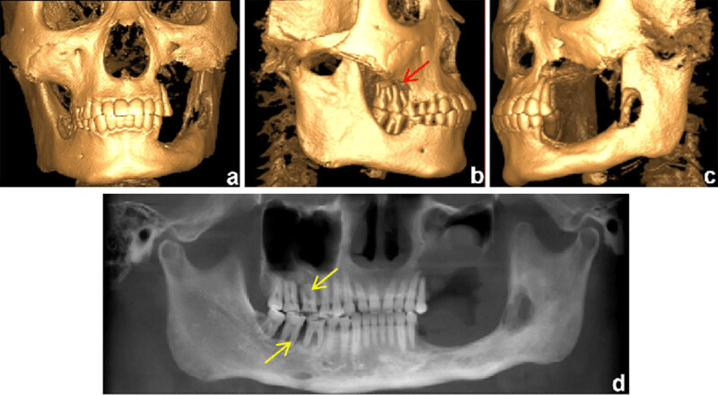
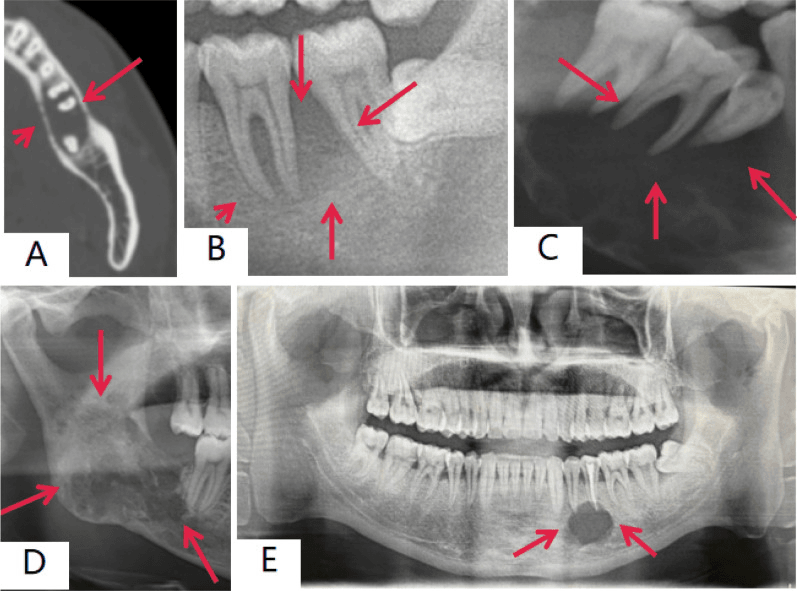
Jaw. Loose teeth, gingival swelling, jaw pain and trismus, with the floating-tooth appearance on radiographs.
Systemic Disease
The Hand-Schüller-Christian triad. The classic triad of multisystem disease:
- Diabetes insipidus, in 25% of multisystem cases
- Exophthalmos, from orbital involvement
- Lytic skull lesions
Risk organs. Involvement of the liver, spleen or marrow is what separates high-risk multisystem disease:
- Liver - hepatomegaly, jaundice and dysfunction; a poor prognostic sign
- Spleen - splenomegaly and hypersplenism
- Bone marrow - cytopenias or pancytopenia
Lung. Cough, dyspnoea and pneumothorax, usually in older children and adults. The smoking-associated adult form has its own section below.
Skin and elsewhere. The skin is involved in 30-40% of multisystem cases, as a seborrhoeic dermatitis-like rash on the scalp and behind the ears or a papular eruption on the trunk and groin. Cervical and axillary lymphadenopathy, hepatosplenomegaly and, in disseminated disease, failure to thrive complete the picture.
LCH-II Randomised Trial — Risk Organs Drive Mortality
- International randomised trial in multisystem LCH: 193 risk patients (risk-organ involvement or age under 2 years)
- 5-year survival 74% (arm A) vs 79% (arm B); disease reactivation 46% in both arms
- Children under 2 years WITHOUT risk-organ involvement had 100% survival
- Risk-organ-positive non-responders at 6 weeks had the highest mortality
- Arm A vs arm B was NULL on every randomised endpoint; the etoposide mortality signal is a subgroup hazard rate of 0.54 whose 95% CI reaches 1.00
Investigations
Radiographs
Skull. A punched-out lytic lesion with well-defined margins. The bevelled edge, with the inner table destroyed more than the outer, is highly specific for LCH in children. A central fragment of bone within the lesion is the button sequestrum, and multiple coalescent lesions give the geographic skull.
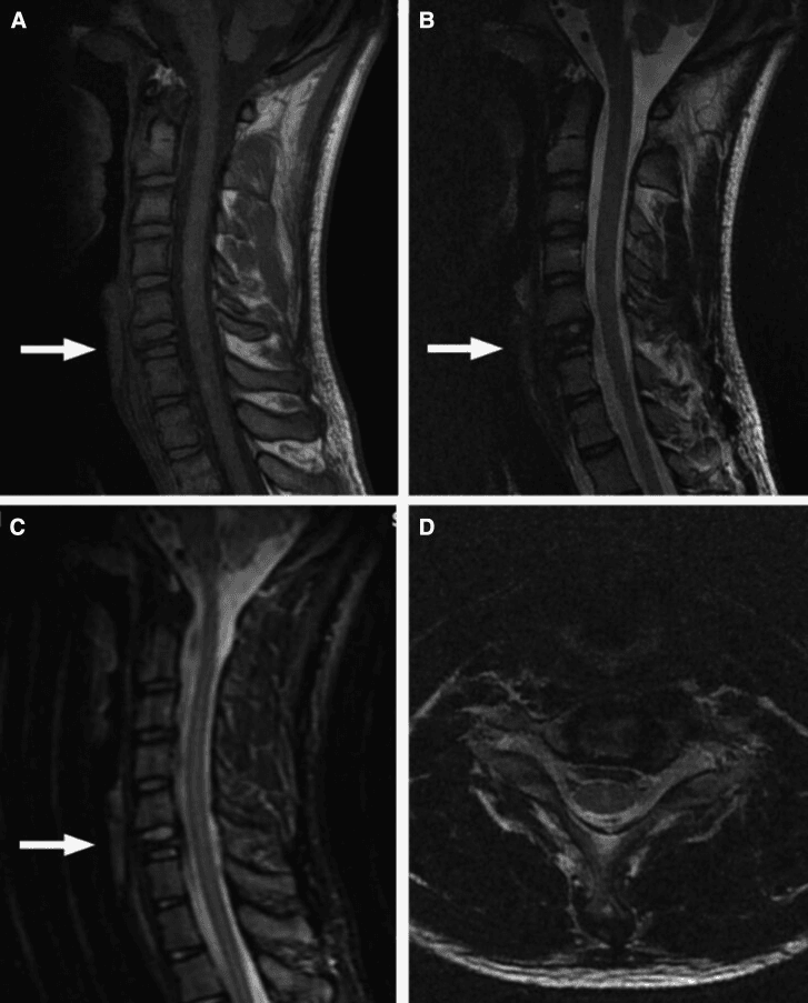
Spine. Vertebra plana is complete collapse of an isolated vertebral body, the pancake vertebra, with the disc spaces preserved and no paraspinal soft-tissue mass. The preserved discs distinguish LCH from infection, where discitis destroys the disc, and from metastasis; the absent mass points away from tumour and infection.
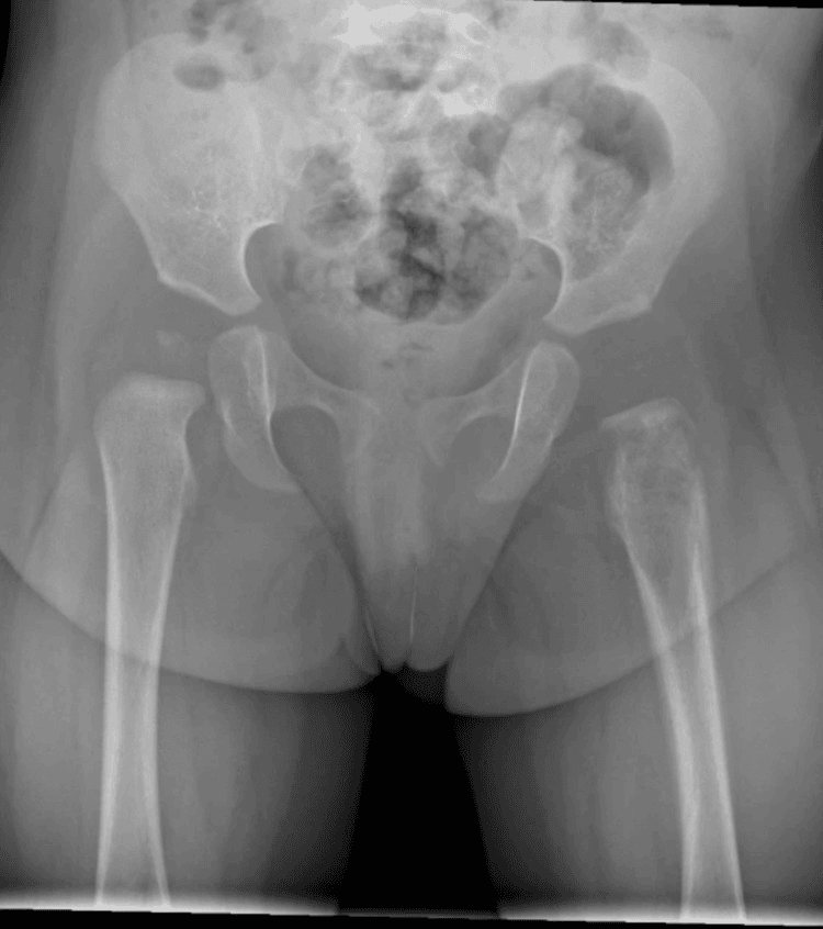
Long bones. A lytic lesion, diaphyseal or metaphyseal, with moth-eaten or permeative destruction. The periosteal reaction may be laminated, solid or onion-skin; a sequestrum can form, and the lesion may present as a pathological fracture.
Advanced Imaging
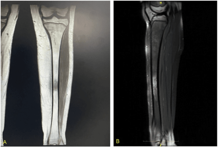
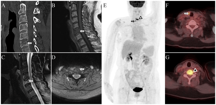
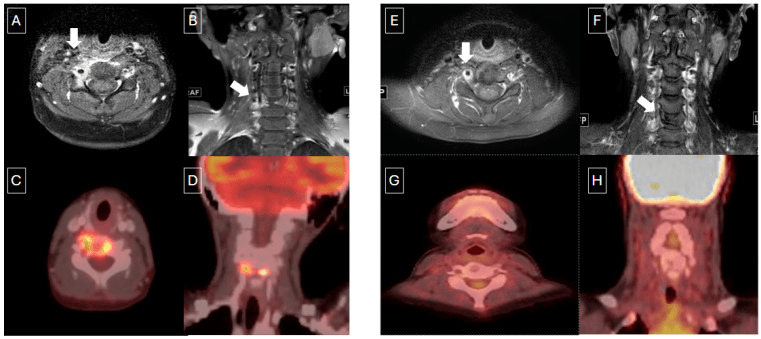
MRI. MRI defines soft-tissue extension and spinal canal involvement, and for a spinal lesion it is essential to rule out epidural extension or instability. The lesion's signal:
- T1: low signal
- T2: high signal, with surrounding marrow oedema
- STIR: bright, with an extensive oedema pattern
- Contrast: marked enhancement (hypervascular)
CT. Superior for cortical bone detail. It delineates skull-base and orbital lesions and vertebral body integrity, and 3D reconstruction helps plan surgery.
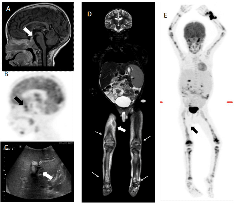
Nuclear medicine. A Tc-99m bone scan shows increased uptake at lesion sites. PET-CT is more sensitive than the skeletal survey for bone lesions and for multifocal disease, detects occult and extraskeletal involvement, and is useful for monitoring treatment response.
Skeletal survey. Mandatory in every case of LCH, because finding multifocal disease changes prognosis and treatment:
- AP and lateral skull, spine and pelvis
- AP chest, including the ribs
- AP long bones, both sides
Laboratory Tests and Staging
Baseline studies. What each is for:
- Full blood count - cytopenias from marrow involvement
- ESR and CRP - often raised but non-specific
- Liver function tests - liver dysfunction; add PT and PTT if the liver is involved
- Renal function - a baseline before chemotherapy
Suspected diabetes insipidus. A water deprivation test showing a low urine osmolality, urinalysis for polyuria and a low specific gravity, and serum and urine osmolality to confirm it.
Staging. Skeletal survey or PET-CT, chest radiograph or CT for pulmonary involvement, and abdominal ultrasound or MRI for the liver and spleen. Consider a bone marrow biopsy if there are cytopenias.
Histopathology
Macroscopic. Tan-grey, granular, friable soft tissue, without gross necrosis or haemorrhage.
Microscopic. Large Langerhans cells with abundant eosinophilic cytoplasm and eccentric, reniform coffee-bean nuclei with nuclear grooves and fine chromatin. Eosinophils are prominent and may form microabscesses, among T lymphocytes, scattered macrophages and occasional multinucleated giant cells. Necrosis may be present in active lesions.
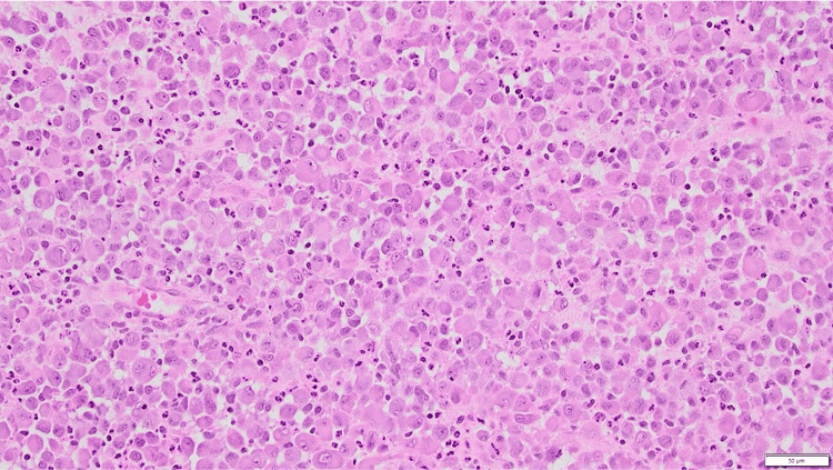
Immunohistochemistry. CD1a and Langerin make the diagnosis:
- CD1a - positive, membranous; the hallmark
- Langerin (CD207) - positive, cytoplasmic; more specific than CD1a
- S100 - positive, nuclear and cytoplasmic
- CD68 - variable
- Cytokeratin - negative, which helps exclude carcinoma
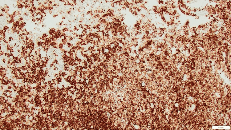
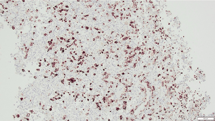
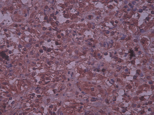
Electron microscopy. Birbeck granules, rod-shaped cytoplasmic inclusions with a "tennis racket" or "zipper" appearance, are diagnostic but not required: CD1a and Langerin are sufficient.
Molecular testing. BRAF V600E mutation analysis, on paraffin-embedded tissue or blood, guides decisions about targeted therapy.
Differential Diagnosis
Infection. Blood cultures, bone biopsy and the clinical course separate it from LCH.
- Osteomyelitis - fever, raised white cell count and CRP, metaphyseal location
- Discitis - disc space narrowing, where LCH vertebra plana preserves the disc
- Tuberculous osteomyelitis - chronic course, epidemiology
Tumour. Biopsy and immunohistochemistry make the distinction.
- Ewing sarcoma - diaphyseal, onion-skin periosteal reaction, soft-tissue mass
- Osteosarcoma - metaphyseal, sunburst periosteal reaction, raised ALP
- Metastases - neuroblastoma in children, carcinoma in adults
- Lymphoma - older children and adults, systemic symptoms
Other histiocytic disorders. Separated by age, site or immunophenotype.
- Erdheim-Chester disease - adults, bilateral femoral and tibial diaphyseal sclerosis
- Rosai-Dorfman disease - massive lymphadenopathy, S100+ CD1a- cells
- Juvenile xanthogranuloma - skin lesions, CD68+ CD1a- cells
Metabolic and other. The remaining mimics.
- Fibrous dysplasia - ground-glass matrix, no Langerhans cells
- Hyperparathyroidism - brown tumours, raised PTH, multiple lesions
- Multiple myeloma - adults, monoclonal protein, plasma cells on biopsy
- lch
- 5-10 years peak
- ewing
- 10-20 years peak
- osteomyelitis
- Any age
- fibrous_dysplasia
- Adolescents/adults
- lch
- Skull, femur, ribs
- ewing
- Diaphysis long bones
- osteomyelitis
- Metaphysis
- fibrous_dysplasia
- Ribs, femur, skull
- lch
- Lytic, punched-out
- ewing
- Permeative, onion-skin
- osteomyelitis
- Lytic, periosteal rxn
- fibrous_dysplasia
- Ground-glass, sclerotic rim
- lch
- Minimal to moderate
- ewing
- Large soft tissue mass
- osteomyelitis
- Abscess possible
- fibrous_dysplasia
- None
- lch
- CD1a+ Langerhans cells
- ewing
- Small blue cells, CD99+
- osteomyelitis
- Inflammatory, organisms
- fibrous_dysplasia
- Fibrous stroma, woven bone
- lch
- May have multisystem
- ewing
- Metastases possible
- osteomyelitis
- Fever, sepsis
- fibrous_dysplasia
- McCune-Albright (rare)
Spinal LCH in Children — Long-Term Follow-Up
- 26 children with biopsy-proven spinal LCH (44 involved vertebrae); 23 followed 2+ years, mean 9.4 years
- Unexpectedly high proportion of cervical lesions; 62% had multifocal skeletal disease
- Severe lesions often produced asymmetric collapse rather than classic symmetric vertebra plana
- Spinal deformity developed in only 4 children; just 2 of the 23 followed (under 9%) required spinal fusion
- All patients were alive and well with resolution of symptoms at latest follow-up
Management
Stage first. The extent of disease and whether the risk organs are involved set the treatment.
- Share of cases
- 60%
- Treatment
- Observation or minimal intervention
- Share of cases
- 20%
- Treatment
- Low-dose chemotherapy if 2 or more lesions
- Share of cases
- Multisystem disease 20%
- Treatment
- Combination chemotherapy
- Share of cases
- Multisystem disease 20%
- Treatment
- Intensive chemotherapy protocols
Observation is a valid first line for the unifocal lesion: half resolve spontaneously within 1-2 years. The alternatives are chosen by symptoms, the bone involved and structural risk.
Observation. For the asymptomatic lesion in a non-weight-bearing bone with no structural risk. Monitor clinically and radiographically every 3-6 months until the lesion heals or stabilises.
Curettage with or without bone grafting. For pain, a weight-bearing bone or structural risk. Intralesional curettage with a local adjuvant (phenol or hydrogen peroxide) and autograft or allograft gives 90% local control and a recurrence rate under 10%; the bone fractures during healing in 5-10%. The approach depends on the site, the cavity is curetted thoroughly, and prophylactic fixation is added if there is structural concern.
Intralesional steroid injection. Methylprednisolone 80-120 mg, for vertebral lesions and inaccessible sites. It is minimally invasive, accelerates healing and is effective in 70-80%, though the injection may need repeating.
En bloc resection. For expendable bones (rib, fibula, clavicle). Local control is near 100%, and diagnosis and cure come in a single procedure.
Radiotherapy. Previously common and now avoided in children, because of the risk of secondary malignancy and growth-plate damage. Its role is very limited, to refractory cases, at 6-10 Gy if absolutely necessary.
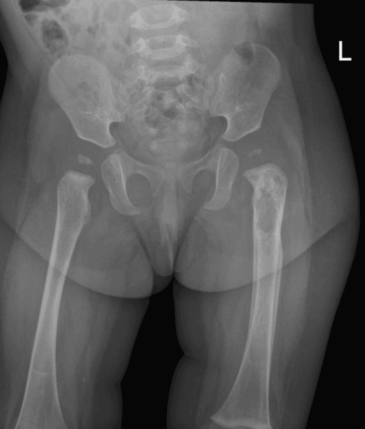
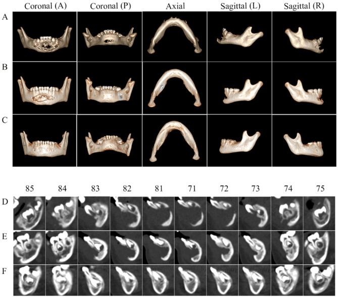
Management Algorithm
The management pathway is determined by disease extent, risk-organ involvement, structural danger and treatment response. Confirm the diagnosis with CD1a and Langerin, stage the skeleton and risk organs, and escalate urgently for cord compromise, instability or liver, spleen or marrow involvement.
Complications and Prognosis
Diabetes insipidus. Develops in 18-25%, from involvement of the hypothalamic-pituitary axis; craniofacial bone lesions and multisystem disease are the risk factors. It presents with polyuria, polydipsia and hypernatraemia, is diagnosed as above, and is managed with lifelong desmopressin (DDAVP).
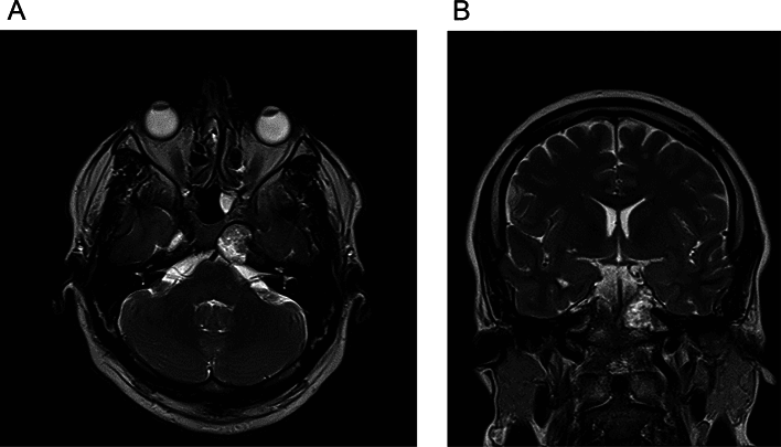
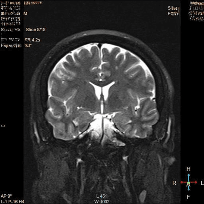
Orthopaedic sequelae. The skeletal legacy of the disease:
- Vertebral collapse and kyphosis, 10-15%
- Limb length discrepancy after femoral lesions
- Malunion or nonunion of a pathological fracture, which is rare
- Joint stiffness after juxta-articular lesions
- Premature physeal closure after metaphyseal lesions
Neurocognitive dysfunction. Affects 10-20% and is associated with multisystem disease: cerebellar involvement (ataxia, dysarthria), behavioural and learning difficulties, and white-matter changes or cerebellar atrophy on MRI. Neurodegenerative LCH, the progressive late CNS syndrome, has its own section below.
Pulmonary. Spontaneous pneumothorax, respiratory failure with diffuse involvement, and progressive fibrosis in chronic disease.
Other. Hearing loss with temporal bone involvement, dental problems with mandibular lesions, sclerosing cholangitis with liver involvement, and growth retardation from pituitary dysfunction.
Prognosis
- Survival
- Near 100%; over 90% resolve spontaneously or are cured, and 50% resolve without intervention
- Reactivation
- Recurrence 10-20%, usually at the same site
- Permanent sequelae
- Minimal; cosmetic deformity rare
- Survival
- 95-100%, good with chemotherapy; disease-free survival 70-80% at 5 years
- Reactivation
- 30-40% (new lesions or progression)
- Permanent sequelae
- 20%, mainly diabetes insipidus and orthopaedic
- Survival
- 90-95%
- Reactivation
- 40-50%
- Permanent sequelae
- 40% (diabetes insipidus, neurocognitive)
- Survival
- 70-80%, significantly worse with a poor initial response
- Reactivation
- 50-60%
- Permanent sequelae
- 60-70%
Favourable factors. Unifocal bone disease, single-system involvement, age over 2 years and a good response to initial therapy.
Unfavourable factors. These predict a worse course:
- Age under 2 years
- Risk-organ involvement: spleen involvement, marrow dysfunction with cytopenias, hepatomegaly or abnormal liver function tests
- Poor response to initial chemotherapy at 6 weeks
- Multisystem disease involving more than 3 organs
- Recurrent or reactivating disease (a 46-58% rate)
- Growth failure and systemic symptoms (fever, failure to thrive)
Age needs reading together with the risk organs. In LCH-II, children under 2 without risk-organ involvement had 100% survival, and risk-organ involvement, not age alone, was the dominant prognostic factor.
Neurodegenerative LCH: the Late CNS Sequela
What it is. Neurodegenerative LCH (LCH-ND) is a progressive, non-mass CNS syndrome that can appear months to many years after the systemic disease has apparently resolved, and is a leading cause of long-term morbidity in survivors. It is distinct from a space-occupying CNS LCH lesion: there is no tumour to resect.
Who is at risk. The strongest associations are craniofacial or "CNS-risk" lesions (orbit, mastoid, temporal, sphenoid, maxilla/zygoma), diabetes insipidus, multisystem disease and BRAF V600E positivity. The lesions that predict diabetes insipidus also predict LCH-ND, and the two often coexist as part of the pituitary-hypothalamic/CNS-risk picture.
Presentation and MRI. Insidious cerebellar signs (ataxia, dysarthria, tremor, dysmetria), pyramidal signs, behavioural and learning decline and eventually cognitive impairment. MRI shows symmetrical T2/FLAIR high signal in the dentate nuclei/cerebellar white matter and basal ganglia, sometimes with cerebellar atrophy. These changes can precede symptoms, so at-risk children are surveilled with MRI.
Mechanism. It is thought to be driven by MAPK-activated myeloid cells seeding the CNS (BRAF V600E has been found in the marrow/blood of affected patients), a neuro-inflammatory/degenerative process rather than direct tumour.
Management. Difficult and not standardised. The options include chemotherapy (cytarabine) and, increasingly, MAPK-pathway (BRAF/MEK) inhibitors, with the hope, unproven, that early targeted therapy may prevent or halt progression. Established neurodegeneration is often irreversible, which is why recognition and surveillance matter.
Pulmonary LCH: the Smoking-Associated Adult Form
A different disease in adults. Isolated pulmonary LCH (PLCH) is largely a disease of young-to-middle-aged adult smokers, typically 20-40 years, with a roughly equal sex incidence. Unlike multisystem childhood LCH, it is often a reactive, polyclonal response to cigarette smoke (though clonal BRAF V600E-positive forms also occur), which is why it behaves differently.
Presentation. Cough and dyspnoea, or an incidental finding. A classic acute presentation is spontaneous pneumothorax from cyst rupture, which can be recurrent. Constitutional symptoms and extrapulmonary disease are usually absent in the isolated pulmonary form.
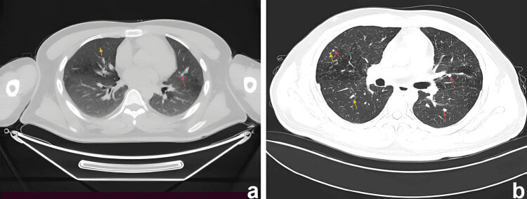
Imaging. HRCT is characteristic: combined nodules and thin-walled cysts with an upper- and mid-zone predominance, sparing the costophrenic angles and lung bases. That is the reverse of the basal predominance of many other cystic and fibrotic lung diseases. The nodules cavitate into cysts over time.
Management. Smoking cessation is first-line and the single most important intervention; it can stabilise or regress the disease. Systemic therapy (such as cladribine, or MAPK-directed therapy in clonal disease) is reserved for progressive disease, and lung transplantation for end-stage fibrosis, with a risk of recurrence, especially if smoking resumes. Recurrent pneumothorax may need pleurodesis.
Guidelines, Registries & Global Practice
Global Epidemiology
- Incidence approximately 4-9 per million children per year; rarer in adults (1-2 per million)
- Peak 1-3 years for multisystem disease; 5-10 years for single-system bone disease
- Slight male predominance (around 1.2-2:1); reported across all populations worldwide
- Bone is the most common organ involved overall (around 80% of paediatric cases)
- Pulmonary LCH in adults is strongly smoking-associated and follows a distinct natural history
Side-by-Side Guidance
- diagnosis
- CD1a + CD207 (Langerin) mandatory; BRAF V600E testing recommended
- first_line
- Vinblastine + prednisone 12 months (LCH-III backbone)
- emphasis
- Risk-organ stratification drives intensity; structured late-effects follow-up
- diagnosis
- Molecular profiling encouraged (BRAF, MAP2K1) at diagnosis
- first_line
- Vinblastine/prednisone; early targeted therapy in refractory MAPK-mutant disease
- emphasis
- Salvage with cytarabine/cladribine; BRAF/MEK inhibitors for refractory disease
- diagnosis
- Biopsy to exclude Ewing sarcoma, osteomyelitis, lymphoma
- first_line
- Observation or intralesional steroid for unifocal bone; curettage if structural risk
- emphasis
- Avoid radiotherapy in children; surgery reserved for instability or neurology
- diagnosis
- HRCT (cysts + nodules, upper-zone), smoking history central
- first_line
- Smoking cessation first-line
- emphasis
- Systemic therapy or transplant for progressive disease
Registry & Trial Networks
- Outcome data are driven by international cooperative trials (LCH-I, LCH-II, LCH-III, LCH-IV) rather than implant/arthroplasty registries
- The Histiocyte Society coordinates global protocol enrolment; regional groups (e.g. NACHO in North America, national paediatric oncology groups in Europe) feed shared datasets
- BRAF/MAPK genotyping is increasingly captured prospectively to stratify targeted-therapy use
High- vs Limited-Resource Practice
- High-resource: routine BRAF/MAP2K1 genotyping, PET-CT staging, access to BRAF/MEK inhibitors and cladribine/cytarabine salvage, long-term endocrine/neurocognitive surveillance
- Limited-resource: diagnosis may rest on morphology plus CD1a/S100 where Langerin and molecular testing are unavailable; skeletal survey substitutes for PET-CT; vinblastine + prednisone remains the affordable, effective backbone; targeted agents often inaccessible
- Universal principles: confirm tissue diagnosis, stage for risk organs, avoid radiotherapy in children, and screen lifelong for diabetes insipidus
Controversies & Areas of Uncertainty
- Reactive vs neoplastic: the discovery of clonal MAPK mutations (BRAF V600E, MAP2K1) reframed LCH as an inflammatory myeloid neoplasm, yet the prominent inflammatory infiltrate keeps the "neoplasia vs immune dysregulation" debate alive.
- Optimal duration of targeted therapy: BRAF/MEK inhibitors produce rapid responses but disease frequently rebounds on cessation, and the ideal duration, role of combination with chemotherapy, and long-term toxicity in children remain undefined.
- Vertebra plana management: the balance between observation, intralesional steroid and bracing is not standardised; most lesions reconstitute, but predicting the minority that deform is imprecise.
- Neurodegenerative LCH: progressive cerebellar/brainstem neurodegeneration can appear years after apparent cure; its pathogenesis, screening interval, and whether early MAPK-directed therapy prevents it are unresolved.
- Role of PET-CT vs skeletal survey: PET-CT is more sensitive for occult and extraskeletal disease, but cost, radiation and access limit universal use, so staging strategy varies by setting.
MCQ Practice Points
Q: What are the diagnostic immunohistochemical markers for Langerhans cell histiocytosis?
A: CD1a positive and Langerin (CD207) positive - these are pathognomonic. Also S100 positive. Cytokeratin negative (differentiates from metastatic carcinoma). Histologically shows Langerhans cells with characteristic coffee-bean nuclei in a background of abundant eosinophils. Birbeck granules (tennis racket shape) on electron microscopy are diagnostic but no longer required.
Q: What is the classic radiographic finding of LCH in the spine?
A: Vertebra plana - complete collapse of a vertebral body with preserved disc spaces. This differentiates LCH from infection (disc destruction) and metastases (variable disc involvement). Most commonly affects thoracic and lumbar spine. Despite dramatic appearance, most cases reconstitute spontaneously and are treated conservatively with observation or bracing.
Q: What is the BRAF mutation significance in LCH?
A: BRAF V600E mutation is present in 50-60% of LCH cases. This establishes LCH as a clonal neoplastic proliferation rather than reactive disorder. Has therapeutic implications - BRAF inhibitors (vemurafenib, dabrafenib) can be used for refractory multisystem disease. Also useful for differentiating from other histiocytic disorders if tissue diagnosis unclear.
Q: How is LCH classified and what determines prognosis?
A: Unifocal (eosinophilic granuloma): Single bone lesion, excellent prognosis (90%+ cure). Multifocal unisystem: Multiple bone lesions, good prognosis. Multisystem without risk organ involvement: Involves bone plus other organs (skin, nodes). Multisystem with risk organ involvement (liver, spleen, bone marrow): Worst prognosis, 70-80% survival. Risk organs define need for systemic chemotherapy.
Q: What is the orthopaedic management approach for unifocal LCH (eosinophilic granuloma)?
A: Conservative management is first-line for most lesions. Options include: Observation alone (many lesions resolve spontaneously), intralesional corticosteroid injection (methylprednisolone), or curettage with bone grafting for large lesions or impending fracture. Surgery indicated only for: neurological compromise, pathological fracture, or diagnostic uncertainty. Radiation reserved for unresectable locations (skull base).
Clinical Decision Scenarios
Practise clinical reasoning and management decisions out loud
“A 7-year-old boy presents with a painless scalp swelling for 2 months. X-ray shows a 3 cm well-defined lytic lesion in the left parietal bone with 'beveled edge' appearance. Parents are very concerned about cancer.”
“A 12-year-old girl presents with 3-month history of mid-thoracic back pain. MRI shows complete collapse of T7 vertebral body (vertebra plana) with preserved disc spaces and no epidural extension. Biopsy confirms Langerhans cell histiocytosis. Parents want aggressive treatment to restore vertebral height.”
“A 6-year-old presents with a limp and thigh pain. Radiographs show a lytic, mildly expansile diaphyseal lesion of the femur with laminated periosteal reaction. The referring team is worried about Ewing sarcoma.”
Must-Know Facts
- DEFINITION: Clonal proliferation of CD1a+ and Langerin+ dendritic cells
- SPECTRUM: Eosinophilic granuloma (unifocal) to Letterer-Siwe (disseminated)
- AGE: Peak 5-10 years, males more than females 2:1
- SITES: Skull 50%, femur 20%, ribs 10%, vertebrae 7% (vertebra plana)
- MOLECULAR: BRAF V600E mutation in 50-60% (therapeutic target)
- PROGNOSIS: Unifocal 90%+ cure, multisystem with risk organs 70-80% survival
Diagnostic Triad
- RADIOLOGY: Lytic lesion, punched-out (skull), beveled edge, vertebra plana with preserved discs
- HISTOLOGY: Langerhans cells with coffee-bean nuclei, abundant eosinophils
- IMMUNOSTAINS: CD1a POSITIVE, Langerin (CD207) POSITIVE, S100 positive, cytokeratin negative
- ELECTRON MICROSCOPY: Birbeck granules (tennis racket) - diagnostic but not required
- STAGING: Skeletal survey or PET-CT (PET more sensitive for multifocal)
- LABS: CBC, LFTs, urinalysis - screen for multisystem involvement
Classification & Prognosis
- UNIFOCAL BONE (60%): Single lesion, greater than 90% cure, observation or curettage
- MULTIFOCAL BONE (20%): 2+ bone lesions, 95% survival, chemotherapy if symptomatic
- MULTISYSTEM NO RISK ORGANS: Bone + skin/LN, 90-95% survival, chemotherapy
- MULTISYSTEM WITH RISK ORGANS: Liver/spleen/marrow, 70-80% survival, intensive chemotherapy
- RISK ORGANS: Liver, spleen, bone marrow (poor prognostic factors)
- DIABETES INSIPIDUS: 18-25% develop (higher with skull lesions), permanent in majority
Management Algorithm
- UNIFOCAL ASYMPTOMATIC: Observation (50% spontaneous resolution) OR curettage ± bone graft
- UNIFOCAL SYMPTOMATIC: Curettage + steroid injection OR en bloc if expendable bone (rib, fibula)
- VERTEBRA PLANA: Steroid injection preferred (NOT surgery), 50-70% reconstitution over 2-5 years
- MULTIFOCAL BONE (2+ lesions): Vinblastine + prednisolone x 12 months
- MULTISYSTEM DISEASE: Vinblastine + prednisolone +/- mercaptopurine x 12-24 months
- REFRACTORY: Cladribine, cytarabine, BRAF inhibitors (if V600E+), MEK inhibitors
Viva Traps to Avoid
- DON'T: Recommend surgery for vertebra plana (reconstitutes spontaneously 50-70%)
- DON'T: Skip skeletal survey - must determine unifocal vs multifocal (changes treatment)
- DON'T: Miss diabetes insipidus screening - ask about polyuria/polydipsia on follow-up
- DON'T: Offer radiation in children - avoid due to secondary malignancy risk
- DON'T: Confuse with infection - LCH vertebra plana has PRESERVED disc spaces
- DO: Always check CD1a and Langerin on biopsy (both must be positive)
- DO: Test BRAF V600E mutation (guides targeted therapy if refractory)
- DO: Involve pediatric oncology for multifocal/multisystem disease
Quick Differentials
- EWING SARCOMA: Age 10-20, diaphyseal, large soft tissue mass, CD99+, small blue cells
- OSTEOMYELITIS: Fever, elevated WBC/CRP, metaphyseal, disc space narrowing (vs preserved in LCH)
- FIBROUS DYSPLASIA: Ground-glass matrix, medullary, no Langerhans cells, any age
- METASTASES: Neuroblastoma (children), history of primary, multifocal
- KEY DISTINGUISHER: Biopsy with CD1a+ and Langerin+ cells confirms LCH
Key Evidence & References
Badalian-Very — Discovery of Recurrent BRAF V600E
- Oncogenic BRAF V600E identified in 35 of 61 archived LCH specimens (57%)
- BRAF V600E tended to occur in younger patients but was independent of disease site or stage
- Langerhans cells stained for phospho-MEK and phospho-ERK regardless of mutation status
- Reframed LCH from a reactive process to a clonal neoplasm of the MAPK/ERK pathway
Héritier — BRAF V600E Predicts High-Risk Disease
- French cohort of 315 children: BRAF V600E present in 54.6%
- BRAF V600E enriched in severe disease — 87.8% of multisystem LCH with risk organs vs 43.9% of single-system LCH (315 patients; the 61-specimen Badalian-Very study on this page found no stage association, but was far too small to detect one)
- Resistance to first-line vinblastine + corticosteroid: 21.9% (mutant) vs 3.3% (wild-type)
- Higher 5-year reactivation (42.8% vs 28.1%) and more permanent sequelae (27.9% vs 12.6%)
Euro Histio Net / Histiocyte Society Guidelines
- Consensus guidelines for diagnosis, work-up, treatment and long-term follow-up of LCH up to age 18
- Mandate CD1a/CD207 (Langerin) confirmation and systematic staging for risk-organ involvement
- Define single-system vs multisystem disease and risk-organ stratification as the basis for treatment intensity
- Recommend structured surveillance for late effects (diabetes insipidus, neurodegeneration, endocrinopathy)