Inflammation | Proliferation | Remodelling
- Similar to tendon healing - three overlapping phases
- Type III collagen first, gradually replaced by Type I
- Never achieves normal properties - healed ligament weaker than original
- Intra-articular ligaments heal poorly (ACL) vs extra-articular (MCL) heal well
- Controlled motion beneficial for ligament healing
- “MCL heals well (extra-articular), ACL does not (intra-articular)
- “ACL fails to heal due to synovial fluid, lack of blood supply, gap
- “Healed MCL regains roughly half to two-thirds of normal tensile strength (animal-derived figure)
- “Similar phases to tendon: inflammation, proliferation, remodelling
Overview
Ligament healing follows similar principles to tendon healing, with important differences that depend on where the ligament sits. Extra-articular ligaments such as the MCL heal reliably. Intra-articular ligaments such as the ACL do not heal spontaneously, and need surgical reconstruction if normal function is to be restored.
Why it matters. Knowing why some ligaments heal and others do not is what guides treatment. MCL injuries are typically managed non-operatively, ACL injuries are reconstructed in active patients, and the biology of healing informs rehabilitation protocols.
Mechanisms - Healing Phases
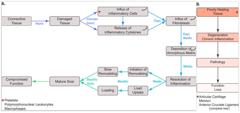
Healing runs through three overlapping phases, similar to those of tendon.
Inflammatory phase (days 0-7). Injury triggers an inflammatory response. A haematoma forms and serves as the scaffold for healing, and inflammatory cells arrive and release cytokines and growth factors. This phase is essential for initiating repair, and it is the step that fails inside the joint, for the reasons compared in the MCL and ACL section below.
Proliferative phase (weeks 1-6). Fibroblasts migrate into the healing zone and proliferate, granulation tissue forms, and synthesis of type III collagen begins, laid down as thin, disorganised fibres. The tissue is highly cellular and disorganised, and its mechanical strength is minimal.
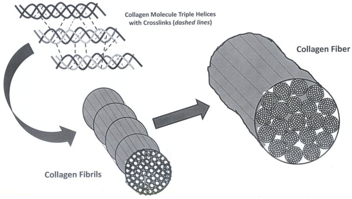
Remodelling phase (week 6 to years). Type III collagen is gradually replaced by thick, organised type I collagen. The fibres align along lines of stress, cross-links form between collagen molecules and cellularity falls. The mechanical properties improve but never return to normal.
The end result is weaker than the original. A healed ligament achieves only part of its normal tensile strength, conventionally quoted as 50-70%, although that band comes from animal (rabbit MCL) studies and no human measurement is cited here. Stiffness is reduced too. An increased cross-sectional area compensates, and functional recovery is possible.
Mechanobiology: Controlled Motion versus Immobilisation
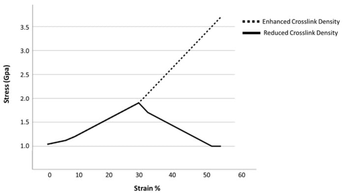
Immobilisation is harmful. Prolonged immobilisation produces a disorganised matrix with randomly oriented collagen, adhesions, reduced matrix turnover, and a loss of tensile strength and stiffness. It also causes resorption and weakening at the bone–ligament insertion, so after a period of immobilisation the insertion becomes the weak link.
Controlled cyclic loading is beneficial. Mechanical stress is a signal. It aligns fibroblasts and collagen fibres along the lines of stress, a soft-tissue analogue of Wolff's law, and promotes larger-diameter fibrils and better cross-linking. Tensile strength and stiffness rise and adhesions are reduced, so loading improves the quality and organisation of the healing scar rather than only its bulk.
The dose matters: an "envelope of function". Too little load gives a weak, disorganised scar, and appropriate controlled load optimises organised healing. Excessive load too early causes gap formation, elongation or re-rupture.
What rehabilitation takes from this. The dose-response is why rehabilitation uses early protected range of motion and graduated functional loading. It is also the rationale for hinged bracing and for internal-brace augmentation, which shares load to protect a repair through the vulnerable phase, though whether the brace also stress-shields the healing ligament remains debated (see Controversies). General bone mechanotransduction is developed in the osteocytes-mechanotransduction topic, and the viscoelastic behaviour of ligament in the viscoelasticity topic.
Differential Diagnosis of Poor Ligament/Soft-Tissue Healing
When a ligament injury fails to recover as expected, the differential is broader than the index injury. Separate a ligament that biologically does not heal, such as the ACL, from a missed associated injury or a systemic cause of impaired healing.
- Key Discriminator
- Synovial environment, gap, retraction
- Confirmatory Clue
- MRI shows discontinuity; persistent pivot shift
- Key Discriminator
- Multidirectional or rotatory instability
- Confirmatory Clue
- Stress radiographs, examination under anaesthesia
- Key Discriminator
- Superficial MCL flips superficial to pes anserinus
- Confirmatory Clue
- MRI; explains a non-healing 'grade II' MCL
- Key Discriminator
- Recurrent giving way, effusions
- Confirmatory Clue
- MRI meniscal/chondral injury, bone bruising
- Key Discriminator
- Generalised poor wound healing
- Confirmatory Clue
- HbA1c, smoking history, nutritional markers
- Key Discriminator
- Generalised hyperlaxity, recurrent injury
- Confirmatory Clue
- Beighton score, family history
Clinical Relevance - MCL vs ACL
The MCL heals. In most cases the MCL heals predictably with conservative management. Its extra-articular position allows a haematoma to form and be contained, and the periligamentous tissues provide a blood supply. The healing MCL is enlarged but functional.
Managing the MCL. Management is protected motion in a hinged brace, with valgus stress protected during healing, followed by rehabilitation. Surgery is rarely needed for an isolated MCL injury. Functional recovery is expected, and most patients return to function.
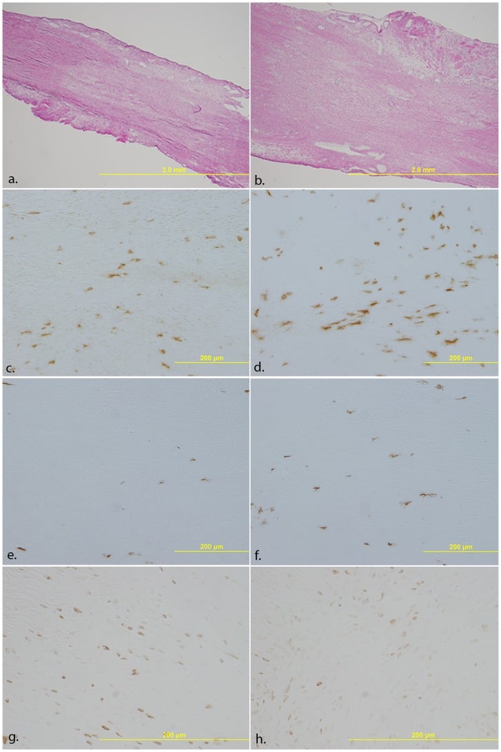
The ACL does not heal spontaneously. Its synovial environment is hostile to healing, and it lacks the capacity for intrinsic repair seen in extra-articular ligaments. The factors that separate it from the MCL are set side by side below.
- Extra-articular (MCL)
- Contained — forms a healing scaffold
- Intra-articular (ACL)
- Dispersed by synovial fluid; no stable clot
- Extra-articular (MCL)
- Retained at injury site
- Intra-articular (ACL)
- Diluted and washed away
- Extra-articular (MCL)
- Ends stay apposed
- Intra-articular (ACL)
- Ends retract, leaving a gap with no scaffold
- Extra-articular (MCL)
- Rich periligamentous vessels
- Intra-articular (ACL)
- Sparse; relies on synovium and fat pad
- Extra-articular (MCL)
- Absent
- Intra-articular (ACL)
- Hostile plasmin-rich, anti-clotting milieu
- Extra-articular (MCL)
- Reliable scar healing; strength recovers only in part
- Intra-articular (ACL)
- No spontaneous healing of complete tears
- Extra-articular (MCL)
- Non-operative (brace, protected motion)
- Intra-articular (ACL)
- Reconstruction (or selected repair/BEAR)
Why reconstruct. Leaving the knee ACL-deficient leads to instability and a risk of secondary meniscal and chondral damage. Reconstruction replaces the ligament's function with a graft, and is required to restore stability in active patients who want to return to pivoting activities.
Repair revisited. ACL repair, by primary suture, internal bracing, or scaffold bridging as in BEAR, is being revisited for proximal or midsubstance tears with good tissue. Reconstruction remains the standard for most complete tears in pivoting athletes. The BEAR II trial and the primary-repair series are in the Evidence Base, and Controversies sets out why they have not displaced reconstruction.
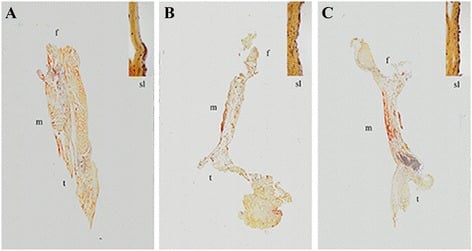
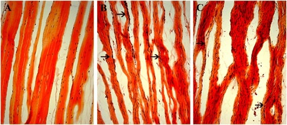
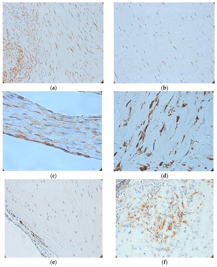
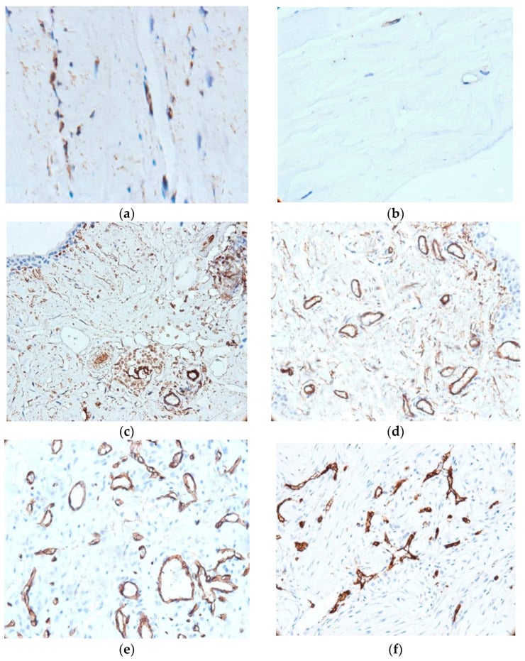
Graft Ligamentisation and Tendon-to-Bone Healing
Because the torn ACL is replaced with a graft, the obvious follow-up question is how the graft heals. A tendon graft does not stay tendon: it remodels into an ACL-like structure through ligamentisation, in overlapping phases.
Early avascular phase (first weeks). The free graft is initially avascular, with central necrosis and falling cellularity. Mechanically it depends on its initial fixation while its intrinsic material strength declines.
Revascularisation and cellular repopulation (around 6 to 12 weeks). Host cells and vessels from the synovium and fat pad invade the graft. This is the window in which the graft is mechanically weakest, the "ligamentisation valley", and it is the biological reason rehabilitation is protected and graduated rather than aggressive in the first few months.
Remodelling and maturation (months to over a year). Collagen realigns, crimp returns and cellularity normalises, and the graft becomes ACL-like. As with native ligament scar it never fully matches the original ACL, with smaller fibrils and incomplete cross-link maturation, and maturation can take well over a year.
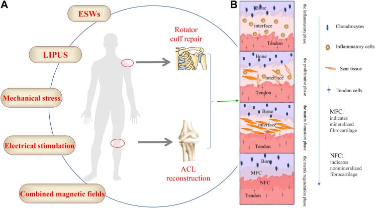
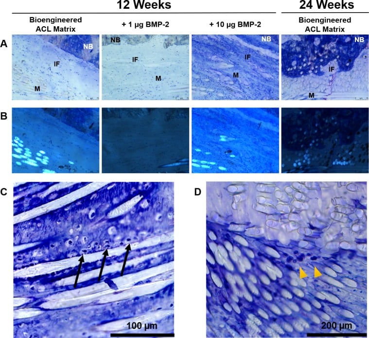
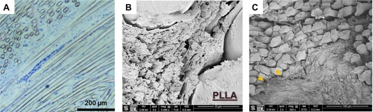
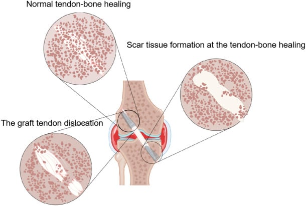
Healing in the tunnel. The graft must also integrate within the bone tunnel, and the interface depends on the graft. Soft-tissue grafts (for example hamstring) heal by an indirect, fibrovascular interface, with Sharpey-like fibres maturing over weeks to months. Bone-block grafts (bone–patellar tendon–bone) achieve faster, more direct bone-to-bone union. This difference is one driver of graft choice and of how early the construct can be loaded. Enthesis and insertion-site biology is developed in the tendon-bone-interface-enthesis topic.
Guidelines, Registries & Global Practice
Global Epidemiology
- ACL injuries occur in roughly 30 to 80 per 100,000 person-years, concentrated in 15 to 45-year-olds and in pivoting/cutting sports; females carry a several-fold higher sport-adjusted risk.
- Isolated MCL injury is the most common knee ligament injury overall and the great majority are managed non-operatively with reliable healing.
Side-by-Side Guidance
- Emphasis
- Evidence-based reconstruction for instability
- Practical Recommendation
- Reconstruction recommended for active patients with instability; repair selected/emerging
- Emphasis
- Shared decision-making, rehab-first pathways
- Practical Recommendation
- Structured rehabilitation; reconstruction if persistent functional instability
- Emphasis
- Individualised, tear-pattern based
- Practical Recommendation
- Recognises a role for repair/augmentation in selected proximal tears; reconstruction default
- Emphasis
- Biology favours non-operative healing
- Practical Recommendation
- Brace and protected motion for isolated grade I-III MCL; surgery for select distal/combined
Registry & Outcome Signals
- National ligament/ACL registries (e.g. Scandinavian and UK datasets) track graft choice, revision and reinjury, informing graft selection and rehabilitation rather than healing biology per se.
- Emerging BEAR and primary-repair data are not yet captured at registry scale, so practice change should remain cautious.
High- vs Limited-Resource Practice
- Well-resourced settings: ready access to MRI, arthroscopic reconstruction, and increasingly repair/BEAR in selected patients, with supervised criteria-based rehabilitation.
- Limited-resource settings: non-operative management and bracing dominate; isolated MCL injuries heal reliably without surgery, and structured rehabilitation is the highest-value intervention where arthroscopy is scarce.
Controversies & Areas of Uncertainty
- Does the ACL truly never heal? The BEAR II trial shows engineered intra-articular healing is possible in selected young patients, but the numerically higher reinjury rate, narrow inclusion (complete midsubstance tears treated within ~45 days), and limited long-term and registry data mean reconstruction stays the default for most pivoting athletes.
- Patient selection for primary repair. Excellent outcomes are confined to proximal avulsion tears with good tissue. There is no validated, reproducible intra-operative grading of tissue quality, so selection remains subjective and operator-dependent.
- Internal bracing — protection or stress-shielding? Suture tape augmentation may protect an early repair, but it could also stress-shield the healing ligament and alter its remodelling; the BEAR II reinjury signal and small repair cohorts leave this unresolved.
- Biological augmentation (PRP, growth factors, scaffolds). Animal data (PDGF-BB, collagen-platelet composite) are encouraging, but human evidence that PRP meaningfully improves ligament healing or outcome remains inconsistent.
- Grade III MCL and combined injuries. Whether high-grade or distal (superficial-MCL avulsion / Stener-like) MCL tears, and the MCL component of combined ACL/MCL injuries, need repair or augmentation rather than bracing is still debated.
MCQ Practice Points
Q: Why does the ACL fail to heal? A: Intra-articular location, synovial fluid disperses hematoma, torn ends retract, poor blood supply. No scaffold forms for healing and growth factors are washed away.
Q: What percentage of normal tensile strength does a healed MCL achieve? A: Roughly half to two-thirds of normal. The healed MCL compensates with increased cross-sectional area, so functional recovery is expected despite inferior tissue quality. Say where the number comes from: the conventional 50-70% band is derived from ANIMAL (rabbit MCL) work, and the review cited on this page reports no human tensile measurement and no percentage of its own.
Q: What are the three phases of ligament healing? A: Inflammatory (0-1 week), Proliferative (1-6 weeks), Remodeling (6 weeks+). Same as tendon healing with Type III to Type I collagen transition.
Exam Viva Scenarios
Practise clinical reasoning and management decisions out loud
“An examiner asks you to explain why the MCL heals with conservative treatment while the ACL does not.”
“The examiner asks you to take them through the phases of ligament healing and explain why a healed ligament never matches the original.”
“A 17-year-old has a complete proximal ACL tear. The examiner asks whether the ACL can ever heal and where primary repair or bridge-enhanced repair fit against reconstruction.”
Healing Phases
- Inflammatory (0-1 week): Hematoma, cells
- Proliferative (1-6 weeks): Type III collagen
- Remodeling (6+ weeks): Type I collagen
MCL Heals Well
- Extra-articular location
- Contained hematoma as scaffold
- Adequate blood supply
- Achieves ~50-70% normal strength (animal-derived figure)
ACL Does Not Heal
- Intra-articular location
- Synovial fluid disperses clot
- Ends retract creating gap
- Poor blood supply
Clinical Management
- MCL: Conservative (brace, protected motion)
- ACL: Reconstruction if stability needed
- Healed tissue never equals normal
- Controlled motion aids healing
Evidence Base
Frank, Hart & Shrive (review)
- Synthesised molecular biology and biomechanics of normal and healing rabbit MCL
- Scar matrix has flaws, smaller-than-normal collagen fibril diameters, and failed cross-link maturation
- These deficiencies explain scar weakness and increased creep despite enlarged cross-sectional area
- Joint motion and systemic hormones (pregnancy) modulate both normal and healing ligament behaviour
Woo et al (growth-factor enhancement)
- Screened growth factors in vitro then applied to a transected rabbit MCL model
- PDGF-BB and EGF best stimulated fibroblast proliferation; TGF-beta1 best stimulated matrix synthesis
- High-dose PDGF-BB significantly increased ultimate load, elongation and energy-to-failure at 6 weeks (dose-dependent)
- Adding TGF-beta1 to PDGF-BB gave no further structural gain
Biercevicz et al (Murray group, porcine)
- Porcine model of ACL reconstruction, collagen-platelet composite augmentation, and bioenhanced primary repair
- T2*-weighted MRI volume predicted healing tissue structural properties (R-squared up to 0.56)
- Combining volume and signal intensity predicted maximum load, yield load and stiffness (R-squared up to 0.73)
- Established a non-invasive surrogate for the strength of a healing ACL repair or graft
Murray et al — BEAR II Trial
- Randomised 100 patients (median age 17) with complete midsubstance ACL tears to bridge-enhanced repair (n=65) vs autograft reconstruction (n=35)
- At 2 years BEAR was non-inferior for IKDC subjective score (88.9 vs 84.8) and AP laxity side-to-side difference (1.61 vs 1.77 mm)
- BEAR gave superior hamstring strength index (98.2% vs 63.2%)
- Reinjury requiring further ipsilateral ACL surgery: 14% BEAR vs 6% ACLR (not significant)
DiFelice & van der List (primary repair, mid-term)
- First 11 consecutive patients with proximal avulsion ACL tears treated by arthroscopic suture-anchor primary repair
- At minimum 5-year follow-up 9 of 10 had IKDC objective grade A and full range of motion
- Mean Lysholm 96, subjective IKDC 92 — excellent durable scores
- Outcomes confined to carefully selected proximal tears with excellent tissue quality
Jonkergouw et al (DiFelice cohort + internal bracing)
- 56 consecutive arthroscopic ACL repairs, 27 with additional internal brace augmentation
- Overall failure rate 10.7% at mean 3.2-year follow-up
- Failure 7.4% with vs 13.8% without internal bracing (not statistically significant)
- 73% achieved objective IKDC grade A in a selected population