Oncological Margins | Enneking Staging | Reconstruction Options | Functional Outcomes
- Wide margin = minimum 2cm bone, cuff of normal tissue (fascia/muscle) for sarcoma
- Enneking staging: Stage I = low grade, II = high grade, III = metastatic
- Endoprosthesis = gold standard for periarticular reconstruction after tumour resection
- Growing prostheses essential in paediatrics to address limb length inequality
- Rotationplasty = biological alternative providing excellent function for distal femur tumours
- “Local recurrence with intralesional/marginal margins approaches 50-100% for high-grade sarcoma
- “Skip metastases occur in 1-5% - whole bone MRI essential before resection
- “Allograft-prosthetic composite combines biological fixation with prosthetic joint
- “Infection rate 10-15% for endoprostheses - higher than primary arthroplasty
Overview and Epidemiology
What it is. Limb salvage surgery is the resection of a malignant bone or soft tissue tumour and the reconstruction of the defect that results, leaving a limb that works. Two conditions have to be met at once: the margin must be oncologically adequate, and the function of the salvaged limb must be better than an amputation would give.
How it became the standard. Amputation was the treatment for extremity sarcoma until effective chemotherapy arrived in the 1970s and 80s; MRI and the modular endoprosthesis completed the change. Limb salvage is now performed in 85-90% of patients with bone sarcoma, and survival is equivalent to amputation when adequate margins are achieved. Multiple studies, including the landmark Rougraff et al. series, show no difference in overall survival or metastasis-free survival, so the choice between salvage and amputation rests on whether an adequate margin can be achieved and what function the limb will have.
Who. The primary bone sarcomas together have an incidence of 8-10 per million each year:
- Osteosarcoma - the most common primary bone malignancy, peak incidence 10-25 years
- Ewing sarcoma - second most common, peak 5-15 years
- Chondrosarcoma - third most common, peak 40-60 years
Where. The sites that most often need limb salvage, as a share of osteosarcoma:
- Distal femur 35-40%
- Proximal tibia 15-20%
- Proximal humerus 10-15%
- Proximal femur 10%
Pathophysiology and Tumour Biology
How a bone sarcoma spreads. The margin is planned around the ways the tumour extends, so the patterns have to be known before the imaging is read:
- Intramedullary spread - osteosarcoma extends along the medullary canal, which is why MRI is essential
- Cortical breakthrough - creates the reactive zone and a soft tissue mass
- Skip metastases - discontinuous intramedullary lesions, in 1-5% of osteosarcoma
- Joint involvement - rare, but occurs through the cruciate ligament insertion or by direct extension
The reactive zone. Around the tumour's pseudocapsule lies a zone of compressed normal tissue, inflammatory cells and oedema that also contains microscopic extensions of the tumour. A dissection that passes through it, a marginal margin, leaves microscopic disease behind in 40-50% of high-grade sarcomas, which is why a wide margin through normal tissue is the minimum acceptable standard.
Response to chemotherapy. Neoadjuvant chemotherapy for osteosarcoma produces necrosis that is graded on the resected specimen. A good response, 90% necrosis or more (Huvos grade III or IV), carries a better prognosis; a poor response prompts consideration of a change to the adjuvant regimen.
- Necrosis
- 0-50%
- Description
- Little effect
- Prognosis
- Poor
- Necrosis
- 51-89%
- Description
- Partial response
- Prognosis
- Intermediate
- Necrosis
- 90-99%
- Description
- Good response
- Prognosis
- Good
- Necrosis
- 100%
- Description
- Complete response
- Prognosis
- Excellent
Classification and Staging
The Enneking surgical staging system is the most widely used classification for musculoskeletal tumours. It combines grade, compartmental status and metastases into a stage that guides both treatment and prognosis: the stage tells you the margin required and whether adjuvant therapy is needed.
- Grade
- Low (G1)
- Site
- Intracompartmental (T1)
- Metastases
- None (M0)
- Treatment Approach
- Wide excision, consider adjuvants
- Grade
- Low (G1)
- Site
- Extracompartmental (T2)
- Metastases
- None (M0)
- Treatment Approach
- Wide excision, margins critical
- Grade
- High (G2)
- Site
- Intracompartmental (T1)
- Metastases
- None (M0)
- Treatment Approach
- Neoadjuvant chemo, wide excision
- Grade
- High (G2)
- Site
- Extracompartmental (T2)
- Metastases
- None (M0)
- Treatment Approach
- Neoadjuvant chemo, wide excision
- Grade
- Any grade
- Site
- Any site
- Metastases
- Present (M1)
- Treatment Approach
- Palliative or aggressive if isolated mets
Compartments. A tumour is intracompartmental (T1) while it stays within its anatomical compartment with the cortex intact, and extracompartmental (T2) once it has breached the cortex or extended into the soft tissues.
Benign tumours are staged too. Enneking's three benign stages run from latent to aggressive, and a stage 3 lesion may need margins similar to a low-grade malignancy:
- Stage 1 (latent) - static and asymptomatic, e.g. fibrous cortical defect
- Stage 2 (active) - symptomatic and growing, e.g. aneurysmal bone cyst
- Stage 3 (aggressive) - locally destructive, e.g. giant cell tumour
Pelvic resections have their own Enneking classification. Each type is named for the region removed, and combined resections are named by their components, so a Type II/III is periacetabular plus pubic:
- Type I, iliac - the ilium, between the sacroiliac joint and the acetabulum
- Type II, periacetabular - the acetabulum; the most challenging, because the weight-bearing hip has to be reconstructed
- Type III, pubic/ischial - the obturator/anterior arch, pubis and ischium
- Type IV, sacral - extension into the sacrum or sacroiliac joint, added to the original three
Reconstruction by type. Type I often needs no bony reconstruction, or an iliosacral fixation or arthrodesis, and function is good; Type III usually needs no bony reconstruction and function is generally good. Type II is the hard problem, with a custom or modular pelvic (hemipelvic) endoprosthesis, an ice-cream-cone (pedestal) prosthesis, a saddle prosthesis, an allograft-prosthetic composite, hip transposition or arthrodesis as the options, and a deliberate flail hip with no reconstruction is a valid choice when the risk of reconstruction is prohibitive. Pelvic resections carry the highest complication rates in limb salvage: infection, wound breakdown, dislocation and nerve injury.
Clinical Presentation and Assessment
How a bone sarcoma presents. Pain that is progressive, worse at night and not relieved by rest, over weeks to months. A palpable mass is present in 50%, and 5-10% present with a pathological fracture. The features that should raise the suspicion of malignancy:
- Night pain
- Progressive pain not responding to simple analgesia
- A mass increasing in size
- Constitutional symptoms, weight loss and fatigue
- Age-size mismatch, a large lesion in a young patient
Do not attribute bone pain in a young person to "growing pains" or sports injury without appropriate investigation. Delayed diagnosis of osteosarcoma remains common - average delay 3-4 months from symptom onset.
Examination. Look at the mass, its location, size and any skin change, and at the limb for muscle wasting, length discrepancy and angular deformity. Palpate for size, consistency, mobility and tenderness and for the relation of the mass to the underlying bone, then check the neurovascular status distally. Examine the range of motion of the adjacent joints, the neurovascular examination in full because a tumour may compress or invade, and the lymph nodes, unusual for bone sarcoma but checked.
Investigations
Investigation Protocol for Limb Salvage Planning
AP and lateral films of the affected bone come first. They show the location, size and matrix of the lesion, cortical involvement and periosteal reaction, and whether there is a pathological fracture. Each sarcoma has its classic look:
- Osteosarcoma - sunburst periosteal reaction, Codman triangle
- Ewing sarcoma - permeative "moth-eaten" destruction, onion-skin periosteal reaction
- Chondrosarcoma - rings-and-arcs calcification, endosteal scalloping
The planning study: T1, T2 and STIR sequences, with gadolinium enhancement for the soft tissue extent. Always request the whole bone, not just the lesion, because skip metastases change the surgical plan, and a skip lesion missed before joint-sparing surgery means an inadequate margin and local recurrence. The MRI also shows joint involvement and how close the tumour lies to the neurovascular bundle. The measurements that plan the operation:
- Distance from the tumour to the joint line
- Intramedullary extent, which sets the level of the bone cut
- Dimensions of the soft tissue mass
- Proximity to the neurovascular bundle
CT complements the MRI with cortical detail and matrix characterisation, gives a 3D reconstruction for complex anatomy such as the pelvis, and guides the biopsy if required. CT of the chest is essential, because the lung is the most common site of metastasis.
Bone scan or PET-CT looks for skeletal metastases; PET-CT is increasingly used for staging and response assessment and may identify occult metastases. LDH and ALP are prognostic markers.
Biopsy is essential for diagnosis before definitive surgery. Core needle biopsy is preferred to open biopsy because it contaminates less, the approach is longitudinal rather than transverse, and an experienced pathologist is essential. At surgery, frozen section confirms the diagnosis and the margins.
The biopsy track is contaminated with tumour cells. Place the biopsy in line with the planned surgical incision so the track can be excised en bloc with the specimen; an incorrectly placed biopsy may compromise limb salvage or require a wider excision. In complex cases, perform the biopsy at, or in communication with, the treating centre.
Management Principles
Setting the margin. The plan is made on the MRI:
- Review the MRI with the radiologist
- Measure the intramedullary extent
- Add a minimum of 2cm to the tumour extent to set the bone cut level
- Plan the soft tissue cuff along the fascial planes
- Assess neurovascular involvement
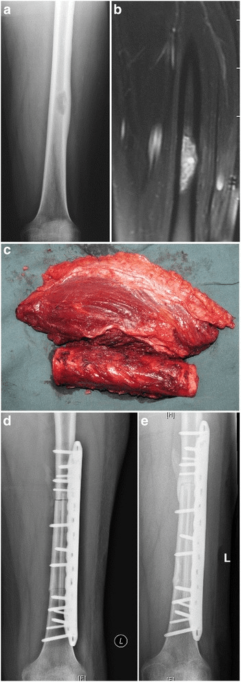
Whether the joint survives. Add the distance from the tumour edge to the joint line to the bone cut you have planned, and you know whether the joint can be preserved (an intercalary resection) or must be resected with the tumour (an articular resection). Joint preservation improves function when it is oncologically safe.
Choosing the reconstruction. The factors that decide it:
- Patient age and growth potential
- Tumour location and the extent of resection
- Expected functional demands
- Available bone stock
- Soft tissue coverage
- Surgeon experience and resources
Planning the prosthesis. Custom or modular; sized from the imaging measurements; stem length and fixation type; and the constraint the joint will need.
Surgical Technique - Reconstruction Options
The workhorse. Modular endoprosthetic reconstruction is the most common reconstruction for periarticular tumours, at the distal femur, proximal tibia, proximal femur and proximal humerus, and the choice whenever immediate stability and early mobilisation are required. It gives immediate stability and weight bearing without donor site morbidity, predictable outcomes, and a modular system that can be customised. The price is mechanical complications, loosening and breakage, infection, a limited lifespan of 70-80% survival at 10 years, and multiple revisions likely in a young patient.
Distal femoral replacement, step by step.
- Incision - extensile anterior or medial approach, excising the biopsy track
- Tumour resection - wide margin including a cuff of normal muscle
- Bone cut - at the level planned from the MRI
- Canal preparation - ream to accept a cemented or press-fit stem
- Prosthesis assembly - the modular system allows length adjustment
- Soft tissue reconstruction - medial gastrocnemius flap for coverage
- Extensor mechanism - suture the quadriceps to the prosthesis if needed
- Closure - layered, over a drain
Technical points. Avoid stripping soft tissue beyond the resection margins. A gastrocnemius flap is essential for coverage at the proximal tibia. Most tumour prostheses use a constrained hinge, and hydroxyapatite collars may improve soft tissue attachment.
Putting it together. The reconstruction follows the site, the age of the patient and what has gone before:
- Preferred Reconstruction
- Modular endoprosthesis
- Alternative
- Allograft-prosthetic composite
- Key Considerations
- Most common tumour site, reliable outcomes
- Preferred Reconstruction
- Extendible endoprosthesis
- Alternative
- Rotationplasty
- Key Considerations
- Must address growth potential and extensor mechanism
- Preferred Reconstruction
- Intercalary prosthesis or vascularised fibula
- Alternative
- Allograft with plate
- Key Considerations
- Preserve joints if possible, biological healing better
- Preferred Reconstruction
- Rotationplasty
- Alternative
- Expandable prosthesis
- Key Considerations
- Multiple lengthening procedures vs single rotationplasty
- Preferred Reconstruction
- Custom hemipelvic prosthesis
- Alternative
- Flail hip (excision only)
- Key Considerations
- High complication rate, consider function vs survival
- Preferred Reconstruction
- Amputation
- Alternative
- Two-stage revision if bone stock allows
- Key Considerations
- Patient safety paramount, functional amputation better than non-functional salvage
Measuring function. The MSTS score is the metric behind every functional figure in this topic and the standard way different reconstructions are compared; it captures patient-perceived function, not just range of motion. Six domains are each scored 0 to 5, giving a maximum of 30, usually expressed as a percentage, and roughly 80% or more is considered excellent.
- Lower limb - pain, function/activity, emotional acceptance, use of supports (walking aids), walking ability, gait
- Upper limb - keeps pain, function and emotional acceptance, and replaces the last three with hand positioning, manual dexterity and lifting ability
Complications
Five ways a tumour prosthesis fails. The Henderson classification sorts endoprosthetic failure into five types and is the frame for any account of the complications. Reoperation for any cause runs at 30% at 10 years, and patients are counselled about it before consent.
- Failure Mode
- Soft tissue: wound dehiscence, flap necrosis
- Incidence
- 5-10%
- Management
- Soft tissue coverage, flap reconstruction
- Failure Mode
- Soft tissue: aseptic instability (dislocation, tendon rupture)
- Incidence
- 10-20% at 10 years (prox tibia)
- Management
- Constraint revision, tendon reconstruction
- Failure Mode
- Aseptic loosening at the bone-prosthesis interface
- Incidence
- 5-10% at 10 years
- Management
- Stem revision
- Failure Mode
- Structural: 3A periprosthetic fracture, 3B implant fracture (stem, body, hinge)
- Incidence
- 5-7%
- Management
- Fracture fixation or component exchange
- Failure Mode
- Deep periprosthetic infection
- Incidence
- 10-15%
- Management
- Two-stage revision or amputation
- Failure Mode
- Tumour progression: local recurrence at the resection margins
- Incidence
- 5-10%
- Management
- Re-resection or amputation
Reading the table. The most common Type 1 failure is the extensor mechanism at the proximal tibia. Type 2 loosening is progressive and presents with pain and a radiographic lucency; Type 3 risk increases with time and with the patient's activity level.
Guidelines, Registries & Global Practice
Global Epidemiology
Primary bone sarcomas are rare worldwide, with osteosarcoma the commonest primary bone malignancy. Large registry analyses (SEER) historically described a bimodal age distribution, but contemporary US data (Kar et al., J Bone Oncol 2024) show the incidence is now unimodal, with a single consistent peak in the second decade of life and no reproducible second peak in older adults (DOI). National-population datasets from high-income settings show low absolute case numbers; Australian national figures report approximately 200 new bone sarcomas annually, with osteosarcoma accounting for roughly one-third. The distal femur remains the single commonest site requiring limb salvage globally.
- Figure
- Osteosarcoma
- Source
- SEER / global registries
- Figure
- Unimodal, peak 2nd decade
- Source
- Kar et al. 2024
- Figure
- Distal femur
- Source
- Multi-series
- Figure
- 85-90%
- Source
- International series
Guideline Comparison
International guidelines are highly concordant: centralised care in a specialist sarcoma centre, multidisciplinary team (MDT) decision-making, image-guided core biopsy with track excision, and limb salvage with wide margins whenever oncologically safe.
- Key Position
- Mandatory referral to a reference sarcoma centre before biopsy; MAP chemotherapy then wide excision for high-grade osteosarcoma
- Evidence Basis
- Clinical Practice Guideline (Strauss et al. 2021)
- Key Position
- Diagnosis and surgery only in designated bone-cancer centres; whole-bone MRI and MDT before definitive surgery
- Evidence Basis
- National service specification, consensus
- Key Position
- Centralised sarcoma services with telehealth and shared-care pathways for patients distant from specialist centres
- Evidence Basis
- Consensus / service model
- Key Position
- Surgical staging (Enneking) and MSTS functional scoring as standard outcome metrics; endoprosthesis the workhorse reconstruction
- Evidence Basis
- Classification / consensus
The ESMO-EURACAN-GENTURIS-PaedCan bone sarcoma guideline (Strauss et al., Ann Oncol 2021) is the most comprehensive international standard, recommending referral to a reference centre before biopsy, neoadjuvant MAP chemotherapy for high-grade osteosarcoma, and wide surgical margins (DOI).
Registry & Access Considerations
The Australian Orthopaedic Association National Joint Replacement Registry (AOANJRR) primarily captures primary and revision arthroplasty; tumour endoprostheses are a distinct subset whose higher revision rates reflect salvage surgery in young patients facing life-threatening disease. For global interpretation, long-term tumour endoprosthesis data are therefore drawn mainly from high-volume sarcoma-centre series (e.g. the Royal Orthopaedic Hospital cohorts) rather than national arthroplasty registries.
Treatment protocols are globally aligned around neoadjuvant MAP chemotherapy and wide resection; the MAP agents (methotrexate, doxorubicin, cisplatin) are widely available in high-income settings. Access for regional and remote patients can carry substantial travel burdens, which strengthens the case for single-procedure options such as rotationplasty where appropriate.
Differential Diagnosis
- Typical Age / Site
- 2nd decade, metaphysis (distal femur)
- Discriminating Features
- Sunburst reaction, Codman triangle, raised ALP/LDH
- Implication for Salvage
- Neoadjuvant MAP then wide resection + reconstruction
- Typical Age / Site
- 5-15 years, diaphysis / flat bones
- Discriminating Features
- Permeative 'moth-eaten' lysis, onion-skin reaction, EWSR1 translocation
- Implication for Salvage
- Chemo + surgery and/or radiotherapy (radiosensitive)
- Typical Age / Site
- 40-60 years, pelvis / proximal femur
- Discriminating Features
- Rings-and-arcs calcification, endosteal scalloping; chemo/radio resistant
- Implication for Salvage
- Wide surgical resection is the mainstay - margins critical
- Typical Age / Site
- 20-40 years, epiphysis/metaphysis
- Discriminating Features
- Eccentric lytic lesion abutting subchondral bone, locally aggressive (Enneking benign stage 3)
- Implication for Salvage
- Often curettage with adjuvant; salvage resection if extensive
- Typical Age / Site
- Over 40 years, axial / proximal long bones
- Discriminating Features
- Multiple lesions, known primary or paraprotein; commonest malignant bone lesion in adults
- Implication for Salvage
- Biopsy and stage before any sarcoma-type resection
- Typical Age / Site
- Any age, metaphysis
- Discriminating Features
- Periosteal reaction with lucent nidus, raised inflammatory markers, may mimic Ewing
- Implication for Salvage
- Biopsy and culture before resection - not a tumour
Clinical Decision Scenarios
Practise clinical reasoning and management decisions out loud
“A 16-year-old male presents with a 3-month history of progressive right knee pain. X-rays show a destructive lesion in the distal femur with periosteal reaction. MRI confirms a 10cm lesion extending to within 4cm of the joint line with soft tissue mass but no joint involvement. Chest CT is clear. Biopsy confirms high-grade osteosarcoma.”
“An 8-year-old girl has been diagnosed with osteosarcoma of the proximal tibia. She has completed neoadjuvant chemotherapy with good response. MRI shows the tumour 3cm from the joint with good response to chemotherapy. Chest CT is clear.”
“A 25-year-old woman had a distal femoral replacement for osteosarcoma 3 years ago. She presents with a 6-week history of increasing knee pain, swelling, and a discharging sinus. Inflammatory markers are elevated. Aspiration grows Staphylococcus aureus.”
Enneking Staging
- Stage I = Low grade (IA intracompartmental, IB extracompartmental)
- Stage II = High grade (IIA intracompartmental, IIB extracompartmental)
- Stage III = Any grade with metastases
- Benign: 1 latent, 2 active, 3 aggressive
Surgical Margins
- Intralesional = through tumour (contamination) - 90% recurrence
- Marginal = through reactive zone - 50% microscopic disease
- Wide = through normal tissue cuff - MINIMUM for sarcoma
- Radical = entire compartment - rarely performed now
- Wide margin: 2cm bone + cuff of normal tissue
Reconstruction Options
- Endoprosthesis = workhorse for periarticular tumours (70-80% 10-yr survival)
- Allograft = biological but high complication (nonunion 20%, fracture 20%)
- APC = allograft-prosthesis composite (soft tissue attachment + joint)
- Vascularized fibula = intercalary defects, paediatric
- Growing prosthesis = paediatric, 15-20 lengthenings needed
- Rotationplasty = excellent function, single procedure, cosmetic concerns
Key Numbers
- Limb salvage rate: 85-90% for bone sarcoma
- Survival equivalent to amputation if margins adequate
- Endoprosthesis infection: 10-15% (vs 1-2% primary TKR)
- 10-year prosthesis survival: 70-80%
- 30% reoperation rate at 10 years (any cause)
- Growing prosthesis complication rate: 50-70% during childhood
Henderson Classification
- Type 1 = Soft tissue (1A wound, 1B instability/tendon)
- Type 2 = Aseptic loosening
- Type 3 = Structural (3A periprosthetic fracture, 3B implant fracture)
- Type 4 = Infection
- Type 5 = Tumour progression
Exam Day Essentials
- Whole bone MRI essential - skip metastases in 1-5%
- Biopsy track must be excised with specimen
- MDT essential for all sarcomas
- Neoadjuvant chemo standard for osteosarcoma
- Rotationplasty underutilized - excellent function in children
- Amputation is NOT failure if margins cannot be achieved
Evidence Base
Limb Salvage vs Amputation Survival (Rougraff Landmark Series)
- 227 patients with non-metastatic high-grade distal femoral osteosarcoma across 26 institutions
- No significant difference in survival or disease-free interval between limb salvage, above-knee amputation, and hip disarticulation
- Local recurrence: 8/73 limb-salvage vs 9/115 amputation vs 0/39 disarticulation
- Limb-salvage patients required additional limb operations more often, but had higher MSTS functional scores
Endoprosthesis Long-term Survival (Jeys/Royal Orthopaedic Series)
- 661 endoprosthetic reconstructions with minimum 10-year follow-up (mean 15 years)
- Implant survival 75% at 10 years with mechanical failure as endpoint; 58% with failure from any cause
- 227 patients (34%) required revision: mechanical failure (116), infection (75), local recurrence (36)
- Limb salvage maintained in 84% at 20 years despite the high revision burden
Rotationplasty Functional Outcomes (Hillmann Gait Analysis)
- 43 patients after rotationplasty for femoral or tibial bone tumour, mean follow-up 6.7 years
- Good functional result: mean MSTS score 23.9/30 (approximately 80%)
- Gait analysis showed a near-normal walking pattern with only a slight limp and lateral trunk lean
- Younger age at operation correlated with better total functional score and walking ability
Paediatric Reconstruction & Growing Prostheses (Groundland Review)
- Endoprosthesis, massive allograft, allograft-prosthesis composite and expandable prostheses are all viable options in the growing child
- Expandable prostheses extend limb salvage even to patients far from skeletal maturity
- Reported high complication and revision burden across the paediatric literature, driven by infection, mechanical failure and loosening
- Authors stress the literature needs outcome reporting stratified by age, anatomical site and reconstruction type
Endoprosthetic Failure Modes (Henderson Classification)
- 2174 patients across five institutions; 534 endoprosthetic failures analysed
- Five failure modes defined: Type 1 soft-tissue, Type 2 aseptic loosening, Type 3 structural, Type 4 infection, Type 5 tumour progression
- Infection was the commonest failure mode in this series; aseptic loosening commonest in the pooled literature
- Failure mode and timing depend significantly on anatomical location - cumulative reporting is misleading
Lifetime Burden of First-Generation Endoprostheses (Grimer)
- 230 patients followed more than 25 years (mean 29.4 years) after endoprosthetic replacement
- Mean 2.7 further operations per patient; only 18% still had the original prosthesis
- Risk of amputation 16% at 30 years; limb salvage maintained in the majority
- Infection risk persisted at roughly 1% per year for the life of the implant (highest at the proximal tibia, 43%)
Surgical Staging of Musculoskeletal Sarcoma (Enneking System)
- Stratifies sarcomas by grade (G), anatomical compartment (T) and metastasis (M)
- Three stages: I low-grade, II high-grade, III any grade with metastases; subdivided A intracompartmental / B extracompartmental
- Defines the four operative margins: intralesional, marginal, wide and radical, relative to tumour and reactive zone
- Remains the foundational surgical staging framework for bone sarcoma decision-making
References
-
Enneking WF, Spanier SS, Goodman MA. A system for the surgical staging of musculoskeletal sarcoma. Clin Orthop Relat Res. 1980;(153):106-120.
-
Rougraff BT, Simon MA, Kneisl JS, et al. Limb salvage compared with amputation for osteosarcoma of the distal end of the femur. A long-term oncological, functional, and quality-of-life study. J Bone Joint Surg Am. 1994;76(5):649-656.
-
Jeys LM, Kulkarni A, Grimer RJ, et al. Endoprosthetic reconstruction for the treatment of musculoskeletal tumors of the appendicular skeleton and pelvis. J Bone Joint Surg Am. 2008;90(6):1265-1271.
-
Henderson ER, Groundland JS, Pala E, et al. Failure mode classification for tumor endoprostheses: retrospective review of five institutions and a literature review. J Bone Joint Surg Am. 2011;93(5):418-429.
-
Hillmann A, Rosenbaum D, Schröter J, et al. Electromyographic and gait analysis of forty-three patients after rotationplasty. J Bone Joint Surg Am. 2000;82(2):187-196.
-
Groundland JS, Binitie O. Reconstruction after tumor resection in the growing child. Orthop Clin North Am. 2016;47(1):265-281.
-
Strauss SJ, Frezza AM, Abecassis N, et al. Bone sarcomas: ESMO-EURACAN-GENTURIS-ERN PaedCan Clinical Practice Guideline for diagnosis, treatment and follow-up. Ann Oncol. 2021;32(12):1520-1536.
-
Kar E, Ammanamanchi A, Yousif M, et al. From bimodal to unimodal: the transformed incidence of osteosarcoma in the United States. J Bone Oncol. 2024;47:100613.
-
Grimer RJ, Aydin BK, Wafa H, et al. Very long-term outcomes after endoprosthetic replacement for malignant tumours of bone. Bone Joint J. 2016;98-B(6):857-864.
-
Mavrogenis AF, Ruggieri P, Mercuri M, et al. Allograft-prosthesis composite for reconstruction of proximal femur bone tumors. Orthopedics. 2010;33(12):888.
-
Benevenia J, Kirchner R, Patterson F, et al. Outcomes of a modular intercalary endoprosthesis as treatment for segmental defects of the femur, tibia, and humerus. Clin Orthop Relat Res. 2016;474(2):539-548.
-
Gosheger G, Gebert C, Ahrens H, et al. Endoprosthetic reconstruction in 250 patients with sarcoma. Clin Orthop Relat Res. 2006;450:164-171.