Benign Adipocytic Tumour | Most Common Soft Tissue Mass | Excellent Prognosis
- Most common soft tissue tumour in adults and the largest single category of benign soft tissue tumours
- Superficial lipomas rarely transform to sarcoma (less than 1%)
- Deep or rapidly growing lipomas require MRI to exclude liposarcoma
- Simple excision curative for superficial lesions; recurrence less than 5%
- Histology shows mature adipocytes without atypia or lipoblasts
- “Deep lipomas require imaging to exclude malignancy
- “Atypical lipomatous tumour (ALT) is well-differentiated liposarcoma by another name
- “Multiple lipomas suggest familial lipomatosis or Madelung disease
- “Intramuscular lipomas: it is the INFILTRATIVE subtype that recurs (19% vs 0% well-circumscribed in Fletcher) - not depth alone
Overview and Epidemiology
Lipomas are benign tumours of mature adipose tissue and the most common soft tissue neoplasm in adults. They are the largest single diagnostic category among benign soft tissue tumours, 16% of an AFIP pathology archive of 18,677 benign lesions and up to roughly half of masses in some clinical referral series, with an estimated incidence of about 1 per 1000 individuals.
The clinical problem. Lipomas are the single most common reason for referral to soft tissue tumour clinics. Most are solitary, slow-growing and superficial (subcutaneous), and present as soft, mobile masses that cause no symptoms. The key challenge is the deep-seated lesion, where a benign lipoma has to be distinguished from well-differentiated liposarcoma (atypical lipomatous tumour). That distinction, with grading, MDM2 amplification and the dedifferentiated end of the spectrum, is developed in full on the liposarcoma page.
Who. Peak incidence is at 40-60 years, with an equal male and female distribution. A minority of patients have multiple lipomas; no series cited on this page quantifies the proportion. The syndromes behind multiple lipomas are set out in their own section below.
Where. Lipomas can arise anywhere adipose tissue is present, and their distribution and behaviour vary by site: 80% are on the trunk and extremities. The table places intramuscular lipomas most often in the thigh; in Fletcher's series they were commonest on the trunk.
- Frequency
- 40%
- Anatomical and clinical considerations
- Subcutaneous fat layer, superficial. Easily accessible, low recurrence
- Frequency
- 25%
- Anatomical and clinical considerations
- Superficial or within muscle compartments
- Frequency
- 20%
- Anatomical and clinical considerations
- Thigh most common, often intramuscular. The infiltrative intramuscular type recurs more often
- Frequency
- 10%
- Anatomical and clinical considerations
- Spindle cell variant common posteriorly. Cosmetically sensitive area
- Frequency
- Under 5%
- Anatomical and clinical considerations
- Deep to abdominal cavity. High risk of being atypical lipomatous tumour
Pathology and Histology
A true neoplasm. Lipomas arise from mesenchymal adipocyte precursors and are made of mature adipocytes identical to normal subcutaneous fat. They are nonetheless true neoplasms rather than simply excess fat: clonal chromosomal aberrations, 12q13-15 rearrangements in 60-70% of cases, set them apart from normal adipose tissue.
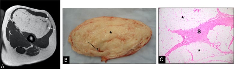
The pseudocapsule. The key histological feature is encapsulation by a thin fibrous pseudocapsule. It gives the surgeon a natural dissection plane, and together with the tumour's soft consistency it allows a lipoma to be enucleated and compressed out through a small incision. The intramuscular type behaves differently, infiltrating between muscle fibres, and deep lesions may compress adjacent neurovascular structures.
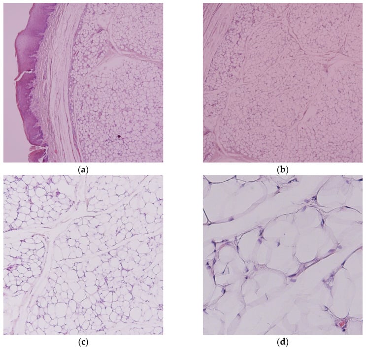
A lipoma shows mature adipocytes with no atypia and no lipoblasts. Well-differentiated liposarcoma shows lipoblasts, nuclear atypia and MDM2/CDK4 amplification (FISH positive). Deep lipomas over 5cm should be biopsied or have MDM2 testing to exclude liposarcoma.
Classification Systems
By depth. Location determines the need for imaging, the surgical approach and the expected recurrence risk.
- Location
- Above muscle fascia
- Characteristics
- 90% of lipomas, easily mobile, typical examination
- Recurrence Risk
- Under 5%
- Location
- Below fascia, not within muscle
- Characteristics
- May compress adjacent structures, MRI needed
- Recurrence Risk
- Not separately reported in the series cited here
- Location
- Within muscle fibres
- Characteristics
- Infiltrates muscles, difficult complete excision
- Recurrence Risk
- 19% (0% if well-circumscribed)
- Location
- Between muscle groups
- Characteristics
- May involve neurovascular bundles
- Recurrence Risk
- 0% in Fletcher's seven intermuscular cases
Morphology, not depth. In Fletcher's series infiltrative intramuscular lipomas recurred in 19% and well-circumscribed ones in 0%. It is the infiltrative subtype that recurs, not depth alone.
By histology. Most lipomas are conventional mature fat. The variant changes the presentation, as with the painful angiolipoma, and the imaging, as with the vascular hibernoma.
- Histology
- Mature adipocytes, thin capsule
- Location Predilection
- Any location
- Behaviour and Treatment
- Most common (80%). Benign, recurrence under 5%. Simple excision
- Histology
- Fat with spindle cells and collagen
- Location Predilection
- Posterior neck, shoulder
- Behaviour and Treatment
- CD34 positive, benign despite cellularity. Simple excision
- Histology
- Floret-like giant cells, bizarre nuclei (degenerative atypia)
- Location Predilection
- Posterior neck, shoulder (older men)
- Behaviour and Treatment
- Benign despite atypia. Excision, must exclude liposarcoma
- Histology
- Fat with vessels (thrombosed capillaries)
- Location Predilection
- Forearm, trunk (young adults)
- Behaviour and Treatment
- Painful variant, often multiple
- Histology
- Brown fat (multivacuolated cells)
- Location Predilection
- Thigh, shoulder, back
- Behaviour and Treatment
- Benign, highly vascular on imaging. Excision if symptomatic
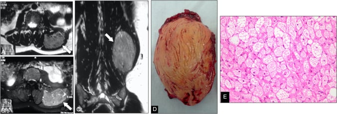
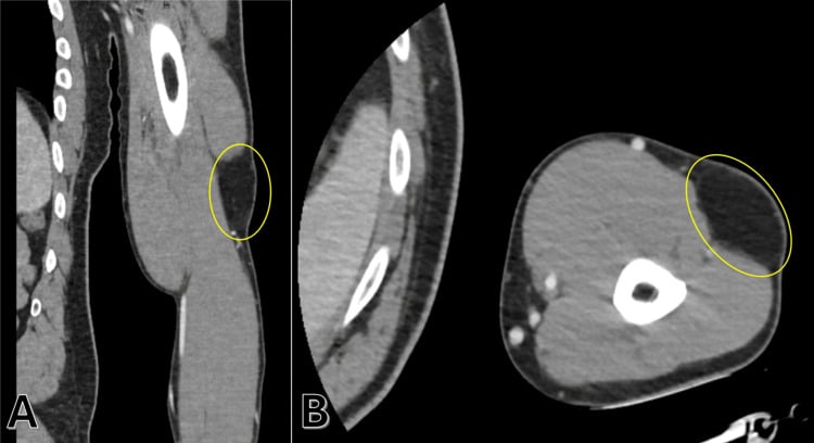
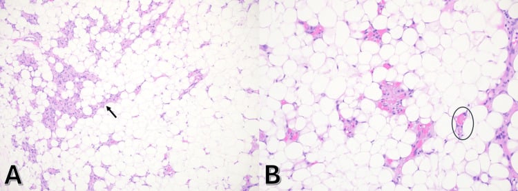
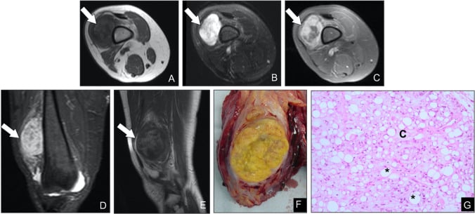
The Benign 'Atypical-Looking' Lipomas: Spindle-Cell and Pleomorphic Variants
One benign family. Spindle-cell and pleomorphic lipoma are two ends of one benign family, with a male predominance (older men) and a predilection for the posterior neck, shoulder and upper back. They share loss of 13q14, including the RB1 gene, and are typically CD34-positive. That molecular fingerprint is quite different from the MDM2/CDK4 amplification (12q13-15) of atypical lipomatous tumour / well-differentiated liposarcoma.
Why they alarm the pathologist. Spindle-cell lipoma adds bland spindle cells and ropey collagen to mature fat, and its cellularity can mimic a spindle-cell sarcoma. Pleomorphic lipoma adds bizarre, hyperchromatic, multinucleated "floret" giant cells, which look atypical enough to be mistaken for pleomorphic or well-differentiated liposarcoma.
How they are told apart. By the absence of MDM2/CDK4 amplification, with RB1 loss and CD34 in support, and not by the atypia itself, so confirm with MDM2 testing before ever labelling such a lesion malignant. Getting this right matters: these are entirely benign lesions, cured by simple marginal excision with essentially no recurrence or metastatic potential. Misdiagnosing one as liposarcoma leads to unnecessary wide resection, radiotherapy and surveillance.
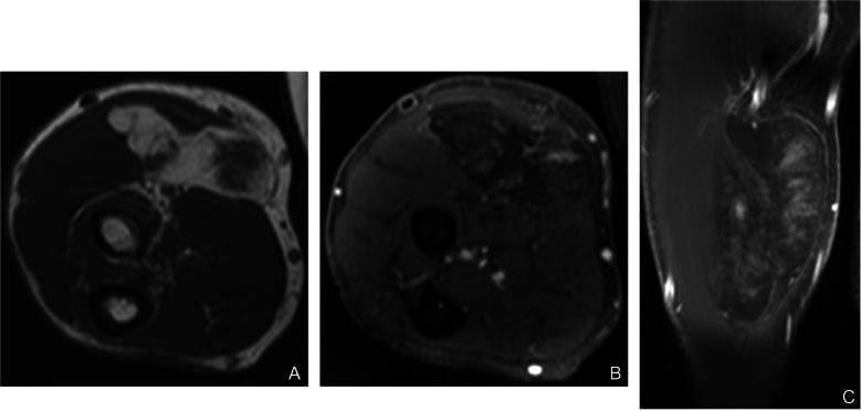
Multiple Lipomas: the Lipomatosis Syndromes
Familial multiple lipomatosis. Autosomal dominant, with many discrete, encapsulated subcutaneous lipomas, tens to hundreds, appearing from early adulthood and typically sparing the head and neck. The lesions are ordinary lipomas and the number, not the biology, is the issue. Excise the symptomatic ones only, and offer genetic counselling when the pattern is autosomal dominant.
Benign symmetric lipomatosis (Madelung disease, Launois-Bensaude). Diffuse, symmetric, non-encapsulated fat deposits around the neck, shoulders and upper trunk (the "horse-collar" or Madelung collar), classically in middle-aged men with a history of alcohol excess. Because the fat is unencapsulated and infiltrative it is not simply enucleated. The important hazard is airway and mediastinal compression, so it is managed by debulking or liposuction with vigilance for the airway. It is distinct from Madelung deformity of the wrist, an unrelated condition with the same eponym.
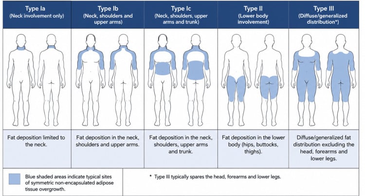
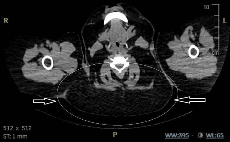
Adiposis dolorosa (Dercum disease). Multiple painful subcutaneous lipomas, usually in peri- or postmenopausal women and often with obesity. The pain, not the mass, dominates, and it is difficult to treat.
The syndromes that carry cancer or overgrowth risk. Gardner syndrome, a variant of familial adenomatous polyposis (APC), combines lipomas or fibromas and colonic polyposis with osteomas, epidermoid cysts and desmoids, and a near-100% colorectal cancer risk. Multiple lipomas with osteomas or dental anomalies should therefore trigger colonoscopy and genetics, and any patient with multiple lipomas is screened for associated conditions such as Gardner syndrome. Proteus syndrome (hamartomatous overgrowth) and PTEN-hamartoma (Cowden) syndrome cause asymmetric overgrowth and multiple lipomas, each with its own tumour-surveillance needs.
Clinical Assessment
History. A painless mass is the most common presentation (80%), usually present for years because growth is slow. Some patients come because of a visible lump, especially on the face or neck, and some with mechanical symptoms from compression of adjacent structures. The questions that matter:
- Rate of growth - rapid growth is concerning for sarcoma
- Pain - lipomas are typically painless, and pain suggests angiolipoma or malignancy
- Family history - multiple lipomas may be familial
- Trauma - some patients report preceding trauma, but no causal link has been proven
Examination. Note the size and location of the mass, look for others (familial lipomatosis) and for skin changes, which are rarely present in lipomas. The characteristic lipoma is:
- Soft and doughy
- Mobile, moving with the skin, not the muscle
- Lobulated
- Non-tender, unless it is an angiolipoma
- Slip sign positive: it slips under the fingers
Depth. The muscle contraction test is the simple test that guides the need for imaging. A superficial lipoma becomes more prominent with the muscle relaxed; a deep lipoma becomes less prominent, or fixed, when the muscle contracts.
These findings warrant MRI to exclude liposarcoma:
- Fixed to deep structures
- Firm or hard consistency
- Rapid enlargement
- Size greater than 5cm
- Deep to fascia
- Associated neurovascular symptoms
LARGELipoma Red Flags (Suspect Liposarcoma)
Hook:LARGE lipomas need imaging - think liposarcoma until proven otherwise!
Investigations
Who needs imaging. Most superficial lipomas under 5cm with a typical examination do not need imaging before excision. Image when the lesion is over 5cm, the examination is atypical, or a deep location is suspected.
Ultrasound is first-line for superficial lesions. A lipoma is a hyperechoic mass with fine internal echoes, in parallel orientation, with a thin or absent capsule and no internal vascularity on Doppler.
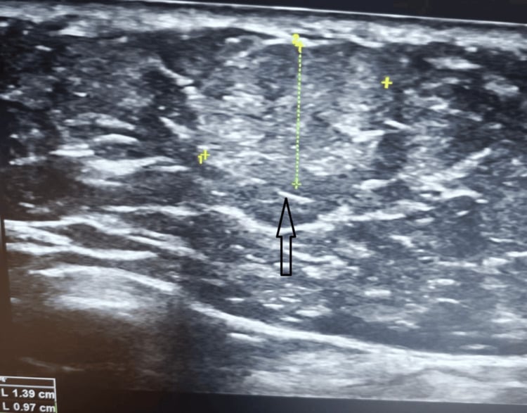
MRI is mandatory for all deep lipomas, to exclude liposarcoma. The benign lipoma is homogeneous and identical to subcutaneous fat on all sequences, bright on T1, and suppresses completely on fat-saturated images. Septa, if present, are thin (under 2mm), and there is no nodular or contrast-enhancing component. Deep lipomas with atypical features require biopsy.
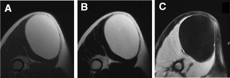
- Benign Lipoma
- Usually under 5cm
- Well-Differentiated Liposarcoma
- Often greater than 10cm
- Benign Lipoma
- Homogeneous fat signal
- Well-Differentiated Liposarcoma
- Heterogeneous, with nodular non-fat areas
- Benign Lipoma
- Thin (under 2mm) or absent
- Well-Differentiated Liposarcoma
- Thick (over 2mm) nodular
- Benign Lipoma
- None
- Well-Differentiated Liposarcoma
- Present in non-fat components
- Benign Lipoma
- Superficial common
- Well-Differentiated Liposarcoma
- Deep (intramuscular, retroperitoneal)
Biopsy. Indicated for:
- A deep lipoma with atypical imaging features
- Size greater than 5cm with thick septations
- Clinical suspicion of malignancy
Core needle biopsy (14-16 gauge) is adequate for most cases, and MDM2 amplification testing distinguishes atypical lipomatous tumour from lipoma. Small superficial lesions are dealt with by excisional biopsy.
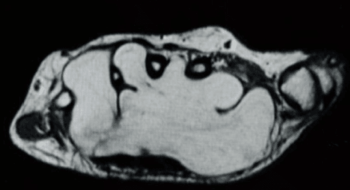
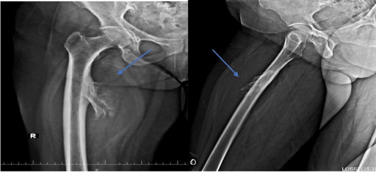
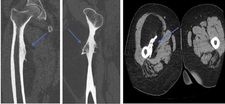
Unusual sites. Deep lipomas can occur in unexpected locations such as the hand, causing compression symptoms, and a lipoma may lie against bone.
Management
Depth, size, symptoms and the imaging decide the operation, and for a confirmed benign lipoma margins are not critical.
- Management
- Observation
- Rationale
- Benign natural history
- Follow-up
- Annual clinical examination or as needed
- Management
- Simple excision
- Rationale
- Curative with low recurrence (under 5%)
- Follow-up
- No routine follow-up needed
- Management
- MRI then excision
- Rationale
- Rule out liposarcoma before surgery
- Follow-up
- Histology confirmation, recurrence monitoring
- Management
- Biopsy then wide excision
- Rationale
- Liposarcoma likely, need margin clearance
- Follow-up
- MDT discussion, oncology referral
Observation. Observation suits the asymptomatic, small (under 5cm) superficial lipoma that is stable over time, in a patient who prefers it. Examine annually, teach the patient the red-flag symptoms (rapid growth, pain), and do not repeat imaging routinely unless symptoms change.
Simple excision is the treatment of choice for the symptomatic superficial lipoma, typically under 5cm, whether the complaint is pain or appearance. Enucleation is adequate once the lesion is confirmed as a benign lipoma. Recurrence is under 5%, the patient goes home the same day, and return to activities is within 1-2 weeks.
Wide excision is recommended for all deep and intramuscular lipomas to reduce recurrence risk, and for any lesion with atypical imaging features or in which liposarcoma cannot be excluded. Aim for marginal to wide margins, 1-2cm if possible. For the infiltrative intramuscular lipoma wide excision is preferred over enucleation, and the patient is counselled about its recurrence risk despite surgery. Deep lipomas have outcomes similar to superficial ones if the margins are clear, and histology is essential to confirm the benign diagnosis.
Surgical Technique
Superficial excision. The steps:
- Position for the lesion: supine with the limb extended for limb lesions, prone or lateral decubitus for back lesions, with adequate lighting and access.
- Anaesthesia. Local anaesthesia (1% lidocaine with adrenaline) infiltrated circumferentially around the lipoma, or a field block for larger lesions. General anaesthesia is rarely needed, and then for deep lesions only.
- Incision directly over the maximum prominence, following skin tension lines where possible, about 50% of the tumour diameter because lipomas compress. Incise through skin and subcutaneous tissue to the pseudocapsule.
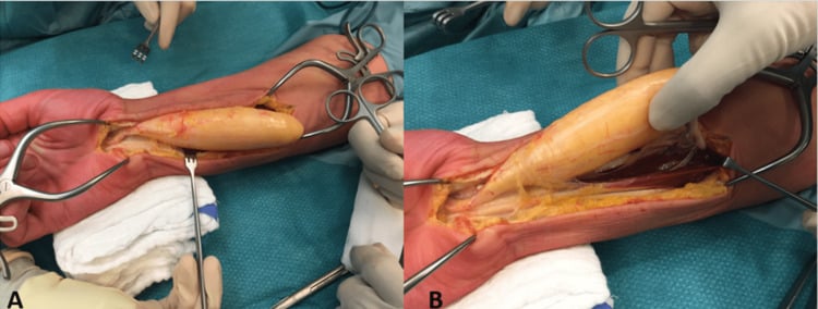
- Enucleation by blunt dissection along the pseudocapsule, with a finger or blunt instrument. The plane is avascular, so the tumour usually shells out with minimal bleeding and sharp dissection is rarely needed. Maintain capsule integrity, which reduces spillage.
- Haemostasis and closure. Inspect the cavity, check the excision is complete and achieve haemostasis. The cavity collapses and leaves minimal dead space, so superficial lesions need no drain. Close the subcutaneous tissue with absorbable sutures and the skin with subcuticular sutures for cosmesis, or interrupted sutures.
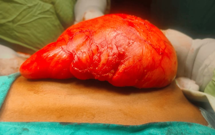
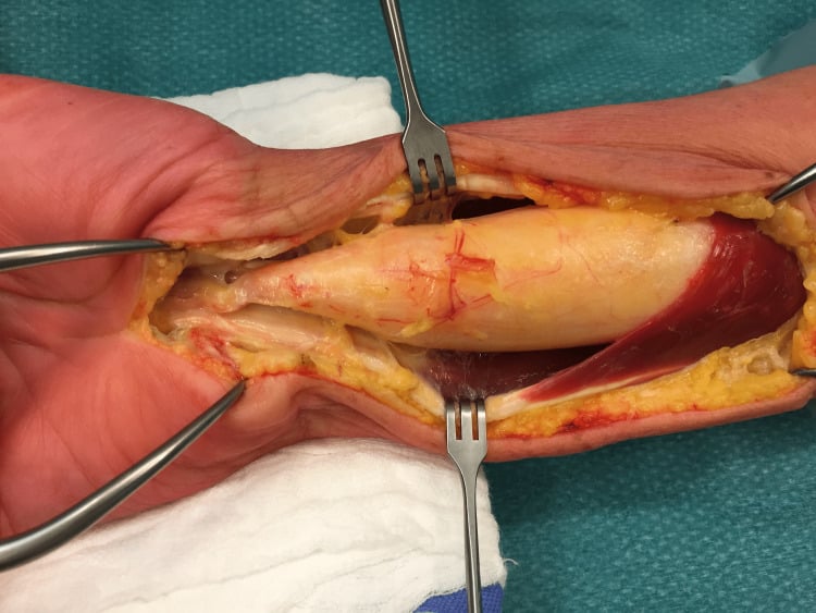
Deep lesions. A pre-operative MRI is mandatory to plan the approach, and the neurovascular structures are identified before the incision. The exposure is wider than for a superficial excision, and a tourniquet is worth considering for limb lesions. Respect the fascial planes and aim for 1-2cm margins if liposarcoma is possible; intramuscular lesions may require muscle resection.
Nerves and closure. Dissect nerves carefully under loupe magnification. Repair the fascia if it was resected, place a drain for a large dead space, close in layers, and send the specimen oriented for margin assessment.
Complications
- Incidence
- Rare (under 1%)
- Prevention
- Haemostasis during dissection
- Management
- Direct pressure, cautery, rarely requires intervention
- Incidence
- 1-2% (higher for deep lipomas)
- Prevention
- Identify nerves preoperatively (MRI), careful dissection
- Management
- Nerve exploration if deficit, neurolysis vs repair
- Incidence
- 5-10% (intramuscular type)
- Prevention
- Wide dissection for infiltrative lesions
- Management
- Re-excision if symptomatic recurrence
Postoperative complications are infrequent after superficial lipoma excision, but monitoring for seroma and infection should be routine.
- Incidence
- 5-10%
- Risk Factors
- Large dead space, inadequate compression
- Management
- Aspiration, compression dressing, rarely requires drainage
- Incidence
- Under 2%
- Risk Factors
- Poor sterility, diabetes, immunosuppression
- Management
- Antibiotics, drainage if abscess forms
- Incidence
- Infiltrative type (Fletcher, 54 cases; rates under Classification)
- Risk Factors
- Incomplete excision, infiltrative type
- Management
- Re-excision with wider margins
- Incidence
- Variable
- Risk Factors
- Location, patient factors, surgical technique
- Management
- Scar revision if significant cosmetic concern
Postoperative Care
The first 48 hours. A pressure dressing for 24-48 hours, ice to reduce swelling, standard analgesia (paracetamol with or without an NSAID) and elevation of the limb where applicable. Keep the wound dry for 48 hours, after which the patient may shower, but no baths for 2 weeks.
The first weeks. Light activities are permitted immediately; avoid heavy lifting or stretching the wound. At the 1-2 week review the sutures come out (7-14 days depending on location), the wound is checked for seroma, haematoma and infection, and the histology is reviewed with counselling on the prognosis it implies. After a superficial excision full activities resume by 2-4 weeks, sport and heavy work at 4-6 weeks, with scar massage to optimise cosmesis. No further follow-up is needed once benign histology is confirmed.
- Postoperative Modification
- Consider drain for 24-48 hours
- Rationale
- Reduce seroma risk
- Postoperative Modification
- Restrict activity for 4 weeks, physiotherapy referral
- Rationale
- Muscle healing, prevent haematoma
- Postoperative Modification
- Document neurovascular status postoperatively
- Rationale
- Medicolegal protection, early detection of deficit
- Postoperative Modification
- MDT referral, possible re-excision
- Rationale
- Atypical lipomatous tumour needs wider margins
Prognosis and Outcomes
Superficial lipomas have an excellent prognosis: simple excision is curative, and the complications are minimal (seroma, scar). Malignant transformation is extremely rare (less than 1%).
Deep subfascial lipomas do excellently once the benign diagnosis is confirmed, though liposarcoma has to be watched for. Their recurrence rate is not separately reported in the series cited here.
Infiltrative intramuscular lipomas do well, but recurrence is common and nerve injury and incomplete excision are more frequent; wide excision is recommended.
Factors associated with higher recurrence:
- Intramuscular or infiltrative subtype
- Incomplete excision (marginal excision for infiltrative lesions)
- Size greater than 10cm
- Deep location with complex anatomy
Guidelines, Registries & Global Practice
Global Epidemiology
Lipoma is the most common soft tissue tumour worldwide, with an estimated incidence of about 1 per 1000 population per year and no consistent geographic or ethnic predilection. Peak presentation is in the 4th to 6th decades with roughly equal sex distribution, and a minority of patients have multiple lesions - no series cited on this page puts a figure on that proportion. Atypical lipomatous tumour / well-differentiated liposarcoma is far rarer (a fraction of the roughly 5 soft tissue sarcomas per 100,000/year) and is concentrated in older adults with deep extremity or retroperitoneal masses.
Society Guidance, Side by Side
- Imaging threshold
- Any deep soft-tissue mass, or superficial mass over ~5cm, growing or painful → urgent imaging
- Referral / diagnosis emphasis
- Refer indeterminate fatty masses to a sarcoma diagnostic pathway before excision
- Imaging threshold
- MRI for any deep or large (over 5cm) soft-tissue mass
- Referral / diagnosis emphasis
- Biopsy and management of suspected ALT/sarcoma at a reference sarcoma centre; MDM2 testing for ambiguous cases
- Imaging threshold
- Cross-sectional imaging (MRI) for indeterminate or deep lesions
- Referral / diagnosis emphasis
- Core-needle biopsy and multidisciplinary planning before resection of indeterminate fatty tumours
- Imaging threshold
- Not an imaging guideline — defines pathology
- Referral / diagnosis emphasis
- ALT and well-differentiated liposarcoma are one MDM2-amplified entity; terminology depends on site/resectability
Across bodies the consensus is consistent: a small, superficial, clinically typical lipoma needs neither imaging nor referral, while a deep or large (over 5cm) or growing fatty mass should be imaged and, if indeterminate, biopsied — ideally within a sarcoma service — before any excision. Differences are mainly in pathway structure (centralised sarcoma units in the UK/Europe vs more variable referral in some systems) rather than the underlying clinical logic. There is no dedicated implant or device registry for lipoma, as management is excisional rather than implant-based.
High- vs Limited-Resource Practice
- MRI readily available for deep or large lesions
- MDM2 amplification (FISH or equivalent) to confirm ALT/WDL
- Sarcoma multidisciplinary teams direct biopsy-first management
- Ultrasound may be the only modality; reliance on clinical red flags
- Histology available but molecular MDM2 testing often not
- Lower threshold for excisional biopsy of accessible lesions; refer deep/retroperitoneal masses where possible
Consent and Documentation
- Key risks to consent for: recurrence (19% if infiltrative, 0% if well-circumscribed, in the series cited on this page), nerve injury (1-2%), seroma, and the possibility that histology returns an atypical lipomatous tumour requiring further surgery.
- Alternatives: observation versus excision; confirm whether the indication is cosmetic or symptomatic
- Pathology: explain that all excised tissue is sent for histology to confirm the benign diagnosis
- Document examination findings including depth assessment (muscle contraction test)
- Clear indication for imaging (size, location, atypical features)
- Informed consent discussing recurrence risk (varies by location)
- If liposarcoma suspected, document referral to sarcoma MDT
- Failure to investigate deep or large lipomas with imaging (missed liposarcoma)
- Inadequate excision leading to recurrence
- Nerve injury during excision of deep lipomas without documentation of preoperative nerve status
Controversies and Areas of Uncertainty
When can MRI alone be trusted? A confidently benign MRI (homogeneous fat, thin septa, no enhancement) has a high negative predictive value, but reader accuracy against MDM2/FISH is only about 73% for indeterminate deep masses. Practice varies on whether an indeterminate deep fatty mass should go straight to biopsy or to a sarcoma centre for re-imaging.
ALT or well-differentiated liposarcoma? The two names describe the same MDM2-amplified entity. The benign-sounding ALT label is preferred for resectable extremity and trunk lesions, while WDL is used for retroperitoneal disease that cannot be widely excised and carries dedifferentiation risk.
Margin width for deep ALT. Marginal excision yields roughly 23% recurrence against 0% for wide resection in extremity ALT, but routine wide resection adds morbidity; in Kito's series, function was equivalent after either (ISOLS/MSTS 98% against 99%). The threshold for accepting a planned marginal margin, near nerves for example, remains debated and is individualised at the MDT.
Non-surgical and minimally invasive options. Liposuction, steroid injection and laser-assisted removal are described for selected superficial cosmetic lipomas. Evidence is limited, recurrence is higher, and no tissue margin or histology is obtained, so they are not used where liposarcoma cannot be excluded.
MCQ Practice Points
Q: What is the key histological feature that distinguishes lipoma from well-differentiated liposarcoma? A: MDM2 amplification - Well-differentiated liposarcoma (atypical lipomatous tumor) shows MDM2 and CDK4 amplification on FISH testing, while lipoma does not. Histologically, lipoma shows mature adipocytes without lipoblasts or significant atypia. Lipoblasts and atypical stromal cells suggest liposarcoma.
Q: What MRI features suggest liposarcoma rather than benign lipoma? A: Thick septations (greater than 2mm), nodular enhancement, heterogeneous signal - Benign lipomas show homogeneous fat signal identical to subcutaneous fat, with thin (less than 2mm) or no septa, and no contrast enhancement. Liposarcomas have thick irregular septa, non-fat soft tissue components, and enhancement.
Q: What is the recurrence rate after simple excision of intramuscular lipoma? A: 19% for the infiltrative type, 0% for the well-circumscribed type in Fletcher's series - the discriminator is morphology, not simply being intramuscular. Infiltrative lesions interdigitate with muscle so complete removal is difficult; wide excision is preferred when feasible. Well-circumscribed intramuscular lipomas and superficial lipomas do well with complete local excision.
Q: What is the most common soft tissue tumor in adults? A: Lipoma - Lipomas are the largest single category of benign soft tissue tumors (16% of an AFIP archive; up to roughly half in some referral series). They are slow-growing, usually superficial, and present as soft mobile masses. Peak incidence is in the 5th-6th decade with equal gender distribution.
Q: What size cutoff prompts imaging for a clinically suspected lipoma? A: Greater than 5cm - Superficial lipomas under 5cm with typical examination findings do not require imaging. Lesions greater than 5cm, deep location, rapid growth, or atypical features warrant MRI to exclude liposarcoma before excision.
Exam Viva Scenarios
Practise clinical reasoning and management decisions out loud
“A 45-year-old woman presents with a 3cm soft, mobile mass on her upper arm that has been present for 2 years. She is concerned about the cosmetic appearance. On examination, you find a non-tender, doughy mass that slips under your fingers and becomes more prominent when she relaxes her arm. What is your assessment and management?”
“A 55-year-old man presents with a 7cm mass in his thigh that has grown over the past 6 months. MRI shows a predominantly fatty mass with some thick septations and small areas of enhancement. What are your concerns and how would you proceed?”
“A 35-year-old man presents with approximately 15 lipomas scattered over his trunk and arms. He has a family history of similar findings in his father. Several are becoming painful. How would you assess and manage this patient?”
Key Epidemiology
- Most common soft tissue tumor (16% of AFIP benign archive; up to ~half in referral series)
- Incidence 1:1000 in population
- Peak age 40-60 years, equal gender distribution
- Deep lesions are a small minority - Fletcher found intramuscular 1.8% and intermuscular 0.3% of adipose tumours
Classification
- Superficial (subcutaneous) = 90% = simple excision
- Deep (subfascial) = need MRI
- Infiltrative morphology (not intramuscular depth) = 19% recurrence = excise the whole lesion
- Variants: spindle cell, pleomorphic (benign despite atypia), angiolipoma (painful)
Red Flags (LARGE Mnemonic)
- Location deep (subfascial/intramuscular)
- Age over 50 (liposarcoma more common)
- Rapidly growing (weeks to months)
- Greater than 5cm (imaging mandatory)
- Enhancement on MRI (suggests malignancy)
Imaging Pearls
- Superficial under 5cm with typical exam = no imaging needed
- MRI mandatory for deep lipomas (rule out liposarcoma)
- Benign features: homogeneous fat signal, thin septa (under 2mm), no enhancement
- Malignant features: thick septa, nodularity, enhancement, heterogeneous signal
Surgical Principles
- Superficial = simple excision (enucleation)
- Intramuscular = wide excision preferred
- Margins not critical if confirmed benign
- Pseudocapsule provides dissection plane
- Large/deep needs MRI first
Complications and Outcomes
- Recurrence 19% for the INFILTRATIVE type and 0% for the well-circumscribed type (Fletcher) - morphology, not depth
- Nerve injury 1-2% for deep lipomas
- Seroma 5-10%, usually managed conservatively
- Malignant transformation less than 1% (extremely rare)
Evidence Base and Key Studies
Distribution of Benign Soft-Tissue Tumours (AFIP Referral Series)
- 18,677 benign mesenchymal lesions from a 10-year AFIP consultation archive
- Lipoma and lipoma variants were the single most common diagnostic category (16%)
- Roughly two-thirds of benign soft-tissue tumours fall into seven diagnostic groups
- Prevalence varies markedly with patient age and anatomical location
MRI of 126 Consecutive Fatty Masses: Lipoma vs Well-Differentiated Liposarcoma
- MRI was 100% sensitive and 100% NPV for detecting well-differentiated liposarcoma
- MRI was 100% specific for the diagnosis of simple lipoma
- But positive predictive value was only 38% — most 'suspicious' masses were benign
- 64% of lesions called suspicious for liposarcoma were actually benign lipoma variants