Mason Classification of Radial Head Fractures
- Classifying the bone and stopping there. Mason type alone never decides treatment — you must add the stability assessment (MCL, LCL, interosseous membrane, DRUJ). "Classify the fracture, then assess stability" is the examiner's mantra.
- Excising a comminuted head in an unstable elbow. The radial head is the primary valgus stabiliser (when the MCL is torn) and the primary longitudinal stabiliser (when the IOM is incompetent) — never excise if the MCL/IOM is deficient or in Essex-Lopresti. Replace it.
- Calling a block on pain alone. The Type II operative trigger is a discrete mechanical block to rotation, not pain — and Type I-vs-II displacement is observer-dependent (even on CT), so corroborate before operating.
- Missing the associated injury. Forgetting to examine the wrist/DRUJ (Essex-Lopresti) or the coronoid + LCL (terrible triad) is the classic fail — exclude the whole TEAM.
- Over-stuffing the arthroplasty. An over-lengthened radial head prosthesis causes capitellar wear, pain and stiffness — size from the excised fragments and check the ulnohumeral joint does not gap on fluoroscopy.
The Mason classification
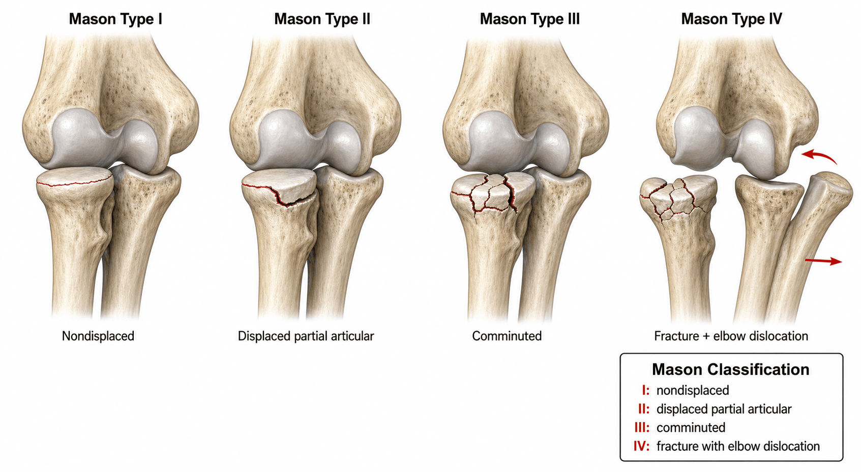
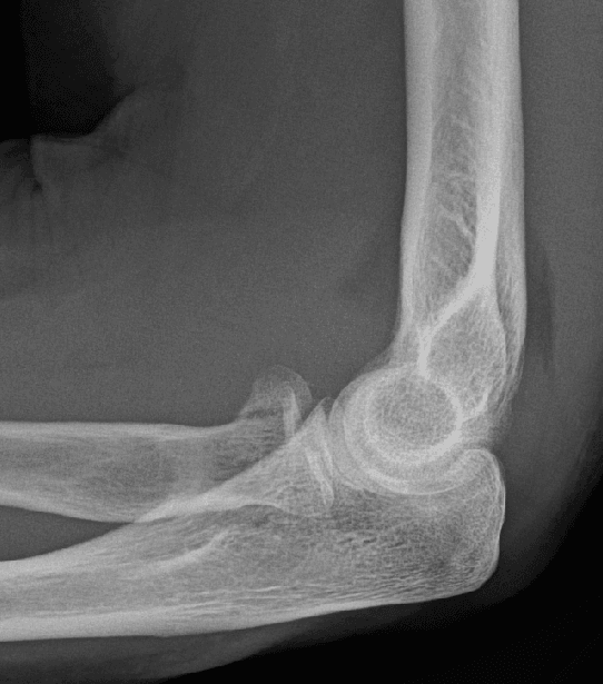
The original Mason classification (1954) had three types; Johnston (1962) added Type IV for any radial head fracture with an associated elbow dislocation. Each type carries distinct management implications.
- Description
- Non-displaced or minimally displaced
- Key feature
- No mechanical block; under 2 mm displacement
- Management principle
- Non-operative: sling for comfort, early active motion within 48 hours
- Description
- Displaced fracture (marginal sector)
- Key feature
- Over 2 mm displacement OR a mechanical block to rotation
- Management principle
- ORIF if reconstructable and blocked; non-operative if minimally displaced and unblocked
- Description
- Comminuted, involving the entire radial head
- Key feature
- No reconstructable fragments; entire head shattered
- Management principle
- Metallic radial head arthroplasty (excise only if elbow/forearm stable)
- Description
- Any Mason type with associated elbow dislocation
- Key feature
- Radial head fracture plus dislocation (often terrible triad)
- Management principle
- Treat both the fracture and the instability; almost always operative
Examine before imaging: passively pronate and supinate the forearm. A discrete hard stop (mechanical block) is the operative indication for a Type II fracture; pain without a block does not mandate surgery. Note that Type I-vs-II displacement is observer-dependent on plain films (and even on CT), so corroborate displacement before committing to surgery.
The Type II decision turns on a true mechanical block, but acute pain and a tense haemarthrosis make forearm rotation unreliable to assess at presentation. The classic technique is to aspirate the elbow haemarthrosis and instil local anaesthetic:
- Where: the lateral "soft spot" — the triangle bounded by the radial head, the lateral epicondyle and the olecranon tip.
- Why: decompressing the haemarthrosis and abolishing pain lets you re-examine pronation/supination accurately — persistent restriction with a firm end-point = a genuine mechanical block (favours ORIF), whereas freely-restored, pain-relieved rotation = no true block (favours non-operative care with early motion).
- It also facilitates early mobilisation in Mason I/II fractures managed non-operatively. This is how you turn "pain without a block does not mandate surgery" into an actual bedside test.
Decision framework — ORIF vs replacement
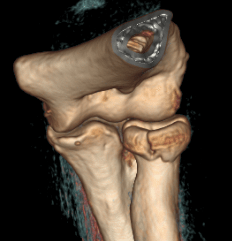
- 1Classify and examine for a blockAssign the Mason type; passively rotate the forearm for a mechanical block. CT (ideally 3D) for displaced/comminuted patterns to define fragment number and size.
- 2Assess elbow & forearm stabilityTest the MCL (valgus), LCL (posterolateral rotatory instability), and the wrist/DRUJ + interosseous membrane (Essex-Lopresti). Stability — not just the articular surface — decides excise vs replace.
- 3Fix the reconstructable, replace the restType II with a block → ORIF (headless screws/mini-plate, under 2 mm step-off). Type III → metallic radial head arthroplasty; excision is acceptable ONLY if the MCL and IOM are intact.
- 4Reconstruct the unstable elbowType IV / terrible triad → replace the head, fix the coronoid, repair the LCL; reassess and add a hinged external fixator if still unstable. Never excise the head here.
- Reconstructable?
- N/A (non-displaced)
- Elbow/forearm stable?
- Typically yes
- Recommended procedure
- Non-operative: short splint, early motion
- Reconstructable?
- Yes (few fragments)
- Elbow/forearm stable?
- Yes
- Recommended procedure
- ORIF — headless compression screws or mini-fragment plate
- Reconstructable?
- Yes, minimal displacement
- Elbow/forearm stable?
- Yes
- Recommended procedure
- Trial of non-operative care; operate only if stiffness persists
- Reconstructable?
- No
- Elbow/forearm stable?
- MCL intact, no IOM disruption
- Recommended procedure
- Radial head excision (safe only if MCL intact and no IOM injury)
- Reconstructable?
- No
- Elbow/forearm stable?
- MCL deficient or IOM disrupted
- Recommended procedure
- Metallic radial head arthroplasty (NEVER excise)
- Reconstructable?
- Usually no
- Elbow/forearm stable?
- No — dislocation ± coronoid/LCL injury
- Recommended procedure
- Radial head replacement + coronoid fixation + LCL repair
Never excise a radial head when the medial collateral ligament is deficient, the interosseous membrane is disrupted, or an Essex-Lopresti injury is present. The radial head is the primary valgus stabiliser of the elbow when the MCL is torn and the primary longitudinal stabiliser of the forearm when the interosseous membrane is incompetent. Excision in these settings causes posterolateral rotatory instability, proximal radial migration, and wrist pain.
Associated injuries & instability patterns
Radial head fractures rarely exist in isolation — always interpret the Mason type in the context of associated injuries.
- Components
- Radial head fracture + coronoid fracture + elbow dislocation
- Significance
- Highly unstable; recurrent dislocation risk
- Management implication
- Radial head replacement, coronoid fixation, LCL repair, hinged fixator if needed
- Components
- Radial head fracture + interosseous membrane disruption + DRUJ injury
- Significance
- Longitudinal forearm instability; proximal radial migration
- Management implication
- Must preserve or replace the radial head; never excise
- Components
- Proximal ulna fracture + radial head fracture (posterior Bado II)
- Significance
- Unstable forearm ring
- Management implication
- Fix the ulna anatomically; address the radial head by Mason type
- Components
- Lateral collateral ligament avulsion with radial head fracture
- Significance
- Posterolateral rotatory instability risk
- Management implication
- Repair the LCL at the time of radial head fixation/replacement
Tenderness over the distal radioulnar joint or interosseous membrane raises the spectre of Essex-Lopresti. Obtain bilateral forearm/wrist radiographs and consider MRI if suspicion is high — missing it and then excising the radial head leads to proximal radial migration and chronic wrist pain.
Surgical technique considerations
- ORIF for Type II: a Kaplan (EDC split) or Kocher (anconeus-ECU) approach depending on fragment location — anterior fragments via Kaplan. Fix with headless compression screws (simple two-part) or mini-fragment locking plates (neck involvement); aim for under 2 mm residual step-off. Plate hardware should sit within the "safe zone" (non-articulating arc).
- Radial head arthroplasty for Type III/IV: a metallic (titanium/pyrocarbon) monopolar or bipolar prosthesis, sized from the excised fragments (articular diameter and neck height). Avoid over-lengthening (over-stuffing), which causes capitellar wear, pain and restricted flexion — the prosthesis should articulate with the capitulum without gapping the ulnohumeral joint on fluoroscopy.
- LCL repair: reattach the lateral ulnar collateral ligament to its origin on the lateral epicondyle (suture anchor or transosseous tunnels) — essential in Type IV/terrible triad, where the lateral approach itself often detaches it.
- Post-operative protocol: early active motion within 48–72 hours; avoid passive stretching/heavy loading for six weeks; a hinged elbow brace if instability persists. The posterior interosseous nerve is at risk in lateral approaches — keep the forearm pronated to translate it away from the field.
Complications
- Complication
- Stiffness / loss of terminal extension
- Note
- The commonest complication after elbow trauma — capsular scarring and prolonged immobilisation cause a flexion contracture; the case for early motion
- Complication
- Heterotopic ossification ± radioulnar synostosis
- Note
- Higher with terrible-triad/fracture-dislocation, associated head injury and delayed/extensive surgery; synostosis blocks rotation
- Complication
- Post-traumatic arthritis / capitellar wear
- Note
- From residual articular incongruity or an OVER-STUFFED (over-sized/over-lengthened) prosthesis abrading the capitellum
- Complication
- Recurrent instability / posterolateral rotatory instability
- Note
- From an unaddressed LCL/ligament injury — or from excising the head in an unstable elbow
- Complication
- Over-stuffing, loosening/osteolysis, painful implant
- Note
- Over-lengthening gaps the ulnohumeral joint and wears the capitellum; size from the excised fragments
- Complication
- Proximal radial migration + valgus instability
- Note
- The penalty for excising when the MCL or interosseous membrane is deficient (Essex-Lopresti) — chronic wrist pain
- Complication
- Posterior interosseous nerve injury
- Note
- Risk in the lateral (Kaplan) approach / from retraction — keep the forearm pronated
- Complication
- Nonunion / malunion
- Note
- Uncommon for the head; neck-level nonunion is the more typical site
The two outcome-defining errors are stiffness from immobilising too long and instability from under-treating the soft tissues. Mobilise early once the construct is stable, and never leave an MCL/LCL/interosseous-membrane injury unaddressed — recurrent instability and proximal radial migration are far harder to salvage than the index fracture.
Guidelines, registries & global practice
- AAOS appropriate use criteria: non-operative treatment for undisplaced fractures, surgery for a mechanical block or instability — ORIF for reconstructable Type II and arthroplasty for unreconstructable Type III.
- BOA/BESS (UK): early mobilisation for Type I; ORIF for displaced/blocked Type II; metallic radial head replacement for comminuted Type III/IV when not reconstructable; excision without replacement only without ligamentous instability.
- Global variation/registries: ORIF rates for Mason II vary with the threshold for defining a mechanical block; registries (Swedish Fracture Register; UK implant data) show increasing use of metallic radial head arthroplasty (pyrocarbon/titanium) — silicone implants are obsolete/abandoned.
- Key principle across guidelines: the decision depends not only on the Mason type but equally on the integrity of the MCL, LCL, interosseous membrane and DRUJ — classify the fracture, then assess stability.
Mnemonics & memory aids
Intact · Indicated · Irreparable · ViolatedThe four Mason types
Hook:Escalation: do nothing → fix → replace → address both fracture and global instability.
TEAMAssociated injuries to exclude
Hook:For every radial head fracture, exclude the TEAM — missing any leads to recurrent instability or longitudinal forearm collapse.
MILD → never exciseWhen NOT to excise the radial head
Hook:If any of MILD is present, REPLACE (don't excise) — excision there guarantees instability or proximal radial migration.
Viva practice
Exam viva
Practise clinical reasoning and management decisions out loud
“A 42-year-old woman falls on her outstretched hand and sustains a radial head fracture. Radiographs show a displaced fracture involving approximately 40 percent of the radial head with a 3 mm articular step-off. Her forearm rotation is limited to 30 degrees pronation and 40 degrees supination with a firm end-point. Classify this fracture and outline your management.”
“A 55-year-old man presents after a fall with a swollen, painful elbow. Radiographs demonstrate a posterior elbow dislocation with a comminuted radial head fracture and a small coronoid tip fracture (Regan-Morrey Type I). After closed reduction, the elbow redislocates in extension at 40 degrees. Discuss the classification and your operative plan.”
Exam cheat sheet
- I undisplaced (early motion); II displaced/blocked (ORIF if reconstructable); III comminuted (replace); IV + dislocation (operative).
- A mechanical block to rotation is the operative trigger for Type II — examine before imaging.
- NEVER excise the head if the MCL or interosseous membrane is deficient (primary valgus + longitudinal stabiliser).
- Terrible triad (Type IV): replace the head + fix the coronoid + repair the LCL; never excise.
- Exclude the TEAM: Terrible triad, Essex-Lopresti, Associated LCL injury, Monteggia variant — examine the wrist for Essex-Lopresti.
- Use metallic implants (silicone obsolete); avoid over-stuffing; Mason typing is observer-dependent (CT/3D helps but doesn't resolve it).
The four types
- Type I: undisplaced/minimally displaced, no block - non-operative, early motion
- Type II: displaced over 2 mm or mechanical block - ORIF if reconstructable
- Type III: comminuted entire head, unreconstructable - replace (excise only if stable)
- Type IV: any type plus elbow dislocation - almost always operative
Decision rules
- Mechanical block to rotation is the operative trigger for Type II
- Never excise the head if MCL or interosseous membrane is deficient
- Terrible triad: replace the head, fix the coronoid, repair the LCL
- Silicone implants obsolete - use metallic (titanium or pyrocarbon); avoid over-stuffing
Must-exclude associated injuries (TEAM)
- Terrible triad (coronoid fracture + dislocation) - check the lateral radiograph
- Essex-Lopresti (IOM + DRUJ disruption) - examine the wrist
- Associated LCL injury - test posterolateral rotatory instability
- Monteggia variant - assess the ulna shaft on forearm films
Evidence Base
Apparently isolated partial articular fractures of the radial head: prevalence and reliability of radiographically diagnosed displacement
- 119 isolated partial radial head fractures classified by the Broberg-Morrey modification of Mason: 85% Mason type 1, 9% borderline 1/2, 6% type 2.
- Intra-observer reliability was excellent (kappa 0.85) but inter-observer only moderate (kappa 0.45).
- Apparently isolated, stable partial fractures are infrequently truly displaced - displacement is likely over-diagnosed.
Interobserver reliability of radial head fracture classification: two-dimensional compared with three-dimensional CT
- 85 orthopaedic surgeons classified 12 radial head fractures (Broberg-Morrey/Mason) on radiographs + 2D vs 3D CT.
- 3D CT gave moderate agreement versus fair for 2D CT - a small but significant improvement.
- Considerable disagreement on classification and treatment persisted even with 3D CT (only fair agreement on treatment recommendations).
According to PubMed, the prevalence of Mason types and the observer-dependence of the type-1-vs-2 displacement threshold come from Doornberg et al. 2007 (DOI), and the (partial) improvement in reliability with 3D CT from Guitton & Ring 2011 (DOI). The four-type system (Johnston's Type IV), the stabiliser role of the radial head, and the terrible-triad/Essex-Lopresti principles are standard, well-established elbow-trauma teaching.