Meary Angle (Talo-First Metatarsal Angle)
- Quoting a bare number. A Meary value without the apex direction is meaningless — apex plantar = pes PLANUS, apex dorsal = pes CAVUS. State the direction every time.
- Measuring on a non-weight-bearing film. The foot must be loaded (standing lateral) — supine films underestimate flatfoot and overestimate cavus.
- Drawing the talar axis through the dome. Use the talar neck-body midpoints, not the curved dome; the first-MT axis uses shaft midpoints.
- Calling a cavovarus foot 'idiopathic'. Every cavovarus foot needs a neurological work-up — Charcot-Marie-Tooth until proven otherwise (nerve conduction + spinal imaging).
- Using Meary in isolation. Pair it with calcaneal pitch and the AP talonavicular coverage angle, and establish flexible vs rigid (corrects on tiptoe / Coleman block).
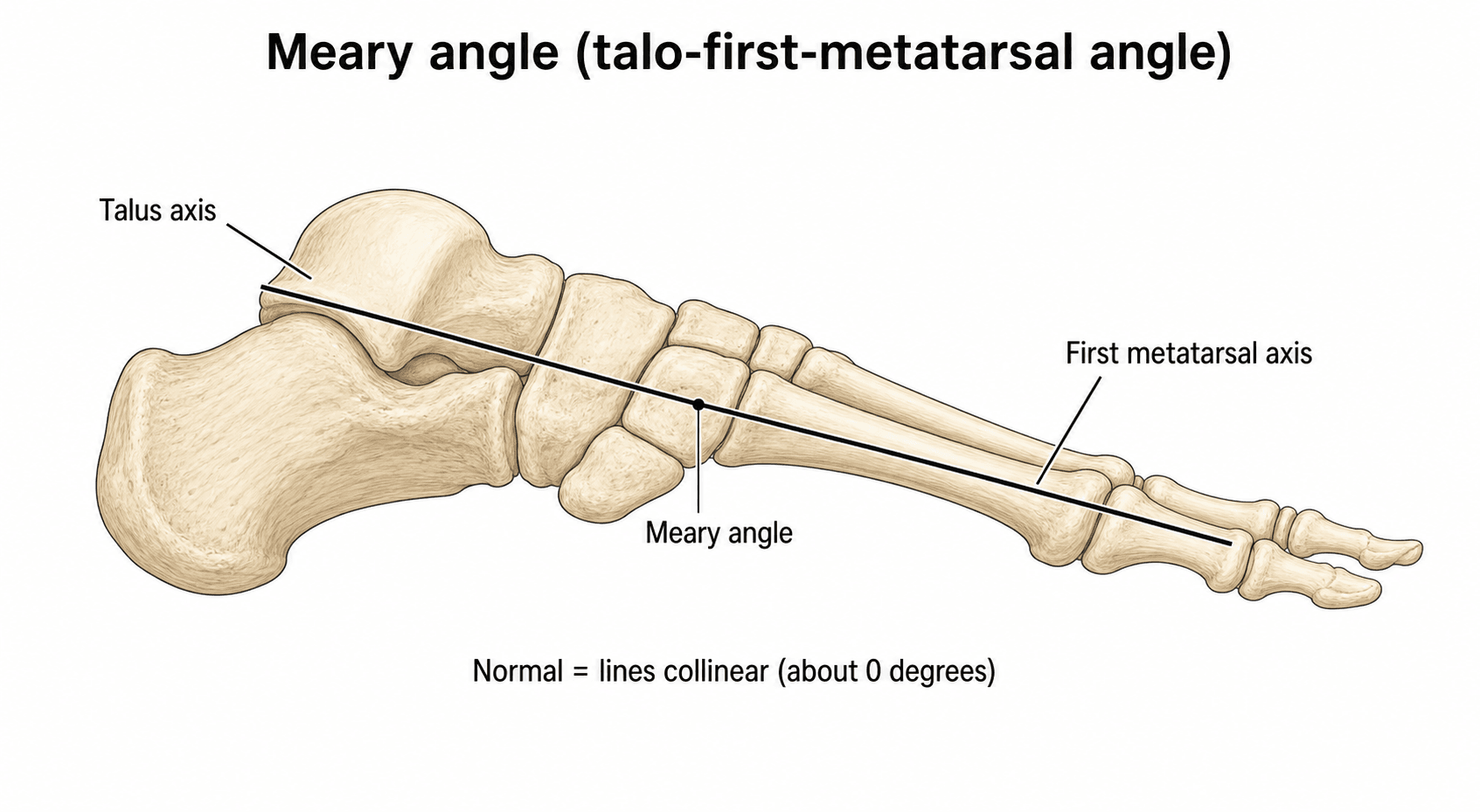
A Meary angle of approximately zero degrees is normal on a lateral weight-bearing foot radiograph. An angle with the apex directed plantarward, greater than about 4°, indicates pes planovalgus; an angle with the apex directed dorsalward, greater than about 4°, indicates pes cavovarus. Always state whether the foot is weight-bearing (it must be) and whether the deformity is flexible or rigid. Examiners expect you to draw the lines: the long axis of the talus and the long axis of the first metatarsal.
How to measure the Meary angle
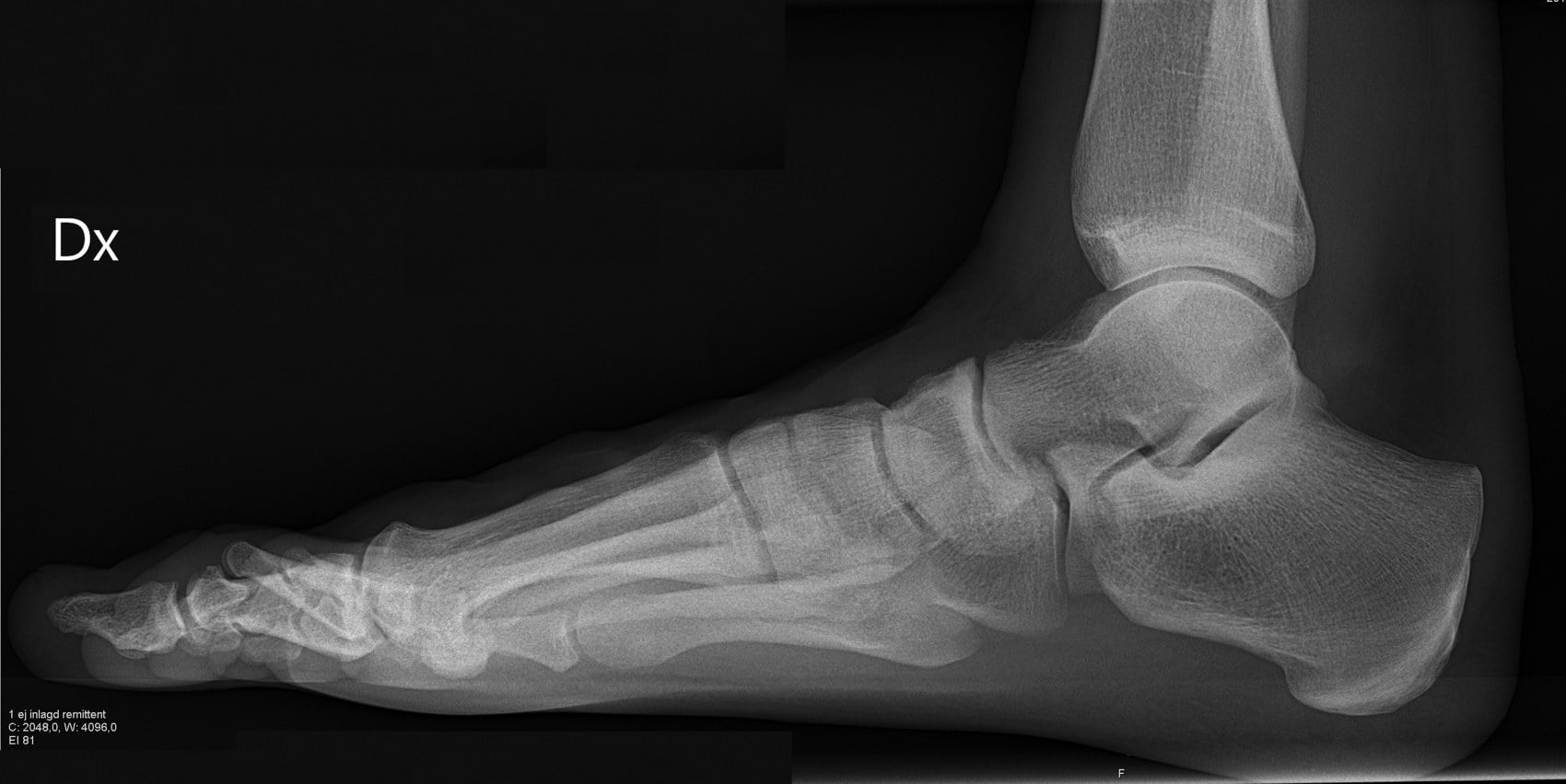
The Meary angle is measured on a true lateral weight-bearing radiograph of the foot — patient standing, beam perpendicular to the foot, centred on the midfoot.
Step-by-step:
- Draw the longitudinal axis of the talus: a line through the midpoint of the talar head and the midpoint of the talar neck-body junction (or a best-fit line along the talar body).
- Draw the longitudinal axis of the first metatarsal: a line through the midpoints of the proximal and distal metaphyseal-diaphyseal junctions of the first metatarsal.
- The Meary angle is the intersection of these two lines at the midtarsal region.
- Normal: the two axes are collinear or nearly so (approximately zero degrees, plus or minus 4°).
- Angle
- Approximately 0° (± 4°)
- Apex direction
- Axes collinear
- Pathology
- Neutral sagittal alignment
- Angle
- Greater than 4°
- Apex direction
- Apex plantar (talar axis angled plantar)
- Pathology
- Flatfoot (flexible or rigid)
- Angle
- Greater than 4°
- Apex direction
- Apex dorsal (talar axis angled dorsal)
- Pathology
- Cavovarus foot
Two lines, find the apexHow to draw the Meary angle
Hook:P-P and D-C: Plantar apex = Planus, Dorsal apex = Cavus.
The commonest error is drawing the talar axis incorrectly. Use the midpoint of the talar neck and the midpoint of the talar body, not the dome — the dome is a curved surface and a line through it does not represent the bone's long axis. The first-metatarsal axis should use the shaft midpoints, not the head or base alone. On exam day, state exactly which landmarks you are using as you draw.
Clinical interpretation
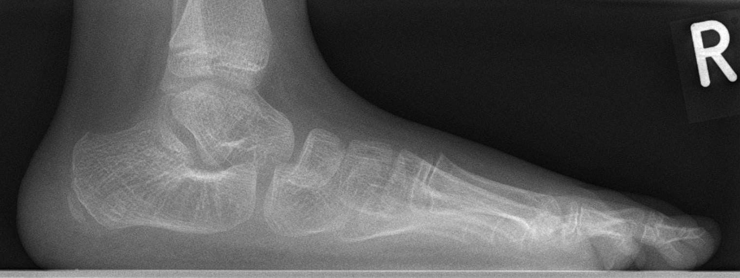
Pes planus (flatfoot). When the apex of the Meary angle points plantarward, the talus is plantarflexed relative to the first metatarsal, indicating loss of the medial longitudinal arch.
- Meary angle
- Apex plantar, over 4°
- Flexibility
- Corrects on tiptoe
- Key associated findings
- Tibialis posterior intact; heel raise restores the arch
- Meary angle
- Apex plantar, over 4°
- Flexibility
- Often rigid in late stage
- Key associated findings
- Too-many-toes sign; weak single heel raise
- Meary angle
- Apex plantar, over 4°
- Flexibility
- Rigid — does not correct
- Key associated findings
- CT shows the bar (calcaneonavicular/talocalcaneal); peroneal spasm
- Meary angle
- Apex plantar, over 4°
- Flexibility
- Rigid
- Key associated findings
- Midfoot arthritis; abduction through the talonavicular joint
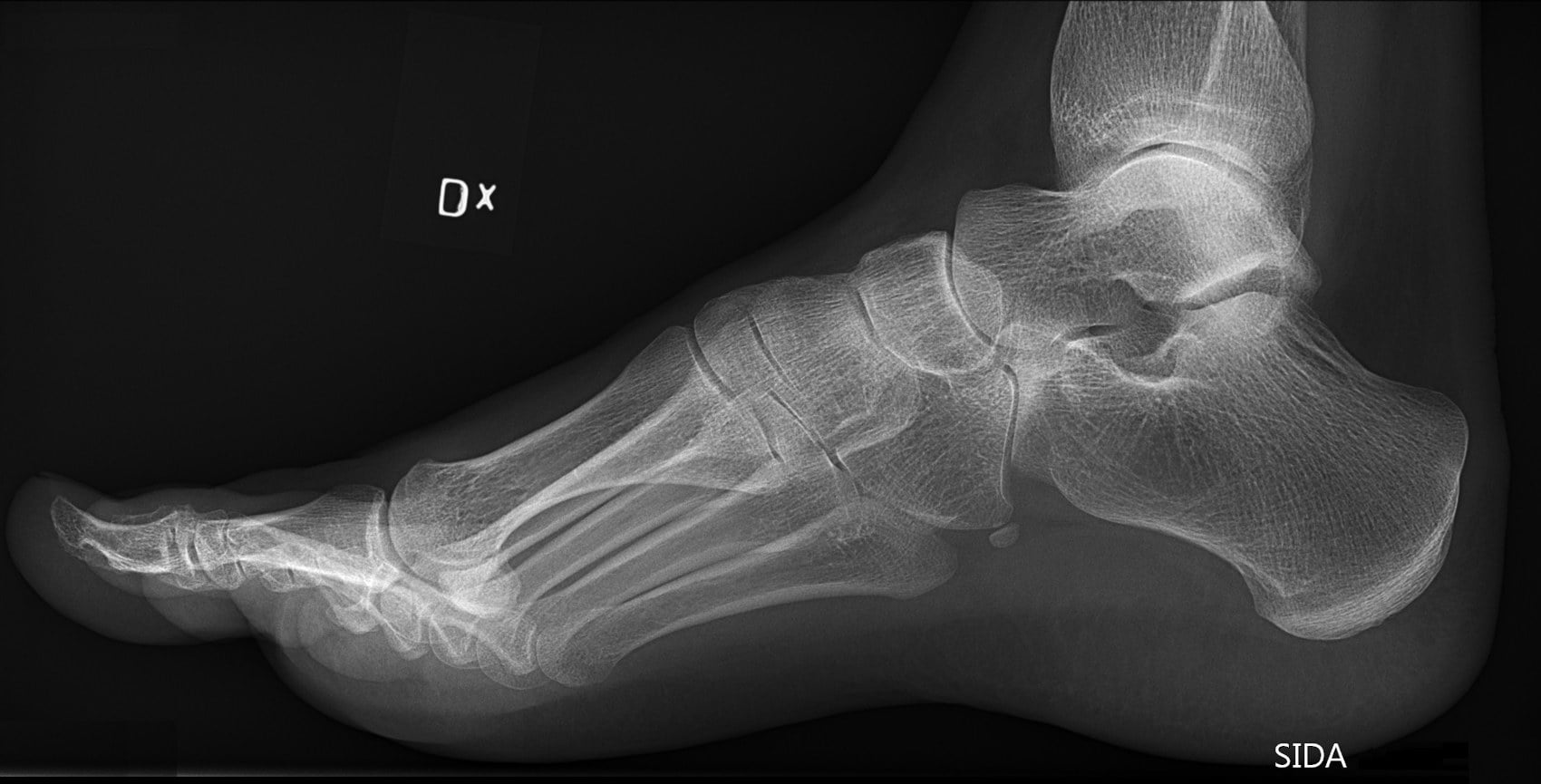
Pes cavus (cavovarus foot). When the apex points dorsalward, the talus is dorsiflexed relative to the first metatarsal — the arch is excessively high.
- Meary angle
- Apex dorsal, over 4°
- Key clinical features
- Progressive peroneal & tibialis anterior weakness, claw toes, inverted heel
- Investigation
- Nerve conduction studies, genetic testing
- Meary angle
- Apex dorsal, over 4°
- Key clinical features
- No neurological cause found; often bilateral
- Investigation
- MRI brain/spine to exclude tethered cord or syrinx
- Meary angle
- Apex dorsal, over 4°
- Key clinical features
- Prior calcaneal or talar fracture malunion
- Investigation
- CT for malunion assessment
Plantar = Planus, Dorsal = CavusApex direction = deformity
Hook:The apex is the arrow pointing at the problem — plantar points at a flat foot, dorsal points at a high arch.
A cavovarus foot with an abnormal Meary angle always warrants neurological investigation — the commonest cause is Charcot-Marie-Tooth disease. Do not label a cavus foot 'idiopathic' until you have excluded a neurological aetiology with nerve conduction studies and spinal imaging.
Relationship to other radiographic angles
The Meary angle is one of several sagittal-plane measures; understanding how they complement each other is essential for the exam.
- Landmarks
- Talar axis vs first MT axis (lateral)
- Normal value
- Approximately 0°
- What it measures
- Sagittal talus-first MT alignment
- Landmarks
- Calcaneal inferior cortex vs floor (lateral)
- Normal value
- 15–30°
- What it measures
- Calcaneal inclination (arch-height surrogate)
- Landmarks
- Calcaneus vs first MT inferior cortices
- Normal value
- Approximately 120–130°
- What it measures
- Overall medial longitudinal arch curve
- Landmarks
- Tibiocalcaneal axis on hindfoot alignment view
- Normal value
- under 5 mm (lateral)
- What it measures
- Coronal hindfoot alignment (not sagittal)
Meary (talus vs first MT) tells you the talus-to-first-metatarsal alignment; calcaneal pitch (calcaneus vs floor) tells you the calcaneal inclination/arch height; the Costa-Bartani angle describes the overall arch curve. All three are read off the one lateral weight-bearing film for a complete sagittal-plane profile. When shown a lateral foot radiograph, draw the Meary angle first (highest yield), then calcaneal pitch, and state the values and interpretation out loud.
Meary is a sagittal measure; the topic keeps citing "the AP talonavicular coverage angle" — here are the transverse-plane measures (read on the AP/dorsoplantar weight-bearing film):
- AP talonavicular coverage angle: the lateral uncoverage of the navicular on the talar head; increased (over about 7°) in planovalgus (forefoot abduction / peritalar subluxation) — the radiographic correlate of the "too-many-toes" sign.
- AP talo–first-metatarsal angle (the AP Meary): the talar axis versus the first-metatarsal axis on the AP view; normal near 0°, with the forefoot abducted (talus medial to the first MT) in planovalgus and adducted in cavovarus / metatarsus adductus.
- AP talocalcaneal (Kite) angle: the talar versus calcaneal axis on the AP view; normal ~15 to 30° (higher in young children); increased in planovalgus (the talar head adducts/plantarflexes, the bones diverge) and decreased/parallel in cavovarus and clubfoot (hindfoot varus).
- With the lateral (sagittal) angles these give the transverse + sagittal picture; add the hindfoot alignment view for the coronal plane.
Surgical decision-making using the Meary angle
The Meary angle directly informs operative planning for flatfoot and cavovarus reconstruction.
- Flexible flatfoot with a large plantar-apex Meary angle: medialising calcaneal osteotomy, flexor digitorum longus transfer, and spring ligament repair; add a lateral column lengthening if the AP talonavicular coverage angle is also abnormal.
- Rigid flatfoot (tarsal coalition): if the angle does not correct on forced dorsiflexion/tiptoe views, resection of a calcaneonavicular coalition may restore flexibility; a large or degenerate talocalcaneal coalition may need subtalar or triple arthrodesis.
- Stage III–IV tibialis posterior dysfunction with degenerative change: triple arthrodesis regardless of flexibility, with the angle quantifying the correction achieved.
- Dorsal-apex deformity, flexible: a dorsiflexion (closing-wedge) osteotomy of the first metatarsal corrects the forefoot-driven hindfoot varus; a lateralising/closing-wedge calcaneal osteotomy addresses the hindfoot.
- Rigid cavovarus with degenerative change: triple arthrodesis correcting the Meary angle to neutral, restoring a plantigrade foot.
- Charcot-Marie-Tooth: tendon transfers (e.g. tibialis posterior to the lateral cuneiform, peroneus longus to brevis, Jones procedure for the great toe) supplement the bony correction; the Meary angle quantifies the bony correction needed.
Common pitfalls in measurement
- Non-weight-bearing films invalidate the Meary angle — the foot must be loaded; supine films underestimate flatfoot and overestimate cavus.
- Malrotation of the lateral film distorts the talar axis; ensure a true lateral (the medial and lateral cortices of the talar dome should overlap).
- Drawing the talar axis through the dome rather than the neck-body midpoint introduces error.
- Not stating the apex direction — a numerical value without "apex-plantar" or "apex-dorsal" is clinically meaningless.
- Using the Meary angle in isolation — flatfoot and cavovarus are three-dimensional; always pair it with the AP talonavicular coverage angle, hindfoot alignment view and clinical examination.
The deformity is three-dimensional, and the coronal (hindfoot) plane completes the assessment this section refers to:
- Clinically: inspect the heel from behind — valgus (heel everted, "too-many-toes") in planovalgus, varus (heel inverted, the "peek-a-boo heel" sign) in cavovarus; test flexibility (the Coleman block for forefoot-driven hindfoot varus).
- Radiographically: the hindfoot alignment view (the Saltzman & el-Khoury long-axial / Cobey view) — a weight-bearing posteroanterior film angled about 20° that profiles the heel beneath the tibia.
- The hindfoot moment arm = the perpendicular distance from the tibial (long) axis to the lowest point of the calcaneus; lateral (valgus) is positive, medial (varus) negative, and normal is small (a few mm).
- It drives the osteotomy choice: a medialising calcaneal osteotomy for hindfoot valgus (planovalgus), a lateralising / lateral closing-wedge calcaneal osteotomy for hindfoot varus (cavovarus) — complementing the sagittal correction the Meary angle quantifies.
Guidelines, registries & global practice
- No single international guideline mandates the Meary angle as the sole criterion for surgery — it is one radiographic parameter used alongside clinical assessment and other angles (calcaneal pitch, talonavicular coverage, hindfoot moment arm).
- Global consensus recognises the Meary angle as a standard sagittal-plane measurement for pes planus and pes cavus; it features in foot-and-ankle textbook chapters and teaching curricula worldwide, with ~4° as the widely accepted abnormality threshold.
- AAOS (US), BOA/BOAST (UK), AO Foundation, EFORT and AOFAS educational materials include the lateral weight-bearing Meary angle in the standard set of foot radiographic parameters for flatfoot and cavovarus assessment and for pre-/post-operative comparison.
- Practice variation exists in whether surgeons routinely report the Meary angle versus relying on calcaneal pitch alone; fellowship-trained foot-and-ankle surgeons more commonly report multiple sagittal parameters.
- No joint registry tracks the Meary angle as an outcome measure (unlike alignment parameters in knee arthroplasty); it is used in research and surgeon databases for pre- and post-operative comparison.
Viva practice
Exam viva
Practise clinical reasoning and management decisions out loud
“A 52-year-old woman presents with progressive flattening of her right foot over 18 months, medial arch pain, and difficulty walking on uneven ground. A lateral weight-bearing radiograph is shown. Describe the radiographic findings and their significance.”
“A 28-year-old man with a known diagnosis of Charcot-Marie-Tooth disease type 1A presents with bilateral high-arched feet, recurrent ankle sprains, and difficulty with foot clearance during gait. His lateral weight-bearing foot radiograph is shown. Interpret the radiograph and outline your management approach.”
Exam cheat sheet
- Talar axis vs first-metatarsal axis on a STANDING lateral foot film; normal ≈ 0° (± 4°).
- Apex plantar = pes PLANUS; apex dorsal = pes CAVUS. State the apex direction every time.
- Draw the talar axis through the neck-body midpoint, not the dome; first-MT axis through shaft midpoints.
- Flexible vs rigid: does it correct on tiptoe / forced dorsiflexion? (coalition = rigid).
- Every cavovarus foot needs neurological work-up — Charcot-Marie-Tooth until proven otherwise; Coleman block test for flexible vs rigid hindfoot varus.
- Pair with calcaneal pitch and AP talonavicular coverage — never use Meary in isolation.
How to measure
- Lateral weight-bearing radiograph of the foot (must be standing)
- Talar axis: line through the midpoint of the talar head and talar neck-body junction
- First MT axis: line through the proximal and distal shaft midpoints
- Intersection angle is the Meary angle — state the apex direction
Normal and abnormal values
- Normal: approximately 0° (± 4°), axes collinear
- Apex plantar over 4°: pes planus (flatfoot)
- Apex dorsal over 4°: pes cavus (high arch)
- Always specify the apex direction — a bare number is meaningless
Clinical decision-making
- Flexible flatfoot: corrects on tiptoe — non-operative or tendon transfer + osteotomy
- Rigid flatfoot (coalition, degenerative): does not correct — resection or arthrodesis
- Cavovarus: always investigate neurologically (CMT most common cause)
- Coleman block test separates forefoot-driven from rigid hindfoot varus
Pitfalls to avoid
- Non-weight-bearing films invalidate the measurement
- Malrotated lateral film distorts the talar axis — ensure a true lateral
- Do not use the Meary angle alone — pair with calcaneal pitch, AP coverage angle, and clinical exam
- Cavovarus foot without neurological work-up is incomplete assessment
Evidence Base
Is Pes Cavus Alignment Associated With Lisfranc Injuries of the Foot?
- The lateral talo-first metatarsal (Meary) angle was measured with high reliability (intrarater ICC 0.93–0.94, interrater ICC 0.91).
- Patients with low-energy Lisfranc injuries had a significantly higher (more cavus) talo-first metatarsal angle than controls (mean difference ~5.7°, p = 0.001).
- Cavus midfoot alignment was more prevalent among Lisfranc-injury patients (association, not causation).
Surgical correction of cavus foot may promote quality of life in patients with Charcot-Marie-Tooth disease: A retrospective study
- Radiological evaluation of CMT cavus correction used the Meary angle alongside calcaneal pitch, talocalcaneal and talo-first metatarsal angles (45 patients, 57 feet).
- At 2-year follow-up most radiographic and functional outcomes improved significantly (median FADI 23→40, SF-12 physical component 26→41, p<0.001).
- Confirms the Meary angle as a standard radiographic parameter for quantifying and tracking cavus deformity and its correction.
According to PubMed, the reliability of the Meary (talo-first metatarsal) angle and its association with cavus alignment come from Podolnick et al. 2016 (DOI), and its use to quantify and track cavus correction from Chen et al. 2025 (DOI). The measurement technique, the ~4° abnormality threshold, and the apex-direction convention (plantar = planus, dorsal = cavus) are standard foot-and-ankle radiographic teaching.