3-5% of ankle sprains | Deltoid 2-3× stronger than ATFL | Commonly with syndesmotic injury | MCS >4 mm diagnostic
- Deep posterior tibiotalar is the strongest band - and the one most often SPARED in supination-external-rotation injury
- Isolated deltoid injury is rare; look for the syndesmosis, the fibula and the posterior tibial tendon
- A torn superficial deltoid does not by itself mean the mortise is unstable - test it
- Deltoid MRI abnormality is present in 72% of chronic LATERAL instability patients who have no medial pain at all
- Success percentages quoted for this condition are conventional; no study cited here measures them
- “Medial clear space normal <4mm on mortise view
- “External rotation stress test for deep deltoid
- “Repair deep layer first in surgery
- “Position ankle neutral dorsiflexion + 5° inversion
Medial Ankle Instability
High-yield exam focus: Understanding deltoid anatomy (superficial vs deep components), recognizing associated injuries (syndesmosis, PTTD, fractures), differentiating medial gutter pain from true instability, identifying when surgical intervention is required, and managing concomitant pathology. Examiners expect discussion of why isolated deltoid injury is rare and what forces/injuries cause deltoid disruption.
Viva Overview Summary
Key Points for Viva
- 3-5% of ankle sprains (much less common than lateral)
- Deltoid is 2-3x stronger than ATFL
- Isolated injury is rare
- Syndesmotic injury (40-60% of deltoid injuries)
- Pronation-external rotation ankle fractures
- Stage 4 PTTD with flatfoot
- Key Point
- 3-5% of ankle sprains
- Key Point
- Deltoid + syndesmosis (40-60%)
- Key Point
- 60-70%
Anatomy and Biomechanics
This page and Medial Ankle Sprains both cover deltoid ligament injury, and they are complementary rather than alternatives. Read this one for chronic deltoid insufficiency: peritalar and rotational instability, deltoid-spring reconstruction, suture-tape augmentation and the flatfoot continuum. Read the companion for the acute problem: sprain grading, the diagnostic-test comparison (ultrasound versus gravity versus manual stress radiography) and repair during fracture fixation.
The deltoid ligament is the primary medial stabiliser of the ankle. It has a superficial and a deep layer, which differ in anatomy and in what they restrain, and it works alongside the spring ligament.
The superficial layer. A fan-shaped sheet with a broad insertion, it arises from the anterior colliculus of the medial malleolus and runs as the tibionavicular, tibiocalcaneal and tibiospring ligaments to the navicular tuberosity, the sustentaculum tali and the spring ligament. Its primary function is restraint of hindfoot valgus; it is the primary restraint to eversion stress and a secondary restraint to external rotation. Its blood supply comes from branches of the medial tarsal artery; it is the layer more commonly injured in isolation, and it has the better healing potential.
The deep layer. Thick, cord-like anterior and posterior tibiotalar bands arise from the intercollicular groove and posterior colliculus and insert on the medial talus at its tubercle. This layer is the critical restraint to external rotation and lateral talar shift, and the deep posterior tibiotalar ligament is its strongest, most consistent band and the most critical for stability. A deep-layer injury indicates a severe force and is often associated with syndesmotic disruption.
The spring ligament. Its superomedial, medioplantar oblique and inferoplantar longitudinal bands support the talar head and maintain the medial longitudinal arch. It is intimately associated with the superficial deltoid through the tibiospring fibres, and the two act together as the tibiocalcaneonavicular complex. Spring ligament injury often accompanies deltoid insufficiency; the combined injury leads to valgus hindfoot and flatfoot deformity, and in chronic cases both may require reconstruction.
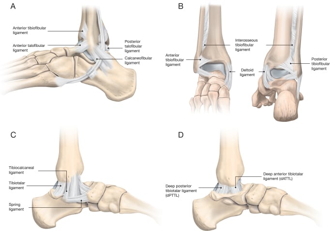
What the complex restrains. The deltoid is the principal restraint to lateral talar translation and external rotation of the talus within the mortise, and a major restraint to hindfoot valgus, resisting 40% of valgus stress. Both layers contribute to resisting valgus tilt of the talus in the mortise.
Position matters. Band engagement is position-dependent: the tibiospring band tightens in plantarflexion, whereas the tibiocalcaneal and deep posterior tibiotalar bands tighten in dorsiflexion.
The deltoid and the syndesmosis. Deltoid competence is essential for syndesmotic stability. An incompetent deltoid permits medial clear space widening and lateral talar shift even with an intact syndesmosis, which is why restoring medial stability can normalise the mortise.
Deltoid Anatomy Summary
Two-Layer Structure
- Tibionavicular, tibiocalcaneal, tibiospring
- Origin: Anterior colliculus
- Function: Restraint to hindfoot valgus
- Anterior and posterior tibiotalar
- Origin: Intercollicular groove
- Function: Restraint to external rotation and lateral talar shift
- Components
- Tibionavicular, tibiocalcaneal, tibiospring
- Function
- Hindfoot valgus restraint
- Components
- Anterior/posterior tibiotalar
- Function
- External rotation, lateral shift
Pathophysiology and Injury Mechanisms
Why isolated injury is rare. The deltoid is 2-3 times stronger than the ATFL, and because of that strength medial ankle instability accounts for only 3-5% of ankle sprains. Most deltoid injuries occur as part of a more complex pattern: with a syndesmotic injury (conventionally quoted at 40-60%), a pronation-external rotation fracture, or stage 4 PTTD.
- mechanism
- Direct valgus force with external rotation (rare)
- associatedInjuries
- Usually none, occasionally spring ligament injury
- incidence
- Less than 5% of deltoid injuries
- treatment
- Conservative management, immobilisation 4-6 weeks
- prognosis
- Good with conservative care (conventionally quoted success 60-70%)
- mechanism
- Pronation-external rotation force
- associatedInjuries
- AITFL disruption (40-60%), IOL injury, fibular fracture
- incidence
- Most common pattern (40-60% of deltoid injuries)
- treatment
- Syndesmotic fixation priority, deltoid may require repair
- prognosis
- Dependent on syndesmosis treatment, deltoid usually heals
- mechanism
- Valgus force with abduction, oblique medial malleolus fracture
- associatedInjuries
- Medial malleolus fracture, lateral ligament injury or fibula fracture
- incidence
- 20-30% of deltoid pathology presentations
- treatment
- ORIF medial malleolus, deltoid repair if tissue quality poor
- prognosis
- Good with anatomic fracture reduction
- mechanism
- Chronic progressive valgus deformity and arch collapse
- associatedInjuries
- PTT rupture, spring ligament attenuation, subtalar arthritis
- incidence
- 10-15% of deltoid presentations
- treatment
- Reconstruction vs fusion based on arthritis presence
- prognosis
- Complex, depends on deformity correction and arthritic changes
Acute injury. Acute deltoid injuries typically result from high-energy trauma with forced valgus and external rotation. The presentation includes significant medial swelling, tenderness over the course of the deltoid, ecchymosis extending to the hindfoot (a clue to a significant medial-side injury) and a positive external rotation stress test; weight-bearing is often impossible because of pain and instability. MRI shows ligament discontinuity with surrounding oedema, and treatment starts by identifying and addressing the associated injuries, particularly syndesmotic disruption.
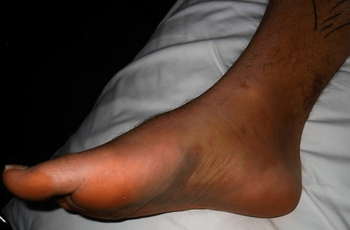
Chronic insufficiency. Chronic deltoid insufficiency develops from an untreated acute injury, from repetitive microtrauma in athletes (gymnastics, soccer), or from progressive deformity in PTTD. It presents with medial ankle pain, subjective instability and valgus hindfoot alignment. MRI shows ligament thickening, scarring or complete attenuation, weight-bearing radiographs may show a widened medial clear space, and treatment has to address the underlying biomechanical abnormality.
Clinical Evaluation
History. Establish the mechanism (valgus force, external rotation), previous ankle injury or instability, and the functional limitation. Pain with cutting or pivoting suggests rotational instability, and a history of flatfoot or PTTD matters in chronic cases.
Inspection. Medial swelling in the acute injury; valgus hindfoot deformity in the chronic one. Assess alignment (hindfoot valgus, arch height, forefoot abduction) and look for pes planus, which suggests concurrent PTTD or spring ligament insufficiency, for an antalgic gait, and for an inability to perform a single heel raise.
Palpation. Work systematically along the medial malleolus, the course of the deltoid (from the anterior colliculus inferiorly), the spring ligament (plantar-medial to the navicular) and the posterior tibial tendon. Where it is tender localises the injury and identifies the associated pathology that also needs treating.
Special tests.
- External rotation stress test - knee flexed 90 degrees, ankle in neutral, external rotation force applied to the foot. Pain and increased rotation compared with the contralateral side indicate deltoid injury.
- Valgus stress test - valgus force to the hindfoot with the ankle in neutral. Increased medial joint opening suggests deltoid insufficiency.
- Kleiger test - external rotation of the foot with the ankle dorsiflexed and the tibia stabilised. Pain at the medial ankle indicates deltoid injury; pain at the syndesmosis suggests combined pathology.
- Single heel raise and resisted inversion - for posterior tibial tendon function. Inability to rise, or no hindfoot inversion during the raise, indicates PTT dysfunction or severe deltoid insufficiency.
Injury Classification
Classification by Pattern
- Isolated deltoid (less than 5%): Direct valgus force
- With syndesmosis (40-60%): Pronation-external rotation
- With fracture (20-30%): Pronation-abduction
- Stage 4 PTTD (10-15%): Chronic progressive
- Grade 1: Intact fibers with edema
- Grade 2: Partial tear
- Grade 3: Complete disruption
- Incidence
- Less than 5%
- Prognosis
- Good (60-70% conservative)
- Incidence
- 40-60%
- Prognosis
- Depends on syndesmosis treatment
- Incidence
- 10-15%
- Prognosis
- Complex, multi-procedure needed
Clinical Examination Summary
Key Clinical Tests
- Knee flexed 90°, ankle neutral
- Externally rotate foot
- Pain and increased rotation = positive
- Apply valgus force to hindfoot
- Compare to contralateral side
- Increased opening = deltoid insufficiency
- Tests PTT function
- Inability or lack of inversion = dysfunction
- What It Tests
- Deep deltoid
- Positive Finding
- Pain, increased rotation
- What It Tests
- Both layers
- Positive Finding
- Increased medial opening
- What It Tests
- PTT function
- Positive Finding
- Unable or no inversion
Investigations
Radiographs. Weight-bearing AP, lateral and mortise views of the ankle, with AP and lateral views of the foot. They show any fracture of the medial malleolus, fibula or posterior malleolus; they give the key measurements (medial clear space, talonavicular coverage angle, lateral talar station); and in chronic cases they show valgus tilt, talar subluxation and arthritic change.
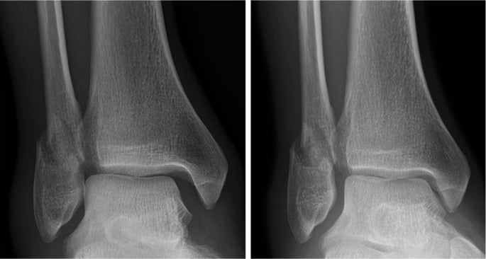
The medial clear space. Measured on the mortise view, it is normally less than 4mm and should equal the superior clear space; the deep posterior tibiotalar ligament maintains it by preventing lateral translation of the talus. Widening beyond 4mm, or asymmetry compared with the contralateral ankle, indicates deltoid insufficiency or syndesmotic injury, and weight-bearing films are essential for accurate assessment.
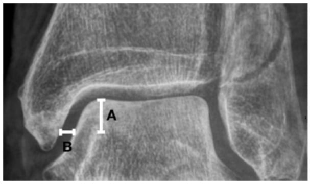
Why stress views. Static non-weight-bearing films can look normal because the talus reduces at rest. A stress or weight-bearing view loads the medial side to unmask a widened medial clear space or talar tilt, and the diagnosis requires stress radiographs showing medial clear space widening (criteria below) or MRI demonstrating ligament discontinuity.
- Manual external-rotation stress view - a mortise radiograph taken while an external-rotation force is applied to the plantigrade foot with the tibia held.
- Gravity stress view - the patient lies with the injured leg dependent (lateral decubitus, ankle unsupported) and gravity supplies the external-rotation/eversion load, giving a mortise view without the examiner's hand in the field. It is about as reliable as the manual test for detecting deltoid incompetence in SER fractures and avoids irradiating the examiner.
- Weight-bearing views - for chronic instability, standing AP/mortise views (and weight-bearing CT) reproduce physiological load and reveal valgus talar tilt and peritalar subluxation better than a supine film.
Reading a stress view. Any one of these indicates deltoid incompetence:
- Medial clear space over 4 mm
- Side-to-side asymmetry over 1 mm against the contralateral ankle
- Medial clear space exceeding the superior clear space
It is the same test that decides whether a supination-external-rotation (SER) fibular fracture is "stable" (deltoid intact) or "unstable" (deltoid torn, needs fixation). The 4 mm cut-off and the side-to-side comparison are pragmatic rather than rigorously validated, and stress technique varies between centres, so interpret the number alongside the clinical stress examination rather than in isolation.
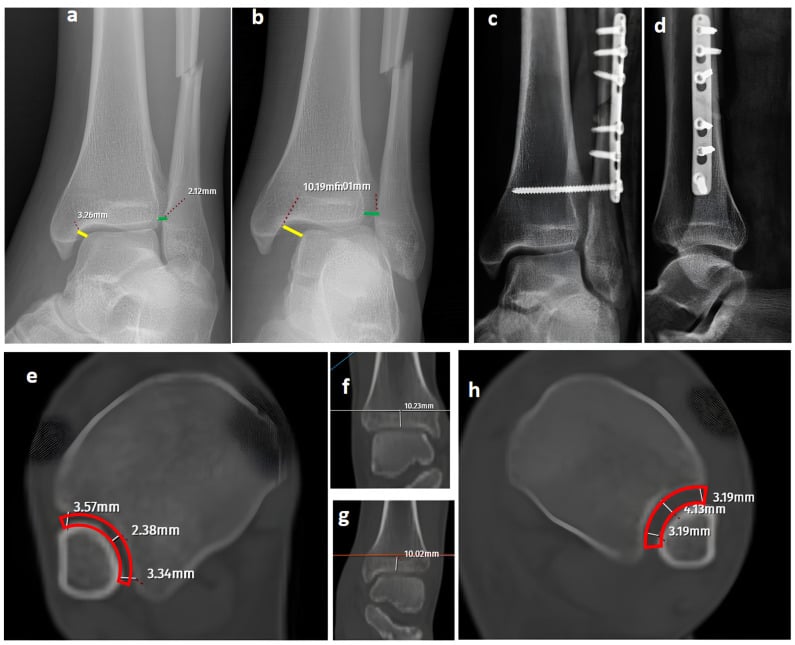
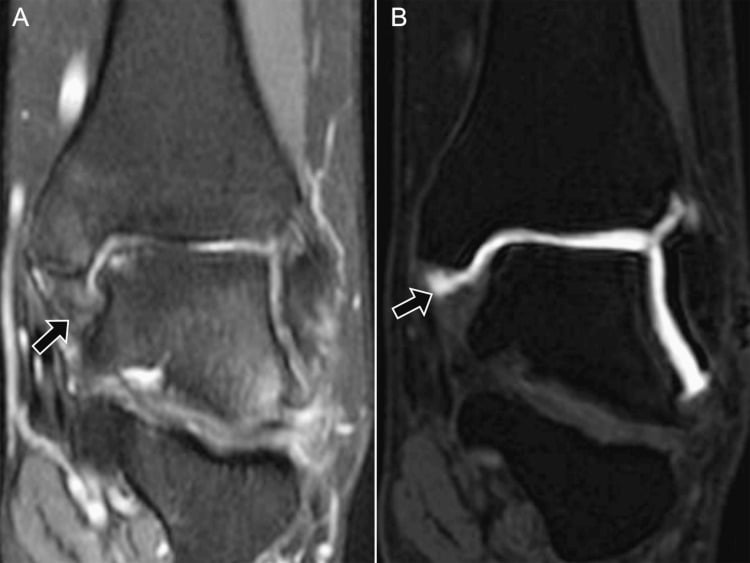
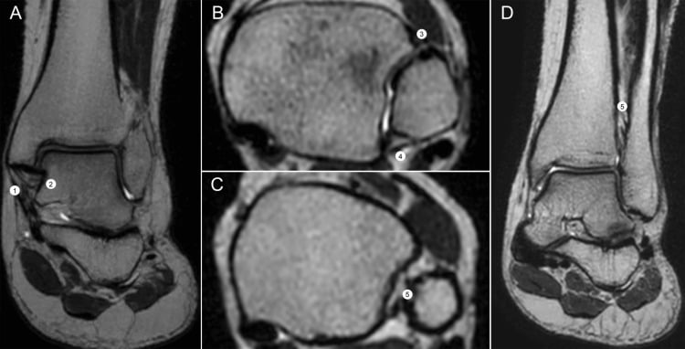
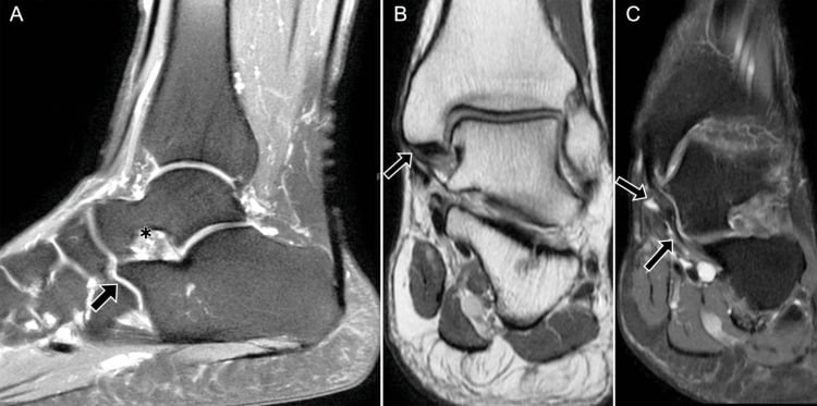
MRI. Indicated for suspected deltoid injury, chronic instability and preoperative planning, using T1, T2 and STIR sequences in coronal, sagittal and axial planes. It shows the associated pathology (syndesmosis, spring ligament, posterior tibial tendon, osteochondral lesions, bone bruising), and in chronic cases ligament thickening, scarring or attenuation without a discrete tear. The deltoid is graded:
- Grade 1 - intact fibres with oedema
- Grade 2 - partial tear
- Grade 3 - complete disruption
CT. For fracture characterisation, preoperative planning and assessment of arthritis. It gives superior bony detail and shows the subtalar joint, and 3D reconstruction helps with complex fracture patterns, at the cost of poor soft-tissue resolution and radiation exposure. Weight-bearing CT is an emerging technology for dynamic assessment of hindfoot alignment.
Viva Imaging Review
Key Imaging Findings
- Medial clear space: Normal less than 4mm
- Greater than 4mm = deltoid insufficiency
- Must compare to contralateral
- Grade 1: Intact fibers with edema
- Grade 2: Partial tear (some fibers intact)
- Grade 3: Complete disruption
- Syndesmosis injury
- Spring ligament attenuation
- Osteochondral lesions
- Key Finding
- Medial clear space greater than 4mm
- Significance
- Deltoid/syndesmosis injury
- Key Finding
- Grade 3 disruption
- Significance
- Complete tear, may need surgery
- Key Finding
- Widening with stress
- Significance
- Confirms instability
Differential Diagnosis
Medial ankle pain with a sense of "giving way" has several mimics. The key task is separating true medial instability (deltoid/spring incompetence with talar tilt or shift) from medial gutter pathology and tendon disorders that cause pain without instability.
- keyFeatures
- Giving way on valgus or external rotation; medial clear space widening on stress or weight-bearing
- examination
- Positive external rotation and valgus stress tests; pain over deltoid course
- imaging
- Medial clear space over 4mm; deltoid discontinuity or attenuation on MRI
- discriminator
- Demonstrable talar tilt or shift is the defining feature
- keyFeatures
- Progressive flatfoot, medial arch pain, fatigue with walking
- examination
- Too-many-toes sign; unable to perform single-heel-raise or no hindfoot inversion
- imaging
- PTT tenosynovitis, split or rupture on MRI; hindfoot valgus on weight-bearing CT
- discriminator
- Tendon, not ligament, is the primary lesion; deltoid stress may be secondary
- keyFeatures
- Activity-related medial pain in runners; no instability
- examination
- Focal bony tenderness at medial malleolus; negative ligament stress tests
- imaging
- Vertical lucency or marrow oedema on MRI; line on radiograph
- discriminator
- Bony tenderness without talar tilt; stress tests negative
- keyFeatures
- Burning, tingling on plantar foot; night symptoms
- examination
- Positive Tinel over tarsal tunnel; sensory disturbance, no mechanical instability
- imaging
- Mass or varix on MRI; nerve conduction abnormalities
- discriminator
- Neuropathic, not mechanical; no ligament laxity
- keyFeatures
- Pain at end-range dorsiflexion, no giving way
- examination
- Tenderness anteromedial joint line; impingement on forced dorsiflexion
- imaging
- Anteromedial osteophytes or soft-tissue thickening; stable mortise
- discriminator
- Positional pain without instability; mortise reduced
- keyFeatures
- Talar head uncoverage, peritalar instability, arch collapse
- examination
- Plantar-medial tenderness to navicular; talar head prominence
- imaging
- Superomedial spring ligament tear on MRI; talonavicular uncoverage
- discriminator
- Often coexists with deltoid injury (TCNL complex) rather than isolated
Conservative Management
Who, and how often it works. Non-operative management is first-line for isolated deltoid sprains without fracture or syndesmotic injury. The conventionally quoted success rate is 60-70%, leaving 30-40% for surgical consideration, but no study cited on this page measures it - the comparative series here all enrol operatively treated fractures.
Weeks 0-3: protection and immobilisation. A CAM boot or short leg cast, non-weight-bearing for grade 2-3 injuries; grade 1 injuries start protected weight-bearing after the first week. Ice, elevation and compression control oedema, with NSAIDs for pain if there are no contraindications.
Weeks 3-6: progressive weight-bearing. Weight-bearing as tolerated in the CAM boot. Gentle ankle range of motion begins, avoiding forced eversion, with isometric strengthening of tibialis posterior and gastrocnemius, continued until daily activities are pain-free.
Weeks 6-12: strengthening and return to activity. The boot gives way to a lace-up ankle brace or taping. Progressive resistance exercises focus on tibialis posterior and the invertors, with proprioceptive training (single leg balance, wobble board) and graded sport-specific work; return to high-level athletics may take 3-4 months.
Critical assessment for syndesmotic injury is required in all deltoid injuries. Failure to identify and treat concurrent syndesmosis disruption leads to chronic pain, instability and post-traumatic arthritis. Perform the syndesmotic squeeze test and the external rotation stress test, and consider MRI if clinical suspicion persists despite negative radiographs.
When to operate. The absolute indications are:
- Acute deltoid disruption with syndesmotic instability requiring fixation
- Deltoid disruption with a medial clear space still widened (greater than 4mm) despite syndesmotic fixation
- Chronic symptomatic instability with functional limitation despite 4-6 months of therapy
- Stage 4 PTTD with deltoid insufficiency and progressive deformity
The relative indications are:
- High-demand athletes with persistent symptoms after 3 months of conservative care
- Deltoid injury with a medial malleolus fracture whose comminution prevents stable fixation
- MRI-confirmed complete deltoid rupture with poor tissue quality in the acute setting
Viva Treatment Review
Management Approach
- CAM boot/cast 4-6 weeks
- Progressive weight-bearing
- Strengthening and proprioception
- Success rate: 60-70%
- Deltoid + syndesmosis requiring fixation
- Widened MCS despite syndesmosis reduction
- Chronic instability failed 4-6 months therapy
- Stage 4 PTTD
- Treatment
- Conservative
- Expected Outcome
- 70-80% success
- Treatment
- Fix syndesmosis, assess MCS
- Expected Outcome
- MCS may normalise
- Treatment
- Reconstruction
- Expected Outcome
- 75-85% success
Surgical Management
Direct Deltoid Ligament Repair
Primary repair is performed when tissue quality is good and the deltoid injury is part of operative ankle fracture or syndesmotic fixation.
Set-up and incision. Supine, with a bump under the ipsilateral hip, a thigh tourniquet, and the foot over the end of the table or on a folded towel for access to the medial ankle. A curvilinear incision of 6-7cm is centred over the medial malleolus, running from just proximal to its tip distally along the deltoid; an existing medial malleolar fracture incision can be used instead.
Approach. Full-thickness skin flaps expose the medial malleolus, the deltoid and the tibialis posterior tendon. The saphenous vein and nerve lie anterior to the incision and are identified and protected; the nerve is at risk if the incision is extended anteriorly.
Assessing the deltoid. Before repairing anything:
- Identify the level of disruption, most commonly at the tibial origin or midsubstance
- Differentiate the superficial from the deep components
- Judge whether the tissue will hold a primary repair or needs augmentation
- Inspect for associated injuries: spring ligament, posterior tibial tendon, medial talar osteochondral lesion
Repair. Debride devitalised tissue conservatively, then:
- Place suture anchors (3.0-3.5mm) at the anatomic deltoid origin on the medial malleolus: the anterior colliculus for the superficial layer, the intercollicular groove for the deep components
- Use high-strength non-absorbable sutures (2-0 or 0)
- Repair the deep layer first, as it is critical for rotational stability
- Repair the superficial layer separately, with slight overlap
- Tie down with the ankle in neutral dorsiflexion and 5 degrees of hindfoot inversion
- Consider suture augmentation with an internal brace in high-demand athletes
Why that position. Neutral dorsiflexion with 5 degrees of inversion, not eversion or valgus, prevents overtightening that would cause stiffness while ensuring adequate tension for stability. The contralateral ankle is the guide to the right position.
The spring ligament. If it is injured (a palpable defect plantar-medial to the navicular, talar head uncoverage on the radiograph), address it at the same sitting with suture anchor repair or reconstruction.
Closure. The deep layer with absorbable suture and the skin with non-absorbable suture or staples, then a well-padded posterior splint with the ankle in neutral and the hindfoot in slight inversion.
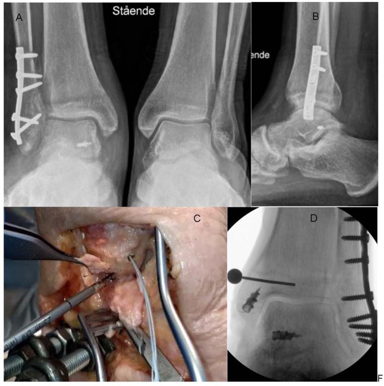
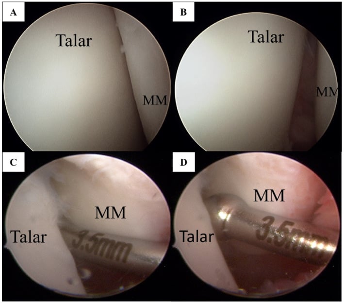
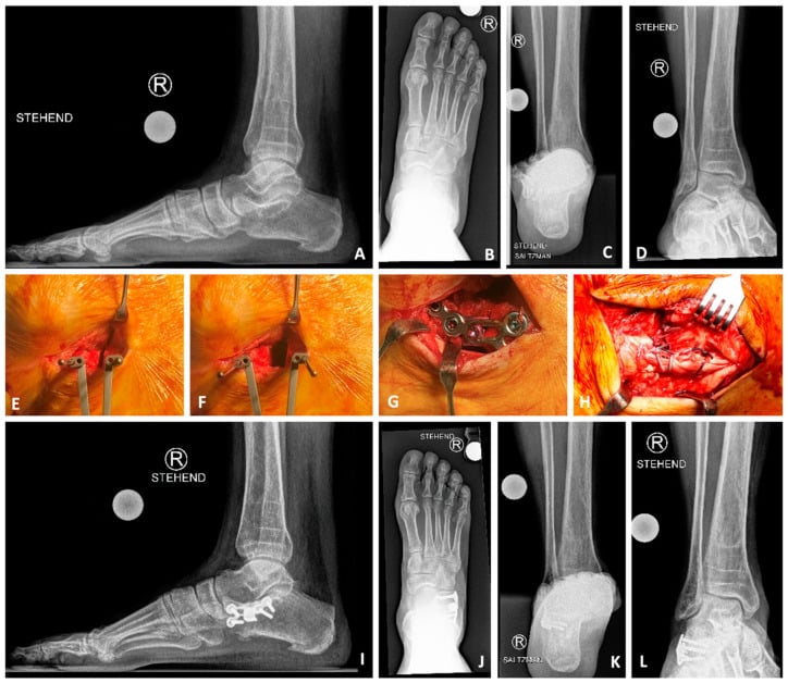
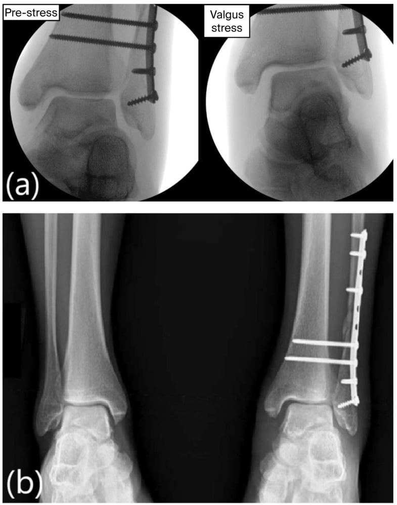
Suture-tape (internal brace) augmentation. A strong, non-absorbable tape such as FiberTape, secured with anchors, spans the repaired or reconstructed deltoid as an independent load-sharing "check-rein", typically from a medial malleolar anchor to a talar (and/or calcaneal or sustentacular) anchor, recreating the deep and superficial vectors. It is not the primary repair. It protects the biological repair or graft while it heals, restrains external rotation and valgus, and is intended to permit earlier, safer rehabilitation, the same rationale as internal bracing of the lateral ligament.
When it is offered. In poor-quality tissue (chronic, diabetic, revision), in high-demand athletes, and to reinforce a repair that would otherwise be marginal. The evidence is largely biomechanical and short-term small series. The tape supplements rather than replaces anatomic repair, over-tensioning it risks stiffness or over-constraint, and so routine use is not established.
Surgical Technique Essentials
Operative Approach
- Medial incision over malleolus
- Protect saphenous nerve (anterior)
- Suture anchors at anatomic origins
- Repair deep layer first
- Autograft (hamstring) preferred
- Bone tunnels in malleolus
- Anatomic recreation of superficial and deep
- Neutral dorsiflexion
- 5° hindfoot inversion (NOT valgus)
- Indication
- Acute, good tissue
- Technique
- Suture anchors to malleolus
- Indication
- Chronic, poor tissue
- Technique
- Autograft through tunnels
Complications
Early Complications
Wound healing problems occur in 5-8%, because the soft-tissue envelope over the medial malleolus is thin. Diabetes, smoking, peripheral vascular disease and steroid use add risk. Prevention rests on careful handling of the skin flaps, a closure without excessive tension and appropriate patient selection. Management ranges from local wound care to debridement and delayed closure for deeper dehiscence.
Saphenous nerve injury presents as numbness or dysaesthesia along the medial ankle and foot, in 10-15%. It is usually iatrogenic, from dissection or retractor placement, and most are neurapraxias that resolve within 3-6 months. Permanent injury occurs in less than 2% but can be debilitating; prevention requires identifying and protecting the nerve during the approach.
Posterior tibial neurovascular injury is rare (less than 1%) but catastrophic. The bundle is at risk during deep dissection for reconstruction or tunnel drilling, and injury presents with plantar numbness, weak toe flexion or vascular compromise. Prevention is through careful dissection and knowledge of the anatomy.
Late Complications
Stiffness and loss of motion affect 15-20% after deltoid reconstruction, more than after lateral ligament procedures. The loss is typically of dorsiflexion (on average 8-12 degrees) and subtalar motion, caused by the length of immobilisation, adhesions or overtightening at repair. Prevention includes early motion after 2 weeks of protection and avoiding excessive tension during reconstruction; treatment is aggressive physiotherapy and occasionally manipulation under anaesthesia.
Persistent instability follows reconstruction in 10-15%. The causes are technical failure (inadequate graft tension, tunnel malposition), unaddressed hindfoot valgus, progression of PTTD, or syndesmotic insufficiency, and it requires thorough re-evaluation with imaging to identify the failure mechanism. Management may require revision reconstruction, calcaneal osteotomy or salvage fusion.
Post-traumatic arthritis develops in 20-30% of patients whose deltoid injury accompanied an ankle fracture or syndesmotic disruption. Initial fracture displacement, the quality of reduction, articular cartilage damage and age over 50 years are the risk factors, and it presents with progressive pain and stiffness over 2-5 years. Treatment ranges from bracing, activity modification and injections to ankle arthroplasty or arthrodesis in severe cases.
Complications Overview
Complication Summary
- Wound healing (5-8%)
- Saphenous nerve injury (10-15%, mostly neurapraxia)
- PTN injury (less than 1%)
- Stiffness (15-20%)
- Persistent instability (10-15%)
- Post-traumatic arthritis (20-30% with fractures)
- Incidence
- 10-15%
- Outcome
- Most resolve 3-6 months
- Incidence
- 15-20%
- Outcome
- May need PT or MUA
- Incidence
- 10-15%
- Outcome
- May need revision
Postoperative Management
Recovery takes longer than after lateral ligament reconstruction because the deltoid carries weight-bearing stability.
Weeks 0-2: protection. A posterior splint or CAM boot with strict non-weight-bearing. Toe wiggling and ankle pumps in plantarflexion and dorsiflexion only, with no eversion and no forced dorsiflexion. Sutures come out at 10-14 days, watching for infection, and pain is managed with multimodal analgesia, ice and elevation.
Weeks 2-6: early motion. Weight-bearing progresses from none to 50% by week 6, in a CAM boot with gradually more time out of it for exercises. Active ankle pumps and gentle inversion-eversion (no forced eversion) begin, with isometric tibialis posterior and gastrocnemius contractions; physiotherapy starts at week 3-4 for oedema control and supervised range of motion.
Weeks 6-12: strengthening. Full weight-bearing in the boot, then a lace-up or Arizona brace. Progressive theraband resistance focused on tibialis posterior, single leg balance and wobble board work, and pool walking, stationary bike and elliptical. Sedentary workers return at 8-10 weeks and manual labourers at 12-14 weeks.
Months 3-6: return to sport. The criteria are full strength, normal range of motion, negative stress tests and passed functional testing, and the progression runs from a walk-jog programme through straight-line running and cutting drills to sport-specific training. An external ankle brace is recommended for sport for 12 months, and a permanent brace may be needed for high-level athletics; 80-90% return to their pre-injury activity level.
Rehabilitation Summary
Post-Op Protocol
Phase 1 (0-2 weeks): NWB, splint, wound care Phase 2 (2-6 weeks): Progress to 50% WB, gentle ROM Phase 3 (6-12 weeks): Full WB, strengthening, proprioception Phase 4 (3-6 months): Return to sport with brace
- Weight-Bearing
- Non-weight-bearing
- Activity
- Splint protection
- Weight-Bearing
- Progress to 50%
- Activity
- Gentle ROM in boot
- Weight-Bearing
- Full weight-bearing
- Activity
- Strengthening, proprioception
- Weight-Bearing
- Sport-specific
- Activity
- Return to play with brace
Outcomes Summary
Expected Results
- 60-70% success for isolated injuries
- 14-week average return to sport
- Higher failure with Grade 3 tears
- Primary repair: 85-90% success
- Reconstruction: 75-85% success
- Combined with PTTD: 70-80% success
- Success Rate
- 60-70%
- Notes
- Isolated injuries only
- Success Rate
- 85-90%
- Notes
- Acute, good tissue
- Success Rate
- 75-85%
- Notes
- Chronic insufficiency
Guidelines, Registries & Global Practice
Global Epidemiology
- Medial-side (deltoid) injury accounts for roughly 3-5% of ankle sprains; isolated deltoid rupture is rare (under 5% of deltoid injuries)
- Deltoid injury commonly accompanies pronation-external rotation and bimalleolar-equivalent (Weber B/C) fractures, with occult deltoid injury found in up to 72% of chronic lateral-instability surgical cohorts
- Adult acquired flatfoot (PTTD) prevalence is approximately 3-10% in adults over 40, the main driver of chronic medial peritalar instability
Side-by-Side Guidance
- Emphasis
- Restore lateral column first; intraoperative stress test of mortise/syndesmosis
- Position on Deltoid Repair
- Selective repair if medial clear space stays widened
- Emphasis
- Stability-driven fixation; recognise occult medial injury
- Position on Deltoid Repair
- Not routine; repair for persistent medial widening
- Emphasis
- Anatomic mortise reduction, early stability assessment
- Position on Deltoid Repair
- Deltoid healing via reduction; repair selectively
- Emphasis
- Increasing interest in direct repair to cut hardware burden
- Position on Deltoid Repair
- Repair a valid alternative to syndesmotic screws
Controversies and Areas of Uncertainty
Medial ankle instability is an area of low-level evidence (mostly cadaveric studies and Level III-IV series). Examiners reward a candidate who can state what is genuinely debated rather than overclaiming.
- Routine vs selective deltoid repair in ankle fractures. Whether to repair the deltoid during fracture fixation remains contested. Comparative series (Woo et al; Whitlock et al) show repair restores a smaller medial clear space and reduces reoperation, but most authorities still favour a stability-driven, selective approach, restore the lateral column and syndesmosis, then repair only if the medial clear space stays widened. No adequately powered randomised trial exists.
- Deltoid repair vs trans-syndesmotic fixation. Some evidence suggests direct deltoid repair gives equivalent function with fewer hardware-removal reoperations than syndesmotic screws, but selection bias limits conclusions and suture-button fixation has changed the comparison.
- Significance of an isolated superficial deltoid tear. Cadaveric band-specific work (Gregersen et al) indicates the deep posterior tibiotalar band is often spared in SER injuries, so a superficial tear does not necessarily mean global deltoid incompetence. Dynamic stress assessment, not static MRI signal, should drive treatment.
- Reconstruction technique and graft choice. No consensus exists on autograft vs allograft, single- vs double-bundle reconstruction, or the role of suture-tape (internal brace) augmentation; recommendations are extrapolated from anatomic and small clinical series.
- Role of weight-bearing CT. Weight-bearing CT is increasingly used to quantify hindfoot valgus and subtle peritalar subluxation, but thresholds defining pathological medial instability are not yet standardised.
- Optimal medial clear space threshold. The widely quoted 4mm cut-off and side-to-side comparison are pragmatic rather than rigorously validated, and gravity/external-rotation stress views vary in technique between centres.
- The two most-repeated numbers on this page are conventional, not cited. "Conservative management succeeds in 60-70%" and "syndesmotic injury accompanies 40-60% of deltoid injuries" appear a dozen times each and are not traceable to any study cited here. None of the seven indexed papers on this page measures either quantity: the comparative series enrol operatively treated fractures, and the prevalence series (Crim) reports MRI findings in surgical candidates for lateral instability. Both figures are reasonable teaching approximations that reflect how these injuries behave, but quote them as convention and be ready to say so if pressed for a source.
MCQ Practice Points
Q: What are the components of the deltoid ligament complex?
A: Superficial deltoid: Tibionavicular, tibiospring (tibiocalcaneonavicular), tibiocalcaneal. Deep deltoid: Anterior and posterior tibiotalar ligaments. Deep deltoid is primary restraint to lateral talar shift; superficial deltoid resists eversion. Injury often associated with lateral malleolus fracture or syndesmotic injury.
Q: What is the relationship between posterior tibial tendon dysfunction and medial ankle instability?
A: Stage II-IV PTTD often involves spring ligament attenuation and medial ankle instability. The spring ligament (calcaneonavicular) is part of medial support complex. PTTD progression leads to hindfoot valgus, forefoot abduction, and increased deltoid ligament stress. Reconstruction must address both tendon and ligament insufficiency.
Q: How is medial ankle instability assessed clinically?
A: Medial talar tilt test: Eversion stress - increased tilt indicates deltoid insufficiency. External rotation stress test: Tests deep deltoid. Compare to contralateral side. Medial clear space on mortise radiograph: greater than 4-5mm indicates deltoid incompetence. Often associated with valgus ankle deformity and lateral ankle impingement.
Q: What is the role of the spring ligament in medial ankle stability?
A: Spring ligament (calcaneonavicular) supports talar head and maintains longitudinal arch. Attenuation allows talar head plantar/lateral subluxation and contributes to flatfoot deformity. Works in conjunction with deltoid ligament and PTTD. Spring ligament reconstruction often required in adult-acquired flatfoot surgery.
Q: What are the surgical options for chronic medial ankle instability?
A: Direct repair: Rarely possible due to attenuated tissue. Reconstruction: Autograft (FHL, peroneus longus) or allograft recreating deltoid anatomy. Medializing calcaneal osteotomy reduces medial ligament stress. Concurrent procedures: Lateral lengthening, spring ligament reconstruction, FDL transfer if PTTD present. Address underlying alignment.
Clinical Decision Scenarios
Practise clinical reasoning and management decisions out loud
“A 32-year-old rugby player sustained an ankle injury during a tackle. Radiographs show a Weber B fibular fracture and widened medial clear space (6mm). Tibiofibular clear space is 7mm. MRI confirms complete deltoid disruption and syndesmotic injury. How would you manage this patient?”
“A 58-year-old female presents with progressive flatfoot deformity and medial ankle pain over 5 years. She has valgus hindfoot, forefoot abduction, and failed conservative management including orthotics and bracing. Weight-bearing radiographs show valgus tilt of talus with medial clear space widening to 6mm. MRI shows complete PTT rupture, spring ligament attenuation, and deltoid insufficiency. No ankle arthritis present. What is your treatment approach?”
“A 26-year-old recreational soccer player reports 8 months of medial ankle pain and a sense of the ankle 'rolling inward' on planting and cutting, following a valgus injury treated as a simple sprain. Plain radiographs are reported as normal. He has failed physiotherapy. How do you evaluate and manage this patient?”
Immediate Answer Opener
- Medial ankle instability from deltoid ligament insufficiency is uncommon (3-5% of ankle sprains) but clinically significant
- Deltoid complex has superficial and deep components; deep layer critical for rotational stability and preventing lateral talar shift
- Isolated injuries are rare; most occur with syndesmotic disruption, ankle fractures, or stage 4 PTTD
- Diagnosis requires medial clear space assessment (pathologic if greater than 4mm), external rotation stress testing, and MRI
- Conservative management succeeds in 60-70% of acute isolated injuries
- Surgical treatment: primary repair for acute injuries with good tissue, or reconstruction using autograft for chronic insufficiency
Anatomy - Superficial vs Deep Deltoid
- Superficial layer: Tibionavicular, tibiocalcaneal superficial, tibiospring ligaments from anterior colliculus - restrains hindfoot valgus
- Deep layer: Anterior tibiotalar, posterior tibiotalar from intercollicular groove
- Deep layer is critical restraint to external rotation and lateral talar translation
- Deep posterior tibiotalar is strongest component and most important for stability
- Spring ligament intimately associated with superficial deltoid, often injured concurrently
Associated Injury Patterns
- Isolated deltoid (less than 5%): Rare, direct valgus force
- Deltoid plus syndesmosis (40-60%): Pronation-external rotation mechanism, most common pattern
- Ankle fractures (20-30%): Pronation-abduction with medial malleolus fracture or deltoid rupture
- Stage 4 PTTD (10-15%): Chronic valgus deformity with progressive deltoid attenuation
- Always assess for associated injuries before treating deltoid in isolation
Clinical Tests and Imaging
- External rotation stress test: Foot externally rotated with tibia stabilized, pain and increased rotation indicates deltoid injury
- Valgus stress test: Medial joint opening compared to contralateral
- Kleiger test: External rotation with ankle dorsiflexed
- Medial clear space on mortise radiograph: normal less than 4mm, pathologic if greater than 4mm or asymmetric
- Weight-bearing radiographs essential
- MRI shows grade 1 (intact with edema), grade 2 (partial tear), grade 3 (complete disruption)
Conservative vs Surgical Management
- Conservative for isolated grade 1-2 injuries: CAM boot immobilization 4-6 weeks, protected weight-bearing, strengthening and proprioception training (60-70% success)
- Surgical indications: Acute deltoid with syndesmotic injury requiring fixation, widened medial clear space despite syndesmotic reduction
- Also surgical: Chronic symptomatic instability failed 4-6 months therapy, stage 4 PTTD with progressive deformity
- Primary repair if acute with good tissue; reconstruction with autograft (hamstring) if chronic or poor quality tissue
Surgical Technique Pearls
- Medial incision protecting saphenous nerve anteriorly, PTN bundle 1.5cm posterior to malleolus
- Suture anchors at anatomic origins: anterior colliculus (superficial), intercollicular groove (deep)
- Repair deep layer first for rotational stability
- Ankle positioned in neutral dorsiflexion and 5 degrees hindfoot inversion during tie-down, NOT eversion or valgus
- Assess medial clear space after syndesmotic fixation before deciding on deltoid repair
- Spring ligament addressed if concurrent injury identified
Postoperative Protocol
- 0-2 weeks: Non-weight-bearing in splint/boot, suture removal 10-14 days
- 2-6 weeks: Progress to 50% weight-bearing in boot, begin gentle ROM avoiding forced eversion
- 6-12 weeks: Full weight-bearing in brace, progressive strengthening of tibialis posterior, proprioceptive training
- 3-6 months: Return to sport with functional testing, external bracing recommended for 12 months
- Timeline longer than lateral ligament reconstruction due to weight-bearing role
Complications and Outcomes
- Early: Saphenous nerve injury (10-15%, usually neurapraxia), wound healing problems (5-8%), PTN injury (less than 1% but catastrophic)
- Late: Stiffness and loss of motion (15-20%), persistent instability (10-15%), post-traumatic arthritis (20-30% with fractures/syndesmotic injuries)
- Outcomes: Primary repair 85-90% success, reconstruction 75-85% success
- Combined procedures with PTTD reconstruction 70-80% success
- Success rates lower than lateral ligament procedures with longer recovery
Evidence Base
Deltoid Repair vs Conservative Management During Ankle Fracture Fixation
Deltoid Repair vs Trans-syndesmotic Fixation for Bimalleolar-Equivalent Fractures
Combined Medial and Lateral Ligament Reconstruction for Chronic Rotational Instability
Prevalence of Deltoid Injury in Chronic Lateral Ankle Instability
Radiographic and Anatomic Definition of the Deltoid Ligament Complex
Deltoid-Spring (Tibiocalcaneonavicular) Reconstruction in Advanced Flatfoot
Band-Specific Anatomy and Tensioning of the Deltoid Ligament in SER Injury
AO Foundation / AAOS Principles for Medial-Side Ankle Injury
Evidence Summary
Key Evidence (this topic's cited studies)
- 78 ankle fractures with deltoid rupture; direct repair gave a smaller final medial clear space than conservative care
- With a concomitant syndesmotic injury, both medial clear space and clinical outcomes were better after repair
- 108 unstable distal fibula fractures; deltoid repair vs trans-syndesmotic vs combined fixation
- Equivalent AAOS-FAM scores; no reoperations after deltoid repair vs 26% (trans-syndesmotic) and 23% (combined), mostly hardware removal
- 81 patients with chronic rotational instability treated with combined medial (deltoid/tibiocalcaneal) and lateral anchor reconstruction
- A deep-deltoid (tibiocalcaneal) lesion accompanied the lateral injury in 67; AOFAS rose and VAS fell significantly, sustained at 12 months
- Key Finding
- Deltoid repair restores a smaller medial clear space, especially with syndesmotic injury
- Level
- Level 3
- Key Finding
- Deltoid repair gives equivalent function with far fewer reoperations than syndesmotic screws
- Level
- Level 3
- Key Finding
- Occult deltoid injury in 72% of chronic lateral instability
- Level
- Level 4
- Key Finding
- Deep posterior tibiotalar band often spared in SER fractures
- Level
- Level 5