Antiresorptive or antiangiogenic therapy | Exposed bone greater than 8 weeks | No prior jaw radiation | Extraction is the common trigger | Prevention beats cure
- Definition: exposed or probeable necrotic bone in the maxillofacial region persisting greater than 8 weeks in a patient with current or previous antiresorptive (bisphosphonate or denosumab) or antiangiogenic therapy, and no history of head and neck radiation
- Risk gradient: intravenous oncology-dose zoledronic acid or denosumab (120 mg monthly) carries the highest risk; oral bisphosphonates for osteoporosis carry very low risk (less than 1 in 10,000 to 1 in 100,000 patient-years)
- Extraction is the classic trigger: the majority of cases follow dental extraction or other invasive procedures in the high-risk group; spontaneous cases occur but are less common
- Staging drives management: stage 1 is usually observation and oral care; stage 2 adds infection control; stage 3 requires surgical resection for cure
- Prevention is everything: dental review and any necessary extractions or periodontal treatment BEFORE starting high-risk antiresorptive or antiangiogenic therapy in cancer patients reduces incidence dramatically
- “When a patient on zoledronate or denosumab needs an extraction, the safest window is before the first dose if possible; if already on therapy, primary closure, antibiotic cover, and chlorhexidine are used but risk is never zero
- “A non-healing extraction socket with exposed bone at 8 weeks in an oncology patient on antiresorptives is MRONJ until proven otherwise - do not assume osteomyelitis or metastasis first
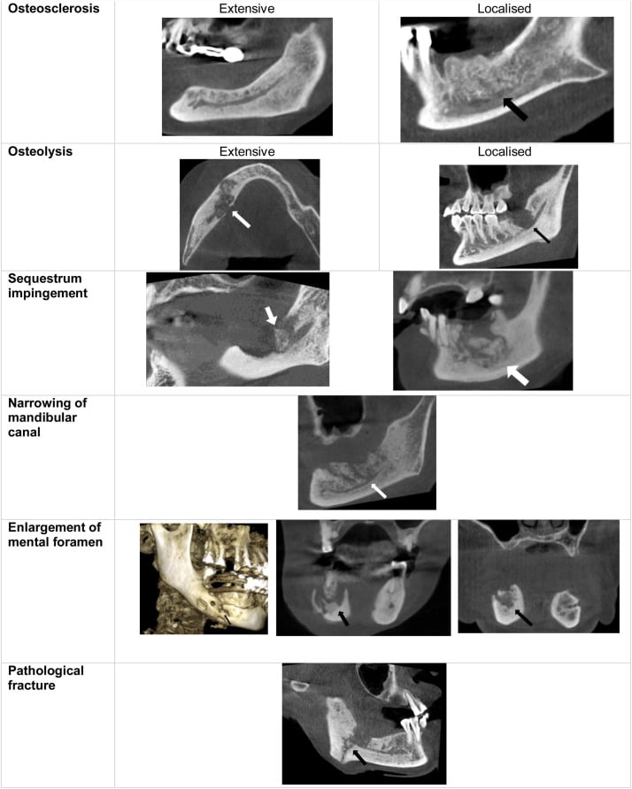
- “CBCT or panoramic radiograph is first line; look for sequestra, moth-eaten lysis, sclerosis, and periosteal reaction; advanced imaging (MRI, bone scan) if extent unclear or to exclude other pathology
- “Drug holiday before extraction is controversial and not supported by strong evidence for MRONJ prevention; the AAOMS position papers emphasise individual risk-benefit rather than routine cessation
Overview
Medication-related osteonecrosis of the jaw (MRONJ) is a rare but serious complication of potent antiresorptive therapy (nitrogen-containing bisphosphonates and denosumab) and certain antiangiogenic agents. It is defined by exposed or probeable necrotic bone in the maxillofacial region that persists for greater than 8 weeks in a patient with current or previous antiresorptive or antiangiogenic exposure and no history of head and neck radiation. That is the AAOMS working definition, and examiners expect it verbatim.
Where it came from. The condition was first recognised with the intravenous bisphosphonates used in oncology (zoledronic acid, pamidronate) and later with denosumab. It also occurs, at much lower rates, with oral bisphosphonates for osteoporosis and with some targeted cancer therapies.
Who gets it. The clinical burden falls almost entirely on patients receiving high-dose intravenous or subcutaneous antiresorptives for skeletal metastases or multiple myeloma: oncology-dose intravenous zoledronic acid (4 mg monthly) and denosumab (120 mg monthly) carry by far the highest risk. In this group the incidence after dental extraction can reach several percent; the pooled meta-analysis in the evidence section puts the proportion at 27.6% in oncology patients, and its authors attribute the risk less to the extraction itself than to pre-existing dental infection and cumulative antiresorptive exposure. In osteoporosis patients on oral agents the absolute risk is orders of magnitude lower, often cited as less than 1 in 10,000 patient-years, although duration of therapy and cumulative dose matter and years of exposure raise the risk even at osteoporosis doses.
Anatomy and Pathophysiology
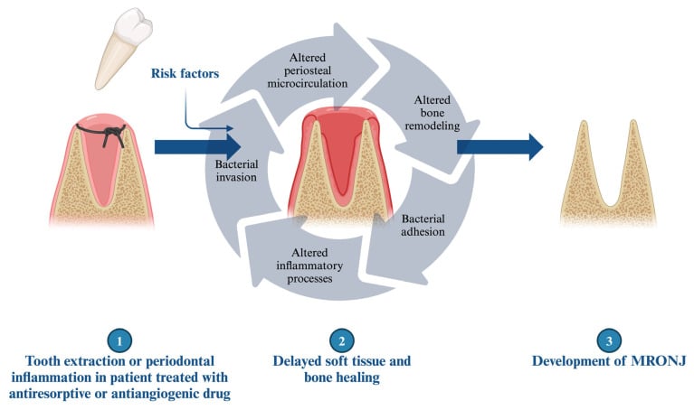
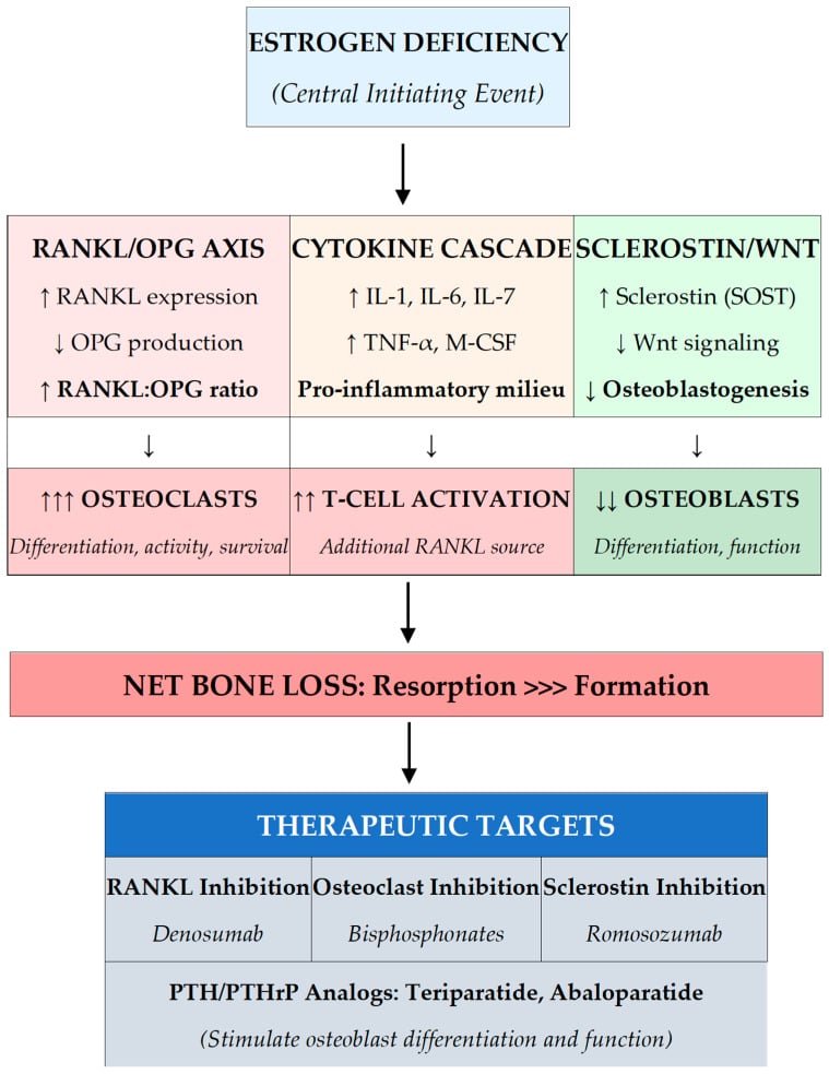
The core failure. MRONJ is a failure of bone repair after trauma in the setting of osteoclast suppression. Amino-bisphosphonates inhibit farnesyl pyrophosphate synthase in the mevalonate pathway; denosumab neutralises RANKL. Both drastically reduce osteoclast number and activity, which in most of the skeleton is the point of the treatment: fewer fractures.
Why the jaw. The jaws take repeated low-level trauma from mastication, periodontal disease and, above all, dental procedures, and the alveolar process that supports the teeth has a high rate of remodelling. An extraction socket heals by a coordinated sequence of inflammation, granulation, woven bone formation and remodelling, in which osteoclasts resorb the damaged bone before osteoblasts lay down new bone. With osteoclast recruitment and function suppressed the remodelling phase stalls, and the devitalised bone is left exposed when the thin mucosa breaks down or after surgical trauma.
Why the mandible. The mandible is affected far more often than the maxilla, roughly 2 to 3:1 or higher in most series, and the posterior mandible (molar and premolar regions) is the classic site: high baseline turnover in alveolar bone, thin mucosa over the mylohyoid ridge, and limited collateral circulation once the inferior alveolar artery is compromised. The inferior alveolar nerve can be involved, producing numbness or paraesthesia when necrosis reaches the canal. Maxillary cases are more likely to present with an oro-antral fistula or sinusitis because of the thin bone and the proximity of the antrum.
What amplifies it. The drug sets the stage; several other factors compound the problem:
- Antiangiogenic effects, seen with some bisphosphonates and with agents such as bevacizumab and sunitinib, reduce vascularity and healing capacity and add risk when combined with antiresorptives
- Local trauma (extraction, ill-fitting dentures, periodontal surgery) creates a wound that cannot epithelialise over non-viable bone
- Secondary infection perpetuates inflammation and bone death; Actinomyces and mixed oral flora are frequently cultured, and periodontal disease and chronic infection raise the odds
- In cancer patients, the underlying disease, chemotherapy, steroids and poor nutrition compound the problem
Spontaneous disease. MRONJ without an obvious dental trigger occurs, especially in the mylohyoid region or over a torus, but extraction or other invasive procedures remain the dominant precipitant in reported series: the majority of cases follow dental extraction, implant placement or periodontal surgery in high-risk patients.
Classification and Staging
The AAOMS staging system, updated in 2014 and clarified further in 2022, is the international standard in examinations and in multidisciplinary discussion. It is clinical-radiographic, and each stage carries its own initial management.
- Clinical Features
- No exposed bone or fistula, but symptoms (pain, tooth mobility, radiographic sclerosis or lysis) or non-specific signs that suggest early disease
- Initial Management Approach
- Optimise oral hygiene, chlorhexidine rinses, address infection, close follow-up; consider drug holiday only if oncologist agrees it does not compromise skeletal protection
- Clinical Features
- Exposed bone or a fistula that probes to bone, present greater than 8 weeks, asymptomatic, no clinical signs of infection
- Initial Management Approach
- Conservative: 0.12 percent chlorhexidine rinses, oral hygiene instruction, avoid further trauma, review every 2 to 3 months; antibiotics only if infection develops
- Clinical Features
- Exposed or probeable bone with pain and clinical infection (erythema, swelling, purulent discharge, or sinus tract)
- Initial Management Approach
- Infection control: culture-directed or empirical oral antibiotics (amoxicillin-clavulanate or clindamycin), antiseptic rinses, pain control, limited superficial debridement of mobile sequestra only
- Clinical Features
- Exposed bone plus one or more of: pathologic fracture, extraoral fistula, osteolysis extending to the inferior border or ramus, or extensive necrosis
- Initial Management Approach
- Surgical: segmental or marginal resection of necrotic bone with margins into bleeding viable bone; reconstruction with plate or free flap as indicated; antibiotics and supportive care
Stage is dynamic. A patient can move from stage 1 to stage 2 with secondary infection, or from stage 2 to stage 1 after successful infection control and sequestration of a small fragment. Document stage at every visit and photograph exposed bone when possible.
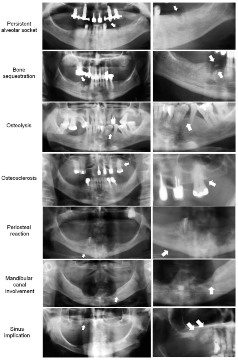
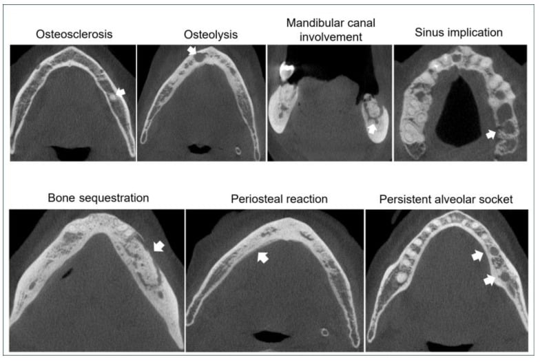
The radiographic vocabulary. Stage 0 is defined partly by radiographic change, and the features below recur across the stages. Naming them on a panoramic film and then on cone beam CT is how staging becomes reproducible.
Clinical Presentation
History. The diagnosis rests on the drug history, so take it precisely: which agent, for which indication, for how long, and what happened in the mouth and when. Timing of any dental event relative to drug exposure is critical.
- Current or previous bisphosphonate (zoledronate, alendronate, risedronate, pamidronate, ibandronate) or denosumab, or antiangiogenic therapy (bevacizumab, sunitinib and similar)
- Oncology indication (bone metastases, multiple myeloma, giant cell tumour) versus osteoporosis or Paget disease
- Recent dental extraction, implant, periodontal surgery or denture trauma
- Pain, swelling, bad taste, halitosis, discharge, or a non-healing socket that has persisted beyond the expected 4 to 6 weeks
- Numbness of the lower lip or chin
Examination. Look for the bone, then for what has happened around it. Always examine both jaws, the full dentition and the neck, because bilateral or multifocal disease occurs, especially with high-dose intravenous therapy.
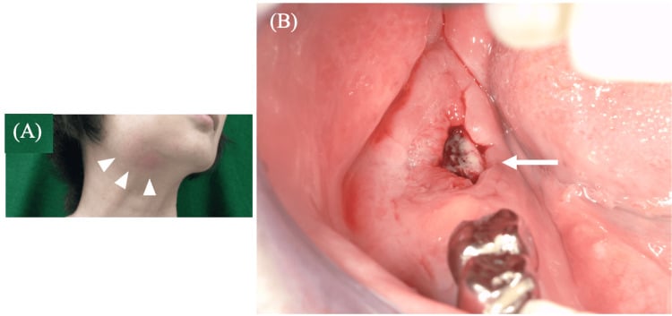
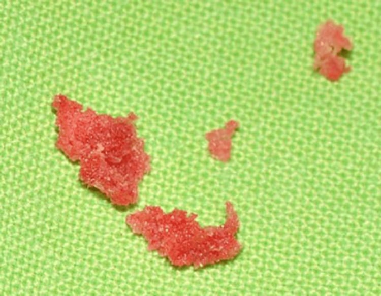
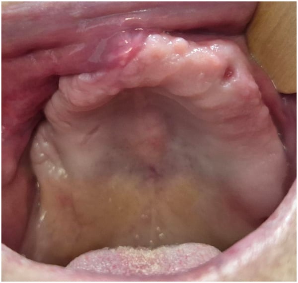
- Exposed bone in the oral cavity, often a yellow-white or grey sequestrum; the surrounding mucosa may be erythematous or rolled
- A fistula, intraoral or extraoral, that can be probed to bone or to a sequestrum
- Signs of infection: pus, swelling, trismus, lymphadenopathy
- Pathologic fracture: mobile mandibular segments, malocclusion, pain on loading
- Extraoral cutaneous fistula or bone exposed through skin in advanced cases
- Dental status: caries, periodontal pocketing, ill-fitting prostheses
Investigations
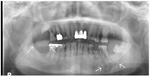
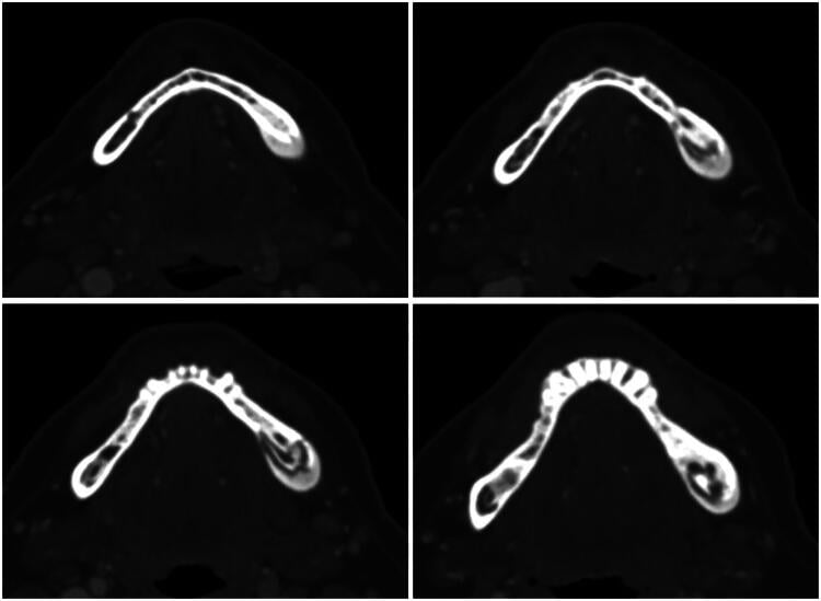
Plain films first. A panoramic radiograph (orthopantomogram) is the first-line study. Look for poorly defined radiolucencies, sclerotic zones, sequestra (dense fragments separated by lucent rims), moth-eaten destruction and periosteal reaction.
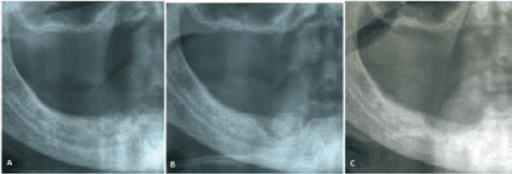
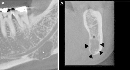
Cross-sectional imaging. Cone beam CT (CBCT) or conventional CT gives better three-dimensional assessment than the panoramic film: extent, cortical perforation, involvement of the inferior alveolar canal, and proximity to the maxillary sinus. MRI is useful when soft-tissue extent or marrow involvement needs clarification, or when ruling out metastatic disease or osteomyelitis. Bone scintigraphy or PET shows increased uptake but is non-specific; it earns its place in selected cases to assess whole-skeleton burden or occult sites.
Blood tests. No blood test diagnoses MRONJ. Check a full blood count and inflammatory markers (CRP, ESR), and albumin or pre-albumin if nutrition is a concern. In oncology patients, coordinate with the medical oncology team on renal function (especially before any further zoledronate) and calcium, since denosumab carries a risk of hypocalcaemia.
Microbiology. Swab or biopsy exposed bone or pus for culture and sensitivity. Actinomyces is frequently isolated, but whether it is the primary pathogen or a coloniser is debated.
Biopsy. Not every case needs one. It is done to exclude metastasis or a primary malignancy, and in pure MRONJ the histology shows necrotic bone with empty lacunae, bacterial colonisation and minimal inflammatory infiltrate; vital bone at the resection margin confirms that the resection was adequate. Always send tissue in stage 3 surgical cases.
Differential diagnosis. The drug and radiation histories separate most of these:
- Metastatic carcinoma (breast, prostate, lung, myeloma), which can coexist with MRONJ
- Osteoradionecrosis, if there is a radiation history
- Chronic osteomyelitis, with no drug history
- Primary bone tumours or osteonecrosis from other causes (rare)
Management
Prevention. Optimising the dentition before the first high-risk dose is vastly more effective than any treatment after exposure has occurred, and it is the only intervention with level-1 impact. Once necrotic bone is exposed, management is staged and largely supportive until sequestra separate or resection is required for complications.
- All patients due to start high-risk intravenous or subcutaneous antiresorptive or antiangiogenic therapy for cancer should have a dental review before the first dose: full examination, radiographs, and treatment of caries and periodontal disease
- Extract non-restorable teeth and complete periodontal therapy, then allow 4 to 6 weeks of mucosal healing before commencing the drug when clinically safe
- Educate the patient to report any non-healing socket, pain or exposed bone immediately, and on the need for lifelong excellent hygiene
- Document the discussion; even oral bisphosphonates carry a small risk, and low risk is not zero risk
Extraction in a patient already on therapy. The safest window has passed, so avoid the wound where you can: endodontic treatment or crown amputation is preferred over extraction when feasible. When extraction is unavoidable it is done with primary closure, perioperative antibiotics and chlorhexidine, and the risk cannot be eliminated.
Stages 0 and 1. Conservative. Chlorhexidine 0.12 percent rinses twice daily, meticulous oral hygiene and regular review, with no elective surgery. Mobile sequestra may be removed if they are easily accessed without further trauma.
Stage 2. Add infection control: empirical or culture-directed oral antibiotics (amoxicillin-clavulanate 875/125 mg twice daily or clindamycin 300 mg three times daily for 7 to 14 days), antiseptic rinses, pain management, and limited debridement of loose bone only. Most cases stabilise; some progress.
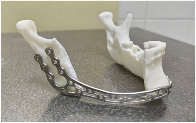
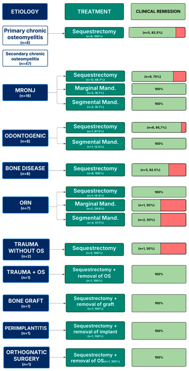
Stage 3. Surgical resection under general anaesthesia; conservative measures will not heal stage 3 disease. All necrotic bone is resected back to bleeding, viable margins: a marginal mandibulectomy if the inferior border is intact, segmental if it is not. The mandible is stabilised with a reconstruction plate, with a free fibula or scapula flap considered for large defects in fit patients. Antibiotics and chlorhexidine continue postoperatively, and long-term follow-up is mandatory.
Drug holiday. Drug holidays before extraction remain controversial. The 2014 and later AAOMS guidance does not recommend routine drug interruption solely for MRONJ prevention in patients who need ongoing skeletal protection; the decision is individualised with the oncologist. For osteoporosis patients on long-term oral therapy a holiday may be considered for other reasons, such as atypical fracture risk, but it is not proven to reduce MRONJ, and it is never a substitute for pre-therapy dental optimisation. Why a holiday behaves so differently between the two drug classes is the subject of the pharmacology section below.
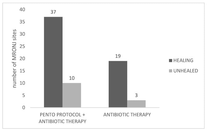
Supportive measures. Nutrition, smoking cessation, glycaemic control in diabetics and management of comorbidities all improve healing potential. Hyperbaric oxygen is not routinely recommended by AAOMS for MRONJ, unlike osteoradionecrosis, because the evidence is weak.
Bisphosphonate versus Denosumab: Pharmacology and Why It Changes Timing
Both drugs suppress osteoclasts and both cause MRONJ, but they do so by different mechanisms with very different kinetics, and that difference is the basic-science reason a drug holiday behaves so differently between them.
- Nitrogen-containing bisphosphonate
- Inhibits farnesyl pyrophosphate synthase in the osteoclast (mevalonate pathway)
- Denosumab
- Monoclonal antibody that neutralises RANKL, preventing osteoclast formation and activation
- Nitrogen-containing bisphosphonate
- YES - binds hydroxyapatite and is incorporated into the skeleton
- Denosumab
- NO - circulates and is not deposited in bone
- Nitrogen-containing bisphosphonate
- Very long skeletal half-life (years) - effect PERSISTS long after stopping
- Denosumab
- Short circulating half-life (about 26 days); bone turnover RECOVERS within months of stopping
- Nitrogen-containing bisphosphonate
- A short holiday does little - the drug lingers in bone, so MRONJ risk persists
- Denosumab
- Effect is reversible, so timing invasive dentistry near the end of the dosing interval (the trough) is rational
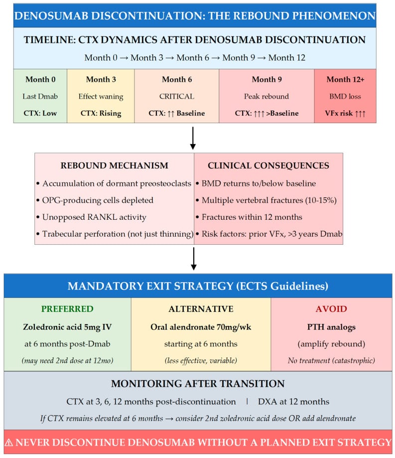
The rebound phenomenon. Denosumab's reversibility cuts both ways. Stopping it without transitioning to a bisphosphonate causes rapid loss of the accrued bone density and a risk of multiple rebound vertebral fractures, so cessation decisions are never the dentist's alone and must involve the prescribing physician.
A short bisphosphonate holiday is largely ineffective; denosumab work is timed towards the trough of the dosing interval; and denosumab is never stopped without a bisphosphonate bridge.
Complications
Local complications are those of the disease in the jaw itself:
- Chronic pain, recurrent infection and halitosis
- Pathologic fracture of the mandible, the most feared stage 3 event
- Oro-cutaneous fistula, oro-antral communication, maxillary sinusitis
- Inferior alveolar or lingual nerve injury, from the disease or from surgery
- Loss of teeth, denture intolerance and severe functional impairment
Systemic. In frail oncology patients, uncontrolled jaw sepsis can contribute to a systemic inflammatory response or delay cancer treatment, and pain on eating leads to malnutrition.
Treatment-related. Surgery brings plate exposure, flap failure, further necrosis at the margins and donor-site morbidity; prolonged antibiotic courses bring antibiotic-associated diarrhoea or resistance. The psychological impact of facial disfigurement or prolonged illness falls on patients already facing cancer.
Late failure. Exposed bone recurs at the resection margin or at distant sites if the underlying drug exposure and risk factors continue.
MRONJ versus Osteoradionecrosis (ORN)
- MRONJ
- Antiresorptive (bisphosphonate or denosumab) or antiangiogenic drug; NO radiation
- Osteoradionecrosis (ORN)
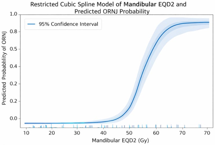
- Head and neck radiotherapy (classically over 60 Gy to the field)
- MRONJ
- Osteoclast suppression - failure to repair and remodel injured bone
- Osteoradionecrosis (ORN)
- Hypovascular-hypocellular-hypoxic tissue (Marx 3-H theory); also a radiation-induced fibro-atrophic process
- MRONJ
- Dental extraction or invasive procedure (or spontaneous)
- Osteoradionecrosis (ORN)
- Dental extraction in the irradiated field, or spontaneous
- MRONJ
- NOT routinely recommended (weak evidence)
- Osteoradionecrosis (ORN)
- Has an established role and evidence base (Marx protocol)
- MRONJ
- Antiseptic and antibiotic cover, staged surgery
- Osteoradionecrosis (ORN)
- Pentoxifylline plus tocopherol (PENTOCLO) targeting the fibro-atrophic process
One question decides it. The two are defined to be mutually exclusive, so the discriminator is the radiation history: an antiresorptive patient with no jaw radiation has MRONJ, and an irradiated patient has ORN even if also on a bisphosphonate. They look alike and share the extraction trigger and the posterior-mandible predilection, which is why examiners ask; the distinction matters because hyperbaric oxygen helps ORN and is not a standard MRONJ treatment.
Clinical Relevance
MRONJ sits at the intersection of oncology, oral surgery and orthopaedic and metabolic bone medicine. Every patient who starts a high-potency antiresorptive for skeletal metastases or myeloma should be viewed as having a lifelong risk to the jaws, and the orthopaedic surgeon or endocrinologist who prescribes these agents must either arrange pre-treatment dental clearance or document that the patient has been counselled and that dental review has occurred or is planned.
The same principles apply to denosumab as to bisphosphonates; at oncology doses the RANKL inhibitor produces a comparable, if not higher, incidence of MRONJ. As more patients receive these therapies for longer and for additional indications (giant cell tumour, osteoporosis with high fracture risk), the absolute number of MRONJ cases will remain clinically relevant even if the per-patient risk is managed. The examination tests the bone biology behind it, risk stratification by drug, dose, route and indication, prevention over salvage, staging language that communicates urgency to the maxillofacial team, and the limits of conservative care.
Guidelines, Registries and Global Practice
-
AAOMS position papers (2007, 2009, 2014, 2022 updates) remain the most widely referenced international guidance. They established the current definition, the four-stage system, and the emphasis on prevention before high-risk therapy. The 2014 paper broadened the term from BRONJ to MRONJ to include denosumab and antiangiogenics.
-
European / international consensus (e.g. from ESMO, MASCC, and national oral medicine societies) aligns closely with AAOMS on staging and the central role of pre-treatment dental assessment for patients starting oncology-dose antiresorptives. Some European guidelines place slightly more weight on drug holidays in selected low-risk scenarios, but evidence remains limited.
-
Denosumab-specific data: oncology-dose denosumab (120 mg monthly) carries a comparable or slightly higher MRONJ incidence than zoledronic acid in head-to-head trials and real-world cohorts. The same prevention and staging principles apply.
-
Registry and cohort evidence: large oncology cohorts and systematic reviews (including the 20-year single-centre series and extraction-risk meta-analyses) confirm that dental extraction is the dominant modifiable risk factor and that structured prevention programs reduce incidence. Absolute risk in osteoporosis patients on oral agents remains very low worldwide.
-
Global practice variation: access to specialist maxillofacial care, CBCT, and reconstructive microsurgery varies. In resource-limited settings, emphasis remains on prevention and conservative infection control; advanced stage 3 disease may be managed with local debridement and antibiotics rather than major resection. No country-specific billing codes are used in guidance.
-
Key practical message: wherever a patient is started on high-potency antiresorptive therapy for cancer, dental review before the first dose is a quality and safety standard across AAOMS, European, and Asia-Pacific recommendations.
Exam Viva Scenarios
Practise clinical reasoning and management decisions out loud
“A 62-year-old woman with metastatic breast cancer on monthly zoledronic acid 4 mg for 18 months requires extraction of three non-restorable lower molars. The oncologist asks how you will manage the risk of MRONJ.”
“A 68-year-old man with myeloma on denosumab 120 mg monthly for 14 months presents with a 10-week history of exposed bone in the left posterior mandible after extraction, now with pain, swelling, and a small amount of pus. Panoramic radiograph shows a 2 cm area of lysis with a sequestrum but the inferior border is intact. How do you stage and manage this?”
“A 55-year-old woman with metastatic lung cancer on zoledronic acid and bevacizumab presents with sudden pain and a mobile mandible after a trivial fall. Examination shows a cutaneous fistula with exposed bone and malocclusion. Radiographs confirm a pathologic fracture through an area of osteonecrosis. What is your approach?”
Evidence
AAOMS Position Paper on Medication-Related Osteonecrosis of the Jaw (2014 Update)
- Updated consensus definition: exposed bone or probeable bone greater than 8 weeks in a patient with antiresorptive or antiangiogenic exposure and no jaw radiation
- Four-stage system (0-3) that directly informs treatment; stage 3 explicitly includes pathologic fracture, extraoral fistula, and extensive osteolysis
- Risk is highest with intravenous oncology-dose bisphosphonates and denosumab; oral agents for osteoporosis carry very low absolute risk
- Dental extraction remains the most common precipitating event; prevention through pre-therapy dental optimisation is the most effective strategy
Diagnosis and Treatment of Medication-Related Osteonecrosis of the Jaws: A Cohort Study Over 20 Years
- Copenhagen ONJ Cohort: 934 MRONJ patients diagnosed 2005-2024 at a single tertiary centre
- Mean age 71.6 years, 68.4% female; commonest antiresorptive indications were osteoporosis, breast cancer, prostate cancer and multiple myeloma
- Conservative management in 18.1% versus surgical intervention in 81.9% of patients
- Tooth removal was the most frequent triggering event (52.9%); demographics and concomitant steroid/immunosuppressant/antiangiogenic use differed across antiresorptive-dose groups
Does Tooth Extraction Increase the Risk of MRONJ in Oncologic Patients? A Systematic Review and Meta-Analysis
- Systematic review and meta-analysis of 7 studies (497 oncology patients) of MRONJ after tooth extraction
- Pooled proportion of MRONJ after extraction was 27.6% (95% CI 16.0-43.3%) in this high-risk oncology population
- No significant association with female sex or chemotherapy; MRONJ patients had a significantly longer duration of bone-modifying-agent therapy (pooled mean difference 8.38 months, 95% CI 2.44-14.32)
- Key nuance: the evidence does NOT support extraction as an isolated, uniformly avoidable trigger - local infection, inflammatory dental disease, extraction timing and cumulative drug exposure appear more consistently associated with risk