ALTR | Pseudotumor | Metal Ion Toxicity | Surveillance and Revision
- Cobalt greater than 7 ppb = concerning, warrants further investigation and surveillance
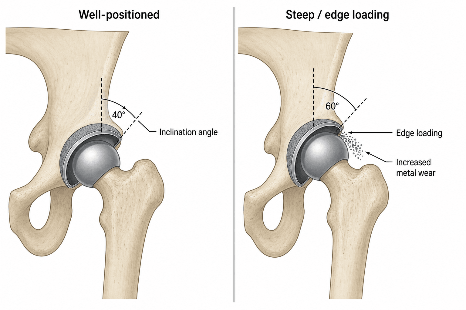
- Cup inclination greater than 55 degrees = edge loading, accelerated wear and ion release
- MARS MRI = Metal Artifact Reduction Sequence for soft tissue assessment
- ALTR = Adverse Local Tissue Reaction (umbrella term for all MoM soft tissue problems)
- ASR recalled 2010 = DePuy ASR hip resurfacing and XL Acetabular System
- “Pseudotumor does not mean malignancy - it is a sterile inflammatory mass
- “Small head MoM THAs have higher failure rates than resurfacing
- “Metal ions can remain elevated for years after revision
- “Revision for ALTR has poorer outcomes than revision for other causes
Overview and Epidemiology
Metal-on-metal (MoM) bearings were introduced as an alternative to metal-on-polyethylene, to reduce wear-related osteolysis and improve longevity, particularly in younger active patients. Adverse local tissue reactions (ALTR), elevated metal ion levels and pseudotumour formation then emerged, use declined significantly, and multiple products were recalled.
The ASR recall. The DePuy ASR Hip Resurfacing System and ASR XL Acetabular System were voluntarily recalled in August 2010 because of higher than expected revision rates. Australian Orthopaedic Association National Joint Replacement Registry (AOANJRR) data showing a 6.4% revision rate at 5 years was instrumental in identifying the problem; the failure rate of the recalled ASR devices is quoted at 12-13% at 5 years.
Scale. Use peaked between 2005 and 2010, before the concerns emerged, and is now near zero for primary THA with very limited resurfacing. An estimated 500,000+ patients worldwide carry a MoM hip and every one of them needs lifetime monitoring. The revision burden is a significant healthcare cost and patient morbidity, and major class action settlements followed globally.
Who does badly, who does not. Pseudotumour is symptomatic in 1-4% of patients. Systemic toxicity is rare but documented, with cardiac, neurological and thyroid effects. At the other end, a well-positioned resurfacing has 95%+ survival at 10 years in experienced hands.
Pathophysiology and Mechanisms
The cascade. ALTR is caused by a combination of wear-generated metal debris and corrosion products from the bearing surface and from modular junctions. In susceptible individuals this triggers a Type IV hypersensitivity reaction, leading to soft-tissue destruction, osteolysis and potentially systemic toxicity.
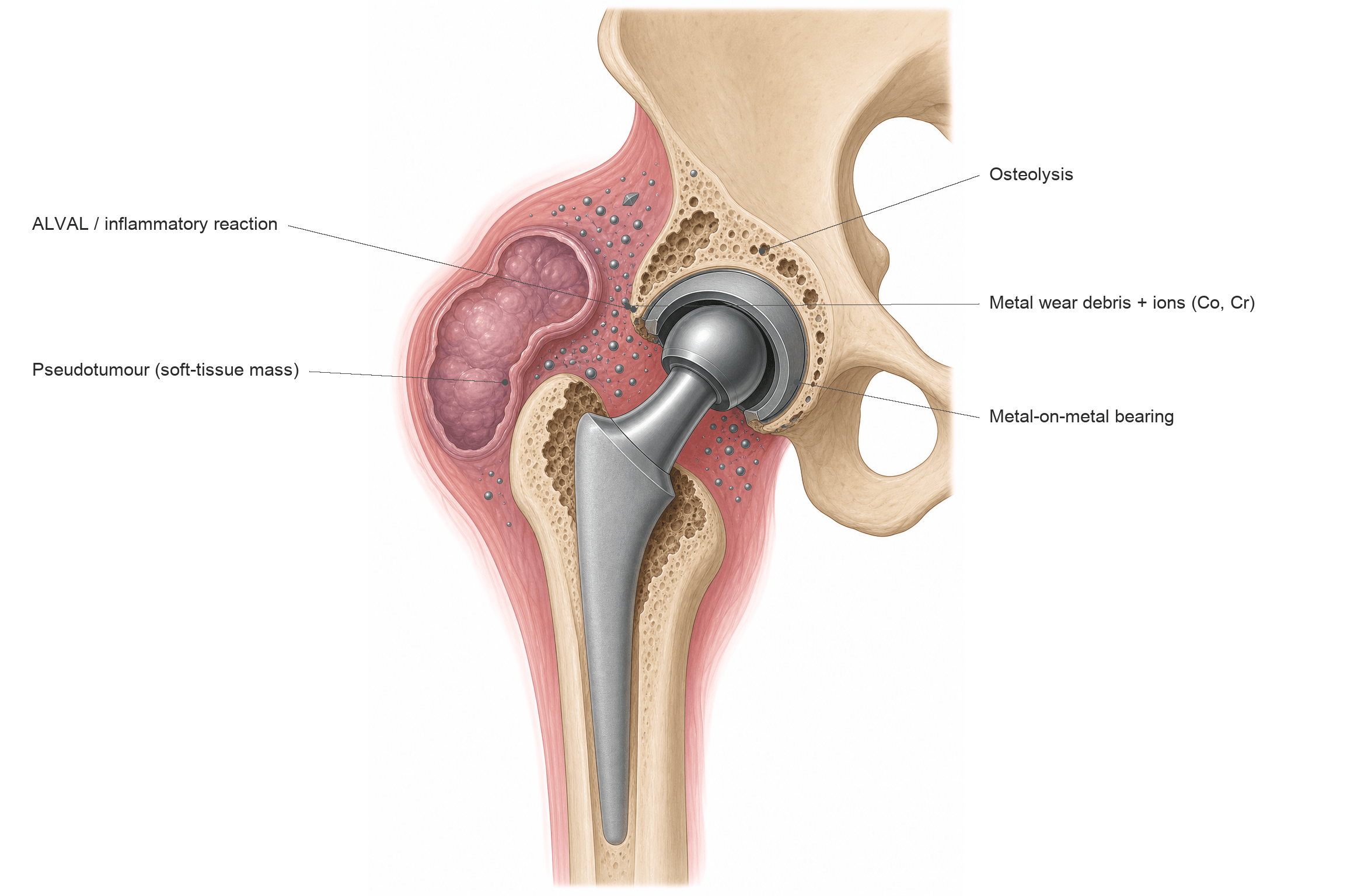
Tribology. A MoM bearing relies on fluid-film lubrication to separate the articulating surfaces. When that film fails, through edge loading, malposition or high activity, metal contacts metal and wear debris is generated.
- Cause
- Cup inclination greater than 55 degrees or low coverage
- Result
- Contact at rim, not centre
- Clinical Significance
- 10-100x increased wear rate
- Cause
- High range of motion, microseparation
- Result
- Linear wear pattern on head
- Clinical Significance
- Visible on explanted components
- Cause
- Modular junction corrosion
- Result
- Metal release from taper
- Clinical Significance
- More common with large heads, long necks
- Cause
- Well-positioned, matched components
- Result
- Minimal wear
- Clinical Significance
- Low ion levels, good outcomes
Cup position. Inclination greater than 55 degrees produces edge loading, with metal-on-metal contact outside the bearing surface; a shallow cup with a low coverage arc does the same. Combined anteversion matters too, so aim for inclination of 40-45 degrees and anteversion of 15-20 degrees. Malposition is the strongest predictor of failure.
Design. Recalled designs such as the ASR carry design-specific higher failure rates. Small-head MoM THAs (less than 50 mm) have higher failure rates than resurfacing, and large heads, long necks and a large taper mismatch make modular-junction corrosion (trunnionosis, in the table above) more common.
The biological response. The characteristic histological finding is ALVAL, the aseptic lymphocyte-dominated vasculitis-associated lesion: perivascular lymphocytic infiltrates, endothelial swelling and tissue necrosis. This pattern distinguishes hypersensitivity from a simple wear-debris reaction and is associated with worse soft-tissue destruction. What that reaction does to the hip, locally and systemically, is set out under Complications.
Classification Systems
Three systems are in use, and they grade different things: Hart grades the MARS MRI appearance, the MHRA categories stratify the patient by symptoms and metal ions, and the Anderson classification grades what the surgeon finds at revision.
Based on MARS MRI appearances of the periprosthetic soft tissue.
- Description
- Fluid only
- MRI Appearance
- Simple effusion, no wall thickening
- Management
- Surveillance, consider aspiration
- Description
- Fluid with synovitis
- MRI Appearance
- Wall thickening, enhancing synovium
- Management
- Close monitoring, consider revision
- Description
- Solid pseudotumour
- MRI Appearance
- Mass lesion, solid component
- Management
- Revision recommended
- Description
- Mixed/destructive
- MRI Appearance
- Complex tissue destruction, muscle atrophy
- Management
- Urgent revision, expect bone/muscle loss
Type 2b and Type 3 lesions have the worst outcomes after revision, and the size of the pseudotumour correlates with abductor damage.
Clinical Assessment
History. The pain is in the groin, lateral hip or thigh and is often different from arthritic pain, and function declines more than expected after an arthroplasty. Ask about swelling, since a large pseudotumour can be a visible or palpable mass, about clicking or clunking that may indicate subluxation or impingement, and about numbness or weakness from nerve compression. The implant details are critical: the date of surgery and the implant type.
Examination. A Trendelenburg gait points to abductor damage, and a palpable mass sits anterolaterally. Range of motion may be preserved despite significant ALTR. Assess leg length for subsidence or dislocation, examine for sciatic or femoral nerve involvement, and look at the skin, where a sinus or discolouration is rare.
Up to 60% of patients with MoM hips may have asymptomatic ALTR on imaging. Symptoms do not correlate well with the severity of soft-tissue damage: local complications frequently progress without proportionate symptoms, and systemic effects appear late. This is why systematic surveillance with imaging is essential regardless of symptoms, with a low threshold for cross-sectional imaging.
Differential diagnosis. A painful MoM hip is not ALTR until the other causes of pain after arthroplasty have been considered, infection above all.
- Distinguishing Features
- Elevated metal ions, characteristic MRI findings
- Investigations
- Metal ions, MARS MRI
- Distinguishing Features
- Raised inflammatory markers, positive culture
- Investigations
- CRP, ESR, aspiration and culture
- Distinguishing Features
- Pain with activity, progressive radiolucencies
- Investigations
- Sequential X-rays, CT
- Distinguishing Features
- Anterior groin pain, pain with hip flexion
- Investigations
- Injection test, CT for cup position
- Distinguishing Features
- Lateral pain, point tenderness
- Investigations
- Clinical diagnosis, ultrasound
Investigations
Metal ions. Measure whole blood cobalt and chromium, not serum: serum underestimates the level by 50%. Cobalt is the more clinically relevant of the two for ALTR; chromium follows a similar pattern. How to act on the number is set out in the next section.
Plain radiographs. An AP pelvis and lateral hip, compared with previous films, for component position (inclination and anteversion, judged against the targets under Pathophysiology), signs of loosening and osteolysis. CT is less useful for soft tissue but shows osteolysis.
MARS MRI. Metal artefact reduction sequence MRI is the gold standard for the soft tissues: it detects fluid collections, pseudotumours, muscle atrophy and osteolysis, and the Hart grade it yields guides management. It should be performed if ions are elevated or symptoms are present.
Ultrasound. The alternative when MRI is unavailable or contraindicated. It is operator dependent but can detect fluid and masses, is less sensitive for muscle atrophy and bone change, and is useful for guided aspiration.
Aspiration. Infection must be ruled out before any revision, and aspiration is essential in every painful MoM hip. Send fluid for microbiology culture with extended incubation, cell count and differential, and synovial alpha-defensin if available.
- Aspirate appearance: ALTR/metallosis produces a cloudy, grey or "metallic"/creamy aspirate that can look like pus.
- Synovial cell count: metal/corrosion particles are miscounted as leucocytes by automated analysers, giving a falsely elevated synovial white-cell count and neutrophil percentage - a manual count is needed, and a falsely high automated count must not be taken as proof of infection.
- Alpha-defensin: can be falsely positive in metallosis/ALTR, reducing its specificity in this setting.
- Serum CRP/ESR: ALTR itself can mildly raise inflammatory markers, so modest elevations are non-specific.
Practical consequence: a painful MoM hip can have biochemistry and an aspirate that look infected when the problem is ALTR - and, conversely, true infection can coexist. Use manual synovial cell counts, multiple culture samples with extended incubation, and the overall picture (ions, MARS MRI, implant) rather than a single automated count, and apply infection criteria (e.g. MSIS/EBJIS) with awareness of these MoM-specific false positives.
Metal Ion Interpretation
- Interpretation
- Normal/optimal
- Action Required
- Annual surveillance
- Interpretation
- Acceptable
- Action Required
- Annual surveillance
- Interpretation
- Borderline elevated
- Action Required
- Increased surveillance, consider imaging
- Interpretation
- Elevated - concerning
- Action Required
- MARS MRI required, 6-monthly review
- Interpretation
- High - significant risk
- Action Required
- Urgent imaging, consider revision
- Interpretation
- Very high - systemic risk
- Action Required
- Revision recommended, systemic assessment
The table above reads as a clean dose-response, but the page's own evidence warns against that reading. Hart's revision series found that nearly 50% of patients revised for a failed metal-on-metal hip had blood cobalt and chromium BELOW the 7 ppb threshold, with low component wear - because much ARMD is a patient-specific Type IV hypersensitivity reaction, not simple dose-related toxicity. So a cobalt of 2-4 ppb marked "acceptable" above does NOT exclude ALTR. Image the symptomatic patient (MARS MRI) regardless of the ion level; the ions guide surveillance, they do not clear a painful hip.
Read the ions as a pattern. Bearing-surface (articular) wear of a MoM bearing releases both cobalt and chromium, classically in a roughly balanced ratio; chromium is partly retained locally as chromium orthophosphate, but both rise in blood. Mechanically-assisted crevice corrosion at the head-neck taper (trunnionosis) preferentially releases cobalt, so a markedly elevated cobalt with relatively normal chromium, a high cobalt:chromium ratio, points to a taper or modular-junction problem rather than the bearing.
ARMD beyond the MoM bearing. ARMD/ALTR also occurs in metal-on-polyethylene THA with a cobalt-chrome head on a corroding taper, especially with large-diameter heads and certain modular-neck stems: an identical pseudotumour/ALVAL reaction driven by taper corrosion, not the bearing. A painful MoP THA with a rising isolated cobalt should prompt the same ARMD workup of ions, MARS MRI and infection exclusion; "it isn't a MoM bearing" does not exclude an adverse metal reaction.
One hip or two. With a unilateral MoM hip, compare the level to population norms. With bilateral MoM hips the levels are additive, so interpret them with caution.
Management Algorithm
Lifelong, regardless of symptoms. All patients with MoM hip implants require lifelong surveillance. Metal ions can rise years after implantation and ALTR can develop in a previously asymptomatic patient, so there is no "safe" time to stop monitoring. Document the surveillance plan in writing to the patient.
The schedule.
- Annual review as a minimum: clinical assessment, metal ion levels (Co and Cr), plain radiographs if any concerns; document symptoms, examination findings and implant function
- Baseline MARS MRI: many centres recommend one for all MoM patients to establish soft-tissue status, repeated if symptoms develop or ions rise
- 6-monthly review if cobalt is above the 7 ppb action level, with new symptoms, or with abnormal imaging; more frequent still if ions are rising rapidly or symptoms are progressing
- Consider revision for progressive ALTR on imaging, cobalt greater than 10 ppb with symptoms, functional decline, neurological compromise, or component failure
Surgical Management
Planning. Review all imaging including the MARS MRI and map the location and extent of the pseudotumour. Assess bone stock on CT if osteolysis is suspected, rule out infection with aspiration, and counsel the patient about the expected outcomes.
In theatre. The principles, in order:
- Extended approach - an extensile exposure may be needed for pseudotumour excision
- Complete debridement - remove all necrotic tissue and metallosis
- Pseudotumour excision - thorough removal of reactive tissue
- Tissue sampling - multiple samples for microbiology and histology
- Bone grafting - address osteolytic defects
- Bearing change - ceramic-on-polyethylene preferred
- Stability assessment - anticipate instability from tissue loss
The bearing. Ceramic-on-polyethylene is preferred for revision. Ceramic-on-ceramic is an alternative with a squeaking risk, and in the NJR analysis in the evidence section a ceramic-on-ceramic bearing was one of the predictors of re-revision. Never use MoM for a revision.
Constraint. A standard articulation if the soft tissues are preserved; dual mobility for abductor weakness, tissue loss or a high instability risk; a constrained liner for severe instability risk. Trial stability intraoperatively before committing to the final construct.
Complications
The complications of a MoM bearing arise from chronic metal-ion and particulate release. They are best grouped as local (the great majority), regional and mechanical, and systemic (rare).
Local soft tissue. The pseudotumour is a sterile inflammatory mass, solid or cystic, and benign: it is not a malignancy. Its histology is ALVAL, and it can cause extensive soft-tissue destruction, including of the abductors.
- Metallosis - black staining of the capsule and soft tissues from deposited metal debris
- Soft-tissue necrosis - destruction of capsule and abductors
- Fluid collections and recurrent effusions
Bone and mechanical.
- Osteolysis - bone resorption around the components
- Aseptic loosening of cup or stem
- Trunnionosis (taper corrosion) in stemmed MoM
- Femoral neck fracture or narrowing in resurfacing
- Instability and dislocation from abductor and capsule loss
Regional. A large pseudotumour can compress the sciatic or femoral nerve, and rarely an expanding mass compresses vessels. Trochanteric bursitis and a Trendelenburg gait disturbance complete the picture.
Systemic. Cardiomyopathy is the most serious, reported at very high cobalt levels. Neuro-ocular toxicity takes the form of cognitive changes and peripheral neuropathy (rare), hearing loss and visual loss; hypothyroidism appears in some case reports; cobalt drives erythropoiesis, producing polycythaemia; and renal metal deposition is of uncertain clinical significance. Complications specific to revision surgery are detailed under Surgical Management.
Guidelines, Registries & Global Practice
Global Epidemiology:
An estimated one million MoM bearings were implanted worldwide during the peak period of roughly 2003 to 2010, the majority in younger, more active patients. Use has since collapsed to near zero for primary arthroplasty. Large national registries were decisive in exposing the problem before manufacturer surveillance, and remain the backbone of outcome data.
Registry Evidence (Side by Side):
- Region
- United Kingdom
- Key MoM Signal
- Resurfacing worse than THR in women at every head size; stemmed large-head MoM high revision rates
- Region
- Australia
- Key MoM Signal
- Early ASR signal (around 6 to 7% revision at 5 years) that contributed to the 2010 recall; small-head MoM THA worse than resurfacing
- Region
- United States
- Key MoM Signal
- Documents sharp decline in MoM use and ongoing revision burden from prior implants
- Region
- Sweden / Scandinavia
- Key MoM Signal
- Confirmed inferior survivorship of large-head MoM versus conventional bearings
Guidelines Compared (Side by Side):
- Surveillance Trigger for Imaging
- Symptoms or whole-blood cobalt/chromium above 7 ppb (about 119 nmol/L)
- Headline Position
- Annual review for all MoM hips; cross-sectional imaging and closer follow-up if symptomatic or ions raised; higher scrutiny for large-head stemmed MoM and ASR
- Surveillance Trigger for Imaging
- Symptoms or rising/elevated metal ions (no single mandated cut-off)
- Headline Position
- Routine ion testing not advised in asymptomatic, well-functioning patients; image and test symptomatic patients; individualised follow-up
- Surveillance Trigger for Imaging
- Symptoms, abnormal exam, rising ions, or suspicious imaging
- Headline Position
- Risk-stratified, symptom-led surveillance; metal ions and MARS MRI reserved for at-risk or symptomatic patients
- Surveillance Trigger for Imaging
- Symptoms or ions above the 7 ppb consensus threshold
- Headline Position
- Aligns broadly with MHRA; emphasises implant-specific risk and lifelong follow-up of high-risk designs
The UK/European approach is more proactive (population surveillance with a 7 ppb action level) whereas the US/AAOS approach is more symptom-led, reflecting differences in registry infrastructure and the absence of a universally agreed ion cut-off. All agree that symptomatic patients and high-risk designs (ASR, large-head stemmed MoM) need metal ions plus MARS MRI, and that monitoring is lifelong.
- DePuy ASR (recalled 2010): ASR Hip Resurfacing System and ASR XL Acetabular System
- DePuy Pinnacle MoM liner option: progressively withdrawn
- Multiple large-head stemmed MoM and other resurfacing systems voluntarily withdrawn worldwide
- Well-resourced settings: structured recall registries, routine whole-blood ICP-MS metal-ion assays, and ready MARS MRI access enable proactive surveillance and earlier revision.
- Limited-resource settings: ion assays and MARS MRI may be unavailable, so practice relies more on symptoms, plain radiographs, and ultrasound; patient tracing after recalls is harder. The principle remains identical - any symptomatic MoM hip warrants infection exclusion and cross-sectional imaging where obtainable, and lifelong follow-up.
Whatever the health system, document surveillance, counselling, and informed consent in writing, and give patients a written surveillance plan - MoM hips require lifelong monitoring with no defined endpoint.
Controversies and Areas of Uncertainty
MoM management remains an area of genuine debate. Examiners reward candidates who can articulate the uncertainty rather than quoting cut-offs as absolutes.
The 7 ppb cobalt/chromium action level is a pragmatic regulatory threshold, not a biological cliff. Up to half of patients revised for ALTR have ions below 7 ppb (Hart 2012), and many with high ions never develop tissue damage. Ion level is one input, never the sole determinant.
Whether ALTR is driven mainly by high-wear particulate toxicity or by a Type IV hypersensitivity (ALVAL) to a normal debris load remains unresolved. The two likely coexist on a spectrum, which is why low-wear, well-positioned implants can still fail in susceptible patients.
UK/European guidance favours scheduled ion surveillance; US/FDA guidance discourages routine testing in asymptomatic, well-functioning hips. There is no randomised evidence that any specific surveillance interval changes outcomes.
Many cystic ALTRs in asymptomatic patients are stable or regress over time (Goldstein 2016), so imaging an abnormality does not mandate revision. Distinguishing lesions that will progress from those that will not is still imperfect.
Not every pseudotumour needs revision, and not every elevated ion level needs surgery. Revision for ALTR carries worse outcomes than primary THA. The decision must integrate symptoms, ion trend, imaging type/size, abductor status, and implant design - never a single number.
Exam Viva Scenarios
Practise clinical reasoning and management decisions out loud
“A 52-year-old man presents with progressive groin and lateral hip pain 6 years after a metal-on-metal hip resurfacing. He was previously very active but has noticed increasing difficulty with walking. Examination shows Trendelenburg gait and limited internal rotation. Investigations show Cobalt 15 ppb and Chromium 12 ppb. Plain films show a well-fixed implant with cup inclination of 52 degrees. What is your diagnosis and management plan?”
“A 48-year-old woman with bilateral ASR hip resurfacings performed in 2008 attends for routine surveillance. She is asymptomatic with excellent function. Metal ions show Cobalt 5 ppb and Chromium 4 ppb. X-rays show cup inclination of 48 degrees on right and 58 degrees on left. How do you manage this patient?”
“A 65-year-old man with a MoM THA from 2009 presents with progressive hearing loss, visual disturbance, cognitive decline, and peripheral neuropathy. Cardiology has found a new cardiomyopathy. Metal ions show Cobalt 180 ppb. What is your diagnosis and emergency management?”
Metal Ion Thresholds
- Cobalt less than 2 ppb = optimal
- Cobalt 2-7 ppb = acceptable, annual surveillance
- Cobalt 7-10 ppb = concerning, MARS MRI
- Cobalt greater than 10 ppb = high risk, consider revision
- Cobalt greater than 20 ppb = systemic risk, revise
Cup Position Risk
- Inclination greater than 55 degrees = edge loading
- Optimal inclination 40-45 degrees
- Optimal anteversion 15-20 degrees
- Malposition strongest predictor of failure
ALTR Classification (Hart)
- Type 1: Fluid only - surveillance
- Type 2a: Synovitis - consider revision
- Type 2b: Solid pseudotumor - revise
- Type 3: Destructive - urgent revision
Investigation Protocol
- Whole blood (not serum) metal ions
- Plain X-rays for position/loosening
- MARS MRI for soft tissue
- Aspiration to rule out infection
- Annual surveillance lifelong
Revision Principles
- Early revision preserves soft tissue
- Complete pseudotumor excision
- Ceramic-on-polyethylene bearing
- Consider dual mobility for instability
- Never MoM for revision
Key Facts
- ASR recalled August 2010
- AOANJRR data key to identifying problem
- Pseudotumor = benign inflammatory mass
- ALVAL = histological pattern
- Small head MoM worse than resurfacing
Evidence Base and Key Studies
Original Description of MoM Pseudotumours
- Case series of 20 hips (17 patients, all women) with soft-tissue masses termed pseudotumours after MoM resurfacing
- Common histology: extensive necrosis and lymphocytic infiltration
- 13 of 20 hips required revision to conventional THA
- Estimated approximately 1% of MoM resurfacing patients develop a pseudotumour within 5 years
ASR Early Failure - Excess Wear and Component Position
- Series of 660 MoM resurfacings/large-bearing THRs; all 17 adverse-reaction revisions (3.4%) were ASR bearings
- Failed hips had significantly smaller components and higher acetabular anteversion
- Significantly higher whole-blood and joint chromium and cobalt levels than asymptomatic patients (all p less than 0.001)
- Explants showed greater surface wear; adverse reaction usually implies high component wear in well-positioned implants
Why MoM Hips Fail - Metal Ions and Cup Inclination (AAOS Exhibit)
- 55 patients revised for failed Birmingham Hip Resurfacing compared with well-functioning controls
- Revised hips had significantly higher cup inclination, smaller head diameter, and higher whole-blood cobalt and chromium (all p less than 0.01)
- Nearly 50% of revised patients had blood metal ions below the 7 ppb threshold and low wear
- Unexplained pain drove 69% of revisions, suggesting patient-specific (hypersensitivity) factors
MRI Classification of Pseudotumours
- MRI classification of periprosthetic masses: Type I thin-walled cyst, Type II thick-walled cyst, Type III predominantly solid
- 33 hips in 25 patients reviewed over 4 years
- Type III solid masses were significantly larger and more often located anterior to the hip
- Symptom severity and revision rates were lowest for Type I and highest for Type III
Evidence Base - Registry, Revision and Toxicity
Registry Failure Rates of MoM Resurfacing (NJR)
- 434,560 primary THRs in the NJR for England and Wales (31,932 resurfacings)
- In women, resurfacing had worse implant survival than conventional THR at every head size
- Predicted 5-year revision in a 55-year-old woman: 8.3% (42mm resurfacing) vs 1.5% (28mm cemented MoP THR)
- Resurfacing matched other options only in men with large femoral heads
Re-revision After ALTR Revision (NJR)
- 2,535 MoM hips revised for adverse reaction to metal debris (ARMD) in the NJR, 2008-2014
- Re-revision in 7.6% of hips; cumulative 5-year implant survival 89.5%
- Predictors of re-revision: high BMI, modular-only revision, ceramic-on-ceramic bearing, and acetabular bone grafting
- Short-term re-revision risk comparable to all-cause non-MoM revision
Systemic Cobalt Toxicity From Metal Hip Prostheses
- Systematic review identifying 18 individual cases of systemic toxicity from metal hip implants
- Reported features: neuro-ocular toxicity (14 cases), cardiotoxicity (11), thyroid toxicity (9)
- Greatest risk was a cobalt-containing revision of a failed ceramic bearing (median blood cobalt 506 ug/L), not primary MoM failure (median 34.5 ug/L)
- Removal of the prosthesis usually lowered cobalt and improved some features, but not always fully
Natural History of Asymptomatic ALTR
- 83 asymptomatic modular MoM THA patients had metal ions and MARS MRI; cystic ALTR found in 26 (31%)
- On repeat MRI a year later, 15 of 19 lesions (79%) decreased in size
- 3 patients became symptomatic and were revised
- All patients had cobalt and chromium below the 7 ppb threshold