Boxer's Fracture | Rotation vs Angulation | Fight Bites | 10-10-30-50 Rule
- Cardinal rule: NO degree of rotation is acceptable - fingers must converge to scaphoid tubercle on flexion without scissoring
- 10-10-30-50 rule for acceptable neck angulation: 2nd=10°, 3rd=10°, 4th=30°, 5th=50° (increases ulnarly due to CMC mobility)
- Interossei cause apex dorsal angulation (head drops volar, shaft points dorsally)
- Fight bite (laceration over MCP) = human bite until proven otherwise - Eikenella corrodens - requires formal washout + Augmentin
- Shortening causes ~7° of MCP extensor lag per 2mm (Strauch) - tolerance ~5mm before functional loss
- “Jahss manoeuvre: Flex MCP + PIP to 90°, push dorsally on proximal phalanx to reduce neck fracture
- “Why 4th/5th tolerate more angulation? CMC joints have 20-30° flexion-extension arc to compensate
- “X-ray CANNOT assess rotation - must check clinically (nail planes, finger cascade, scissoring)
- “Never suture a fight bite wound tightly - leave open for drainage
Metacarpal Fractures
Overview and Epidemiology
Metacarpal fractures account for roughly 18-44% of all hand fractures (Kollitz), and about 88% involve the non-thumb metacarpals. The fifth metacarpal neck, the boxer's fracture, is the most common pattern. Incidence peaks in young males aged 10-29, and the common mechanisms are a punch, a fall or a crush.
Scope. This page covers the non-thumb metacarpals. Related pages: Bennett's Fractures and Rolando's Fractures for the thumb metacarpal base, which behave quite differently because of the abductor pollicis longus deforming force and the saddle joint; Phalangeal Fractures for the digit distal to these injuries and the same rotation rules; and Scaphoid Fractures for the other hand fracture that is missed on the initial radiograph.
Anatomy and Deforming Forces
The bones. There are five metacarpals, the first for the thumb and the second to fifth for the fingers, and each has a head, neck, shaft and base. The extensor tendons run dorsally over the MCP joint, and the sagittal bands stabilise the extensor over the metacarpal head.
The carpometacarpal joints. The second and third CMC joints are rigid, fixed pillars. The fourth and fifth are mobile, with a 20-30° arc of flexion and extension that allows compensation for a fixed deformity (pseudoclawing).
The deforming force. The interossei originate from the metacarpal shafts; they flex the MCP joint and extend the IP joints. In a fracture they flex the distal fragment, producing apex dorsal angulation: the head drops volarly into the palm and the knuckle loses its prominence.
How much angulation is acceptable. Tolerance increases from radial to ulnar because the mobile fourth and fifth CMC joints compensate. For neck fractures remember the 10-10-30-50 rule: 10° for the index, 10° for the middle, 30° for the ring and 50° for the little finger. Texts vary, and the wider limits in the table below are also quoted, as is 40-50° for the boxer's fracture.
- CMC Mobility
- Rigid
- Angulation Tolerance
- 10-15°
- CMC Mobility
- Rigid
- Angulation Tolerance
- 10-15°
- CMC Mobility
- Mobile
- Angulation Tolerance
- 30-40°
- CMC Mobility
- Mobile
- Angulation Tolerance
- 50-70°
Shortening. A shortened metacarpal loses tension on the extensor apparatus, and the MCP joint cannot fully extend. Strauch measured about 7° of extensor lag for every 2 mm of shortening, and shortening greater than 4-5 mm produces an extensor lag.
Classification
By location. The anatomical classification is by location, and each site has its usual treatment.
- Pattern
- Intra-articular (MCP joint)
- Treatment
- Often need ORIF
- Pattern
- Most common (Boxer's); apex dorsal angulation
- Treatment
- Usually conservative
- Pattern
- Transverse, oblique, spiral or comminuted
- Treatment
- Assess rotation carefully
- Pattern
- Intra-articular (CMC joint) or extra-articular
- Treatment
- May need surgery if displaced
By fracture pattern. The geometry of a shaft fracture tells you which deformity to expect and which implant holds it.
- Stability and Deformity Risk
- Prone to apex dorsal angulation; stable in rotation
- Fixation Preference
- Plate or IM screws
- Stability and Deformity Risk
- Intermediate stability; shortening, rotation
- Fixation Preference
- Lag screws
- Stability and Deformity Risk
- Prone to rotation and shortening
- Fixation Preference
- Lag screws or plate
- Stability and Deformity Risk
- Unstable; shortening
- Fixation Preference
- Often need surgery: plate +/- bone graft
Special patterns. Three carry names:
- Boxer's fracture: fifth metacarpal neck with apex dorsal angulation
- Reverse Bennett (baby Bennett): intra-articular fracture of the fifth metacarpal base
- Hamate hook fracture: associated with fourth and fifth metacarpal base injuries
Open fractures. Gustilo-Anderson applies to open metacarpal fractures. Always assess for a fight bite (tooth penetration), which is a contaminated wound requiring washout.
Clinical Presentation and Examination
Inspection. Look for swelling over the dorsum of the hand and loss of knuckle prominence where the head is depressed.
Rotation, the cardinal rule. No degree of rotation is acceptable. A radiograph cannot show it, so it must be assessed clinically:
- Ask the patient to make a fist
- The fingertips should all point to the scaphoid tubercle
- Look for scissoring or overlap
- Compare the fingernail planes, which should be parallel, and compare with the contralateral hand
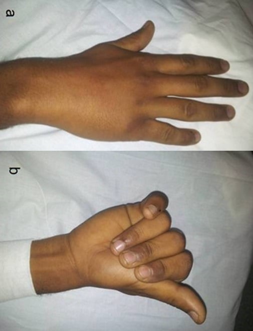
Neurovascular. Check sensation in the territory of the digital nerves and the perfusion of each finger.
The fight bite. Any laceration over an MCP joint in a young male is a human bite until proven otherwise, and should be assumed to be an infected one (Eikenella corrodens). The tooth penetrates the joint capsule with the MCP flexed and drags bacteria in; when the finger extends, the tract seals.
WASHFight Bite Management
Hook:WASH the fight bite properly - don't believe the glass story
Investigations
Radiographs. PA, lateral and oblique views of the hand, assessing all five metacarpals systematically. On each film define:
- Location: head, neck, shaft or base
- Angulation: apex dorsal is the most common
- Shortening, compared with the adjacent metacarpal
Rotation cannot be assessed on the radiograph; it is judged clinically, as above.
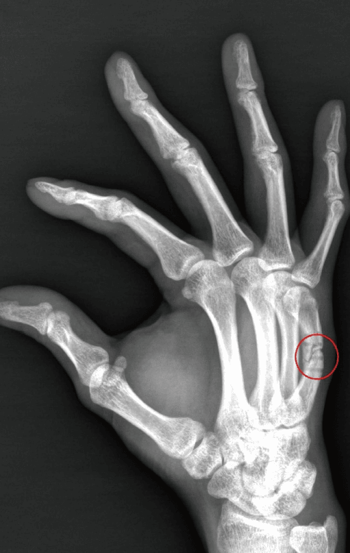
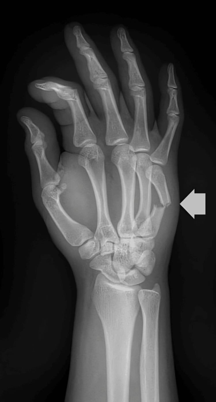
Special views. Two views answer specific questions about the MCP joint and the head.
- Indication
- MCP collateral ligament injury
- What It Shows
- Collateral ligament avulsions
- Indication
- Metacarpal head assessment
- What It Shows
- Articular surface fractures
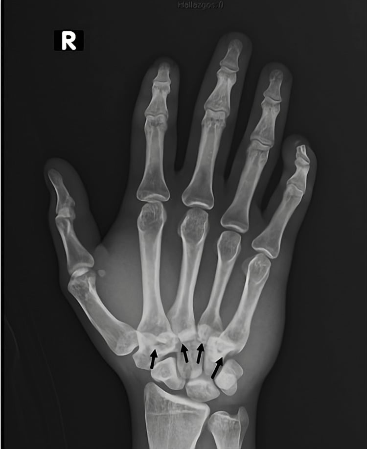
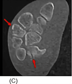
CT. CT shows CMC joint involvement in complex base fractures, characterises intra-articular head fractures, and is used for preoperative planning before ORIF.
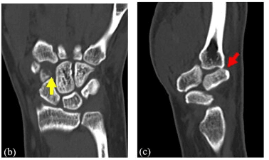
Differential Diagnosis
The dorsal hand injury is rarely a true diagnostic dilemma, but the exam tests whether you can separate the metacarpal fracture from look-alikes that change management.
- Distinguishing features
- Apex-dorsal angulation, lost knuckle prominence, punch mechanism
- Why it matters
- Usually conservative; high angulation tolerance
- Distinguishing features
- Laceration over MCP, often a denied bite, joint signs/pus
- Why it matters
- Surgical emergency: washout, never close
- Distinguishing features
- Thumb CMC pain, intra-articular base fracture-dislocation
- Why it matters
- Needs anatomic reduction +/- fixation
- Distinguishing features
- 5th CMC, base displaced by ECU pull
- Why it matters
- Often unstable - pinning/ORIF
- Distinguishing features
- Carpometacarpal step-off, missed on PA, seen on lateral/oblique
- Why it matters
- Easily overlooked; needs reduction
- Distinguishing features
- Extensor subluxation, no fracture on X-ray, painful MCP
- Why it matters
- Soft-tissue injury - splint vs repair, not a fracture
- Distinguishing features
- Tenderness distal to MCP, finger-level deformity
- Why it matters
- Different alignment and rehab principles
Management
The decision. A stable fracture with acceptable angulation for its ray, no rotational deformity and a closed injury (no fight bite) is treated conservatively. The operative indications below mark the fractures that are not.
Conservative treatment. The splint follows the ray:
- Buddy strapping for stable shaft fractures: simple, and allows early motion
- Ulnar gutter splint for the fourth and fifth metacarpals
- Radial gutter splint for the second and third metacarpals
The intrinsic-plus position (position of safety) is usually used for immobilisation, but some evidence suggests buddy taping alone allows earlier return to work for boxer's fractures. Immobilise for 3-4 weeks, with an X-ray at 1 week to check the position.
Reduction. The Jahss manoeuvre reduces a neck fracture. Flex the MCP and PIP joints to 90° and apply dorsal pressure on the proximal phalanx, which pushes the metacarpal head dorsally.
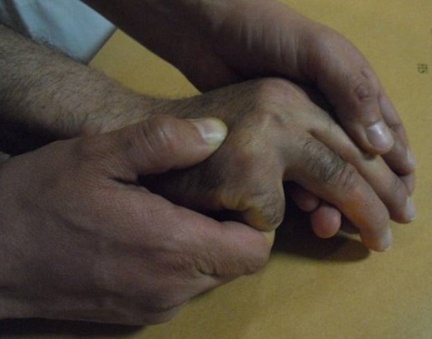
Operative indications. The indications for surgery:
- Rotational deformity of any degree (the cardinal rule)
- Angulation beyond the limit for the ray (for example, greater than 50° at the fifth neck)
- Shortening greater than 5 mm (relative)
- Open fractures, including fight bites
- Multiple metacarpal fractures
- Displaced intra-articular fractures of the head or base
ROMISurgical Indications
Hook:ROMI goes to theatre - Rotation, Open, Multiple, Intra-articular
- First-Line
- Buddy tape or ulnar gutter
- Surgical Option
- Rarely needed
- First-Line
- Surgery indicated
- Surgical Option
- K-wires or plate
- First-Line
- Often surgery
- Surgical Option
- Lag screws or plate
- First-Line
- Assess stability
- Surgical Option
- K-wires or ORIF
Surgical Technique
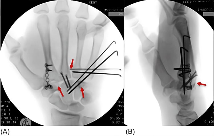
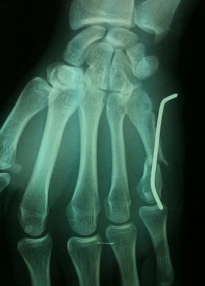
K-wires. Placed retrograde down the medullary canal, or transversely to pin the fractured metacarpal to its neighbour. The technique is minimally invasive, and the wire is removed later.
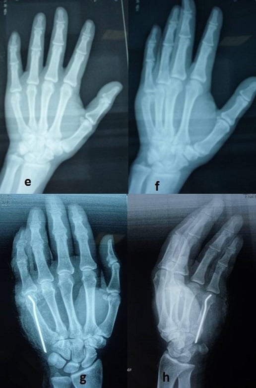
Plates and screws. ORIF is for shaft fractures and unstable patterns, and a plate suits transverse and comminuted fractures that need absolute stability. It gives an anatomical reduction, at the risk of tendon adhesions and prominent hardware.
Lag screws. Lag screws suit long oblique and spiral fractures (greater than 2x diameter).
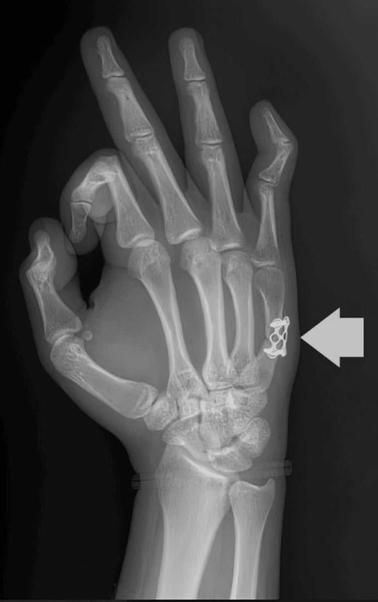
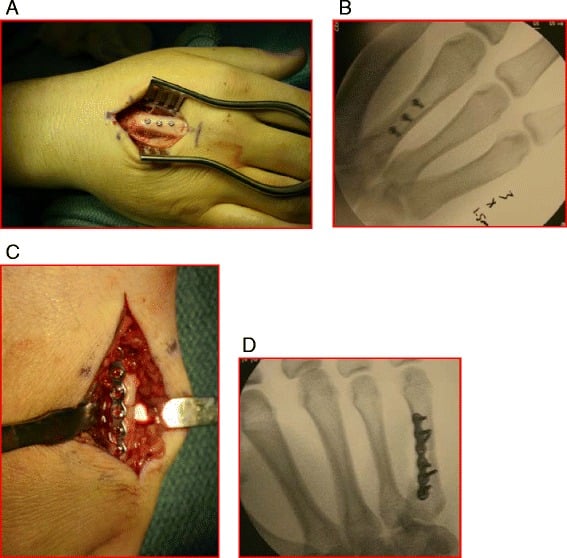
Intramedullary screws. A headless compression screw down the canal is a newer technique for neck and shaft fractures.
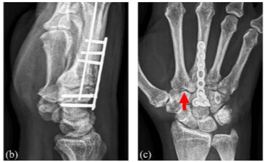
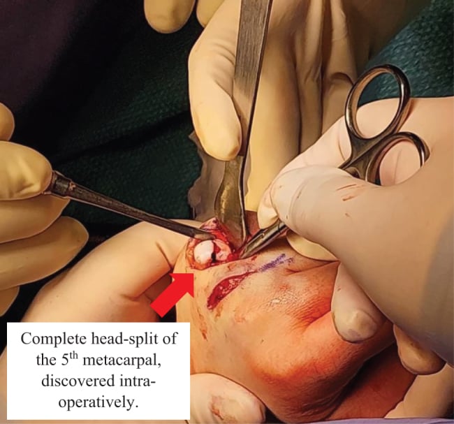
Metacarpal Head Fractures: the Intra-articular Pattern
The head fracture is the intra-articular pattern that often needs ORIF, and it behaves very differently from the far commoner neck and shaft fractures.
Why they matter. Metacarpal head fractures are uncommon, of the order of one in a hundred hand fractures, but they involve the small, highly congruent MCP joint. They carry a real risk of stiffness, articular incongruity and post-traumatic arthritis. The index metacarpal head is the most frequently involved, and a laceration over the joint should raise the suspicion of a fight bite with cartilage penetration.
The blood supply. The metacarpal head has a largely terminal, dorsally entering blood supply, which is why a comminuted head fragment can be devascularised. Comminuted head fractures therefore carry the highest risk of avascular necrosis.
Pattern recognition. The McElfresh and Dobyns scheme is descriptive rather than prognostic, but it maps directly onto treatment:
- Epiphyseal fractures
- Collateral-ligament avulsion fractures
- Oblique (sagittal, unicondylar) fractures
- Vertical (coronal-plane) fractures
- Comminuted fractures
- A boxer's neck fracture extending into the head
- Fractures with bone loss
- Fractures associated with joint or soft-tissue loss
Management. Treatment follows displacement and comminution:
- Undisplaced: brief immobilisation followed by early protected motion, to preserve the articular cartilage and prevent MCP stiffness
- Displaced (articular step-off of about 1 mm or more, or an unstable unicondylar sagittal or coronal split): open reduction and internal fixation, because an incongruent MCP joint leads to stiffness and early arthritis. Headless compression screws, mini-fragment screws, K-wires or bioabsorbable pins aim for anatomic joint restoration and a construct stable enough for early motion
- Severely comminuted: internal fixation may be impossible, and these are often better served by distraction external fixation than by attempting rigid fixation of small devascularised fragments. Ligamentotaxis maintains the joint space and allows motion; osteochondral reconstruction or arthroplasty is reserved for salvage
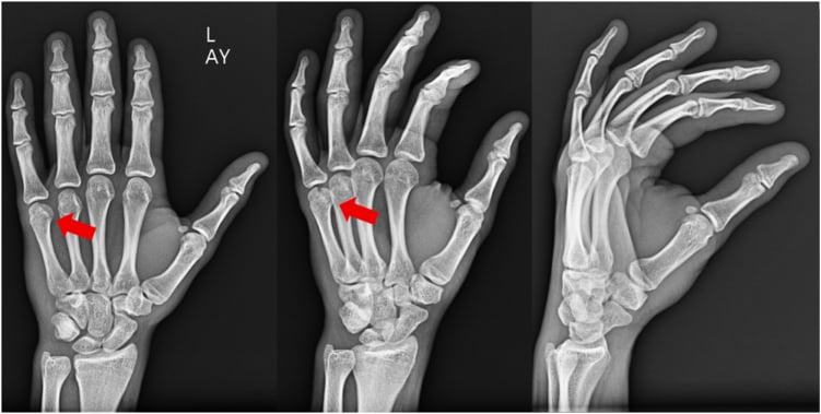
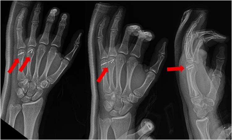
Why Multiple and Border-Ray Fractures Are Unstable
Neighbours as splints. Each metacarpal is tethered to its neighbours proximally by the interosseous muscles and distally, at the level of the heads, by the deep transverse metacarpal (intermetacarpal) ligament, which links the volar plates of the index-to-little MCP joints. Intact adjacent metacarpals therefore act as internal splints, resisting shortening, angulation and rotation at a fracture in the ray between them. This is why an isolated central (middle or ring) metacarpal shaft fracture is one of the most stable fractures in the hand, and is usually treated non-operatively.
Multiple fractures. When two or more adjacent metacarpals are fractured, the splinting buttress is lost on both sides. The injury is inherently unstable and far more likely to shorten, angulate and rotate, which is why a double or triple metacarpal fracture is fixed and why any suspicion of instability lowers the threshold for operative fixation.
Border rays. The index and little finger are held by the intermetacarpal ligament on only one side. Isolated second and fifth metacarpal fractures are therefore less well splinted, and displace and rotate more readily than the well-protected central rays.
Complications
- Effect
- Loss of knuckle prominence, pseudoclawing
- Management
- Accept if functional, Osteotomy if severe
- Effect
- Scissoring, weak grip
- Management
- Corrective osteotomy (Derotation)
- Effect
- Inability to fully extend MCP
- Management
- Shortening greater than 4-5mm decreases extensor tension
- Effect
- Tendon adhesions, joint contracture
- Management
- Early ROM, stable fixation
Rotational malunion. The conventional teaching figure is that every 5° of rotation at the metacarpal produces about 1.5cm of fingertip overlap, an approximation rather than a measured value from the series cited on this page. Correct it with a dorsal-approach derotation osteotomy and plate fixation.
Stiffness. Stiffness from tendon adhesions is the most common post-surgical complication. Stable fixation and early protected motion mitigate it.
Hardware. Dorsal plates cause adhesions and prominence; K-wires cause pin-site infection and migration.
Postoperative Care
Immobilisation, 0-3 weeks. Protect the fracture in an ulnar or radial gutter splint, elevate the hand to reduce swelling and avoid heavy loading. Start active finger range of motion if the fixation is stable.
Mobilisation, 3-6 weeks. K-wires come out at 4-6 weeks. Wean from the splint, begin active range-of-motion exercises and refer to hand therapy.
Strengthening, 6-12 weeks. Progressive grip strengthening, functional exercises and a return to light activities.
Why move early. Stable fixation allows early range of motion, which reduces stiffness and adhesions and gives better functional outcomes.
- Immobilisation
- Immediate ROM
- Return to Work
- 1-2 weeks light duties
- Immobilisation
- 3-4 weeks
- Return to Work
- 4-6 weeks
- Immobilisation
- 4-6 weeks to removal
- Return to Work
- 6-8 weeks
- Immobilisation
- 1-2 weeks splint
- Return to Work
- 8-12 weeks manual
Return to sport. Contact sport at 8-12 weeks, boxing at 12-16 weeks minimum, and protective splinting may be needed initially.
Outcomes
Overall. Outcomes are excellent for most metacarpal fractures, with a 95% union rate with appropriate treatment. Stiffness is the main complication, especially after surgery.
Boxer's fracture. Function is excellent even with radiographic malunion, and the loss of knuckle prominence is cosmetic only. Surgery is not superior to conservative treatment for typical patterns, and has a higher complication rate without clear benefit. The Cochrane review (Poolman, 2005) found conservative treatment effective, with no difference between splinting and buddy taping.
Shaft fractures. Outcomes are good if rotation is corrected. The risk of stiffness is higher with plate fixation; K-wires are associated with less stiffness but give less rigid fixation.
What decides the result. Correcting rotation is paramount, early motion improves outcomes, and the patient's occupation and demands matter.
- Union Rate
- Greater than 95%
- Main Concern
- Cosmetic only
- Union Rate
- Greater than 95%
- Main Concern
- Pin complications
- Union Rate
- Greater than 95%
- Main Concern
- Stiffness, adhesions
- Union Rate
- 90-95%
- Main Concern
- MCP arthritis
Guidelines, Registries & Global Practice
Global Epidemiology
- Metacarpal fractures are 18-44% of all hand fractures; the hand accounts for ~20% of all skeletal fractures presenting to emergency departments worldwide.
- Strong young-male preponderance (peak 10-29 years), reflecting interpersonal violence/punch and sports mechanisms; an alcohol-related, weekend, urban pattern is reported across high- and middle-income settings.
- The fifth metacarpal neck (Boxer's fracture) is the single most common pattern; non-thumb metacarpals comprise ~88% of metacarpal fractures.
- Position on Boxer's & shaft fractures
- Non-operative for most isolated closed neck/shaft fractures with acceptable alignment
- Emphasis
- Rotation and intra-articular displacement drive surgery
- Position on Boxer's & shaft fractures
- Functional treatment and early motion for stable Boxer's fractures
- Emphasis
- Avoid over-immobilisation; hand-therapy access
- Position on Boxer's & shaft fractures
- Operative options (lag screw, plate, antegrade IM nail/screw) defined by pattern and stability
- Emphasis
- Anatomic reduction of rotation; absolute vs relative stability
- Position on Boxer's & shaft fractures
- Conservative-first; IM techniques increasingly favoured when fixation needed
- Emphasis
- Minimise stiffness, early rehabilitation
There is broad international agreement (and no major guideline disagreement) on the core principles: zero tolerance of rotation, ray-dependent angulation thresholds, and conservative-first management of stable fractures. Differences are largely in surgical technique preference rather than indications.
Controversies and Areas of Uncertainty
Angulation thresholds are eminence-based, not evidence-based. The widely quoted 10-10-30-50 (or 10-20-30-40) figures vary between texts. The Cochrane review (Poolman) found no high-quality data defining a precise cut-off, and many patients tolerate angulation well beyond classic limits with good function.
The best non-operative method is unsettled. Poolman found no regimen (buddy taping, soft wrap, ulnar gutter, functional brace) superior to another. Practice varies from immediate mobilisation to 3-4 weeks of splinting.
IM screw, K-wire or plate. Antegrade headless IM screws give excellent union and motion (Beck), but the evidence is largely Level III-IV, and the concerns include articular cartilage violation at the entry point and cost. No adequately powered RCT defines the optimal implant.
Shortening tolerance. Strauch quantifies the extensor lag, but the clinically acceptable limit, commonly cited as about 5 mm, is extrapolated, and MCP hyperextension may compensate.
Fight-bite antibiotic duration and washout setting. There is universal agreement on washout plus amoxicillin-clavulanate. The optimal antibiotic duration, and whether minor early injuries can be managed without theatre, remain debated.
Conservative Treatment of Fifth Metacarpal Neck Fractures (Cochrane)
- Five randomised/quasi-randomised trials, 252 participants, comparing functional treatment with immobilisation
- No single non-operative regimen was statistically superior to another in result
- Trials were of limited quality and size; validated hand function was not reported in any study
Rotational Deformity Following Metacarpal Fracture
- Prospective series of 91 patients with 98 metacarpal fractures
- A quarter had minor rotation under 10 degrees; only 5 had more, and just 2 needed operative correction for rotational instability
- Rotation must be assessed with an end-on view of the fingernail, as MCP joint motion is often restricted after fracture
Effect of Metacarpal Shortening on the Extensor Mechanism
- Cadaver model (9 hands), 2nd and 5th metacarpal shaft fractures shortened in 2 mm increments to 10 mm
- Average of 7 degrees of MCP extensor lag produced for every 2 mm of metacarpal shortening
- MCP hyperextension capacity may clinically compensate for some of this lag
Human Bite Wounds and Eikenella corrodens
- Clenched-fist (fight-bite) injuries to the hand carry far higher infection and complication rates than bites elsewhere
- Infections are polymicrobial; Eikenella corrodens is the characteristic pathogen of human bites
- Hand bites warrant aggressive irrigation/debridement and beta-lactam plus beta-lactamase-inhibitor cover
Intramedullary Screw Fixation of Metacarpal Fractures
- Systematic review of 9 studies, 169 metacarpal fractures (74% small finger; mostly neck fractures)
- Radiographic union in 100% of reported cases; mean MCP flexion 86 degrees and grip 96% of contralateral
- No serious complications; only minor complications including asymptomatic hardware removal
Metacarpal Fractures: Treatment and Complications
- Metacarpal fractures comprise 18-44% of all hand fractures; the fifth finger is most commonly involved
- Around 88% of metacarpal fractures involve the non-thumb metacarpals; most are simple, closed and stable
- Persistent controversy and limited high-level evidence to define the optimal treatment algorithm
MCQ Practice Points
Q: What is the maximum acceptable angulation for a fifth metacarpal neck fracture (boxer's fracture) treated non-operatively?
A: Up to 70 degrees of apex dorsal angulation is acceptable for fifth metacarpal neck fractures due to the compensatory motion at the 4th and 5th CMC joints (30-40 degrees of flexion-extension). The fourth metacarpal accepts up to 40 degrees, the third metacarpal 15 degrees, and the second metacarpal (index) only 10-15 degrees because the 2nd and 3rd CMC joints have minimal motion. Rotational deformity is never acceptable and always requires correction.
Q: What is the indication for surgical fixation of metacarpal shaft fractures?
A: Surgical indications include: Rotational malrotation (any degree - clinical scissoring), angulation exceeding acceptable limits (varies by ray), multiple metacarpal fractures, open fractures, intra-articular fractures with displacement, and shortening greater than 5mm (causes extensor lag). Spiral fractures are prone to rotational deformity while transverse fractures are prone to angular deformity. Lag screws are ideal for long oblique/spiral patterns; plates for transverse/short oblique/comminuted patterns.
Q: How do you clinically assess for rotational malalignment in metacarpal fractures?
A: Finger cascade test: With the MCP joints flexed, all fingers should point toward the scaphoid tubercle. Scissoring: Overlapping of fingers during flexion indicates malrotation. The conventionally quoted rule is that each 5 degrees of rotational deformity at the metacarpal produces roughly 1.5cm of digital overlap at the fingertip - a teaching approximation rather than a measured figure, and not from Royle's series, which found clinically significant rotation in only 5 of 98 fractures. Compare tenodesis effect (passive wrist extension causes finger flexion) to the contralateral hand. Rotational deformity is the most poorly tolerated malunion and requires correction. Judge it on the end-on view of the nail plates, because MCP movement is often restricted after fracture and a composite fist will mislead you.
Q: What is the difference between a Bennett's fracture and a Rolando's fracture?
A: Bennett's fracture: Intra-articular fracture-dislocation at the thumb CMC joint with a single volar-ulnar fragment attached to the AOL while the metacarpal shaft subluxates radially and proximally due to APL pull. Rolando's fracture: Comminuted intra-articular fracture at the same location with T or Y pattern (at least 3 fragments). Both require anatomic reduction. Bennett's is typically fixed with K-wires or screw, while Rolando's may need plate fixation or external fixation for severe comminution.
Q: What is the reverse Bennett fracture and how is it managed?
A: Reverse Bennett fracture is an intra-articular fracture-dislocation at the fifth CMC joint with a volar-ulnar fragment remaining attached to the hamate while the metacarpal base displaces dorsally and proximally (pulled by ECU). Also called a baby Bennett. Treatment follows similar principles to thumb Bennett's: closed reduction and percutaneous pinning if anatomic reduction achieved, or ORIF for irreducible or significantly displaced fractures. Maintain reduction with splinting in slight flexion.
Exam Viva Scenarios
Practise clinical reasoning and management decisions out loud
“A 22-year-old rugby player presents with a swollen right hand after punching a wall. X-ray shows a 5th metacarpal neck fracture with 40 degrees of volar angulation. There is no rotational deformity clinically. How do you manage this?”
“A 30-year-old presents 5 days after a fall onto his hand. X-rays show a short oblique fracture of the third metacarpal shaft with minimal displacement and approximately 15 degrees of apex dorsal angulation. However, on clinical examination, when he makes a fist, you notice that the middle finger crosses over the index finger and the fingernail plane is rotated compared to the adjacent fingers. What is your management?”
“A 24-year-old presents to the emergency department 3 days after 'cutting his hand on glass' at a bar. He has a 1cm laceration over the dorsum of his right 3rd MCP joint that he cleaned himself. The wound now has purulent discharge, the MCP joint is swollen and erythematous, and he has pain and restricted motion. He is febrile at 38.5 degrees. X-rays show soft tissue swelling but no fracture or gas. What is your assessment and management?”
Acceptable Angulation
- Index/Middle: less than 10-15 degrees (Rigid)
- Ring: less than 30-40 degrees
- Little: less than 50-70 degrees (Mobile)
Operation Indications
- ANY Rotation (fingers scissor)
- Open fracture
- Multiple fractures
- Intra-articular step-off
Fight Bite
- Laceration over MCP joint
- Eikenella corrodens
- Formal washout required
Evidence Base
Key Studies
- Conservative treatment effective for Boxer's fractures
- No difference between immobilisation methods
- Surgery adds risk without clear benefit for standard patterns
- Rotation tolerance: Zero degrees acceptable
- 5° rotation ≈ 1.5cm fingertip overlap (conventional teaching figure)
- Shortening greater than 5mm causes extensor lag
- Key Finding
- Conservative = surgical for Boxer's
- Clinical Impact
- Avoid unnecessary surgery
- Key Finding
- K-wire biomechanics effective
- Clinical Impact
- K-wires are reasonable fixation
- Key Finding
- Review of treatment options
- Clinical Impact
- Algorithm-based approach
References
- Ali A, et al. Biomechanical stability of intramedullary K-wire fixation of metacarpal neck fractures. J Hand Surg Br. 2005.
- Kollitz KM, et al. Metacarpal fractures: treatment and complications. Hand (NY). 2014.