Inherited Metaphyseal Dysplasias | Spared Epiphyses & Spine | Not Rickets
- MCD affects metaphyses only — epiphyses and spine are spared (key vs SED)
- Schmid type: most common, AD, COL10A1, normal biochemistry — must differentiate from rickets
- Jansen type: constitutive PTH1R activation (H223R commonest) gives the full triad of HIGH calcium, LOW phosphate and a PTH that is normal or undetectable - the low phosphate is as characteristic as the high calcium and is what separates it from primary hyperparathyroidism. Inheritance is autosomal dominant but most reported cases are sporadic de novo mutations
- McKusick (cartilage-hair hypoplasia): AR, immune deficiency, lymphoma risk, Hirschsprung association
- Biochemistry is NORMAL in all types except Jansen (hypercalcaemia) — the critical discriminator from rickets
- “Metaphyseal dysplasia + normal biochemistry = NOT rickets, think MCD
- “Schmid = COL10A1, Jansen = PTH1R, McKusick = RMRP
- “McKusick type has EXTRA-SKELETAL features (hair, immunity, gut, malignancy)
- “Spine and epiphyses SPARED in all MCD types — if involved, consider SED instead
Overview and Epidemiology
The metaphyseal chondrodysplasias are a group of genetically heterogeneous inherited skeletal dysplasias in which endochondral ossification at the metaphyses is abnormal. The defects lie in genes that control endochondral ossification at the growth plate, so the metaphyses ossify irregularly while the epiphyses and spine stay normal. All types cause short stature of varying severity.
Why they matter. They are frequently misdiagnosed as rickets, or as child abuse (metaphyseal fractures), because the radiographic appearances overlap. Correct identification hinges on recognising the pattern of isolated metaphyseal involvement with normal biochemistry and spared epiphyses and spine. Each subtype then carries its own genetics, systemic features and management.
Who. The three types are very unequal in frequency:
- Schmid type is the most common; the exact incidence is unknown and it is likely under-diagnosed
- Jansen type is extremely rare, with fewer than 30 reported cases worldwide
- McKusick type, cartilage-hair hypoplasia, has its highest prevalence in the Finnish (approximately 1 in 23,000) and Old Order Amish populations because of founder effects
- Overall MCD prevalence is approximately 1 in 50,000 to 1 in 100,000 live births
Pathophysiology
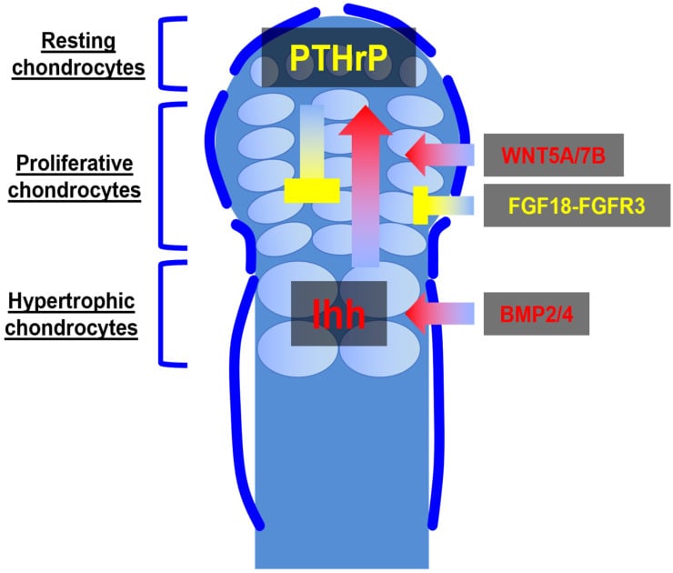
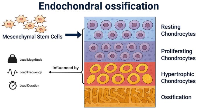
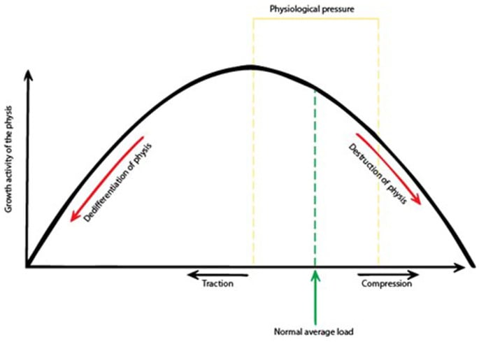
The growth plate. The physis has distinct zones: resting, proliferative, hypertrophic and calcification. The metaphyseal chondrodysplasias disrupt it at the zone of hypertrophy and the transition from cartilage to bone, and each type gets there by its own route:
- Schmid: type X collagen, the product of hypertrophic chondrocytes, is defective and matrix mineralisation is impaired
- Jansen: constitutive PTH1R signalling disrupts chondrocyte differentiation
- McKusick: the RMRP gene affects ribosomal RNA processing, impairing chondrocyte proliferation
Despite the different mechanisms, all three produce the same pattern of metaphyseal irregularity with preserved epiphyses.
- Schmid Type
- COL10A1 (chromosome 6q21)
- Jansen Type
- PTH1R (chromosome 3p21)
- McKusick / CHH
- RMRP (chromosome 9p13)
- Schmid Type
- Type X collagen (hypertrophic chondrocyte matrix)
- Jansen Type
- PTH/PTHrP receptor (chondrocyte differentiation)
- McKusick / CHH
- Mitochondrial RNA-processing endoribonuclease
- Schmid Type
- Autosomal dominant
- Jansen Type
- Autosomal dominant
- McKusick / CHH
- Autosomal recessive
- Schmid Type
- Hypertrophic zone (matrix mineralisation)
- Jansen Type
- Proliferative to hypertrophic transition
- McKusick / CHH
- Chondrocyte proliferation (all zones)
- Schmid Type
- None (skeletal only)
- Jansen Type
- Hypercalcaemia (constitutive receptor activation)
- McKusick / CHH
- Hair, immune system, gut, haematopoietic
Why the epiphyses are spared. The epiphysis develops through a secondary ossification centre that is mechanistically separate from the physis. The MCD gene defects primarily disrupt the physis, the primary growth plate between metaphysis and epiphysis, and not the epiphyseal ossification centre itself, which is why the metaphysis is abnormal and the epiphysis beside it is normal.
Why the spine is normal. For Schmid type the evidence gives the cleanest answer: type X collagen is expressed only by hypertrophic chondrocytes in regions of endochondral ossification, so the disease follows that expression and the vertebrae are untouched (Warman, in the evidence section). Whatever the type, platyspondyly suggests SED or a mucopolysaccharidosis, not MCD.
Classification and Types
Three types share the metaphyseal pattern and differ in gene, inheritance, severity and what lies outside the skeleton. In one line each: Schmid is the common, purely skeletal type; Jansen is the rare, severe type with hypercalcaemia; McKusick is the recessive type in which the skeleton is the least of the problem.
Schmid Metaphyseal Chondrodysplasia
Genetics. Autosomal dominant. COL10A1 on 6q21 encodes the alpha-1 chain of type X collagen, and the mutations are typically missense in the C-terminal NC1 domain.
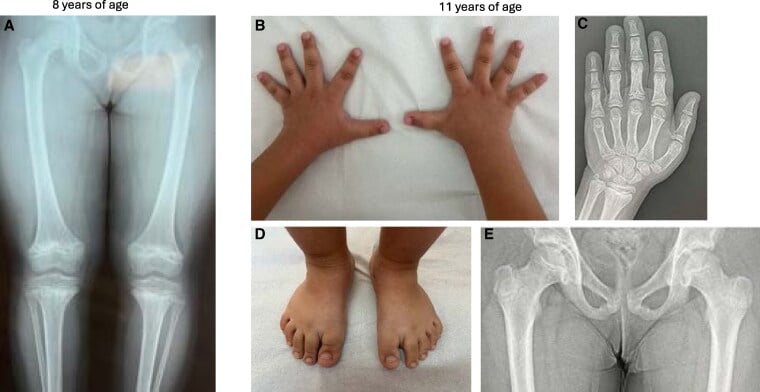
Presentation. Often between 2 and 5 years, when weight-bearing begins and the bowing becomes apparent; familial cases may be detected earlier. The child has coxa vara, genu varum, anterior femoral bowing and a waddling gait, with widened metaphyses and flared ankles and wrists. Short stature is mild to moderate, with a final height typically of 135-155 cm.
What is not there. Biochemistry is completely normal, with calcium, phosphate, ALP and vitamin D all within the reference range, and the condition is purely skeletal: no extra-skeletal features, normal intelligence, normal lifespan. That normal biochemistry is the discriminator from rickets.
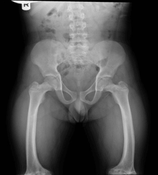
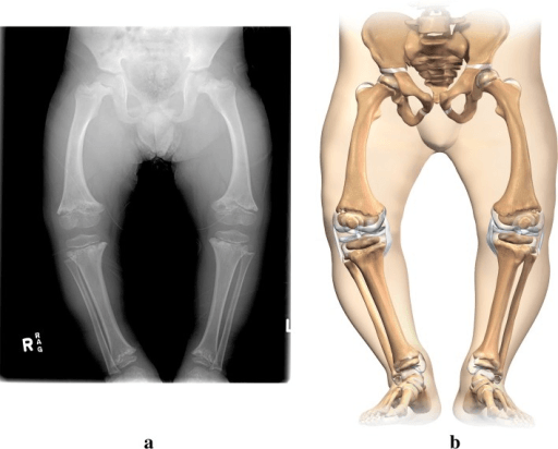
The Schmid/Jansen/McKusick trio are metaphyseal chondrodysplasias (defective endochondral ossification), but examiners also expect the non-chondrodysplastic metaphyseal dysplasias caused by failure of metaphyseal remodelling/modelling - recognised by the Erlenmeyer flask deformity (under-tubulated, widened, flask-shaped metaphyses, classically of the distal femur):
- Pyle disease (metaphyseal dysplasia): autosomal recessive (SFRP4), with gross Erlenmeyer flask widening of the metaphyses but little or no skull involvement and often surprisingly mild symptoms (genu valgum, fragility) - the "metaphyses look dramatic, the patient is well" entity.
- Craniometaphyseal dysplasia (CMD): autosomal dominant (ANKH) - Erlenmeyer flask metaphyses plus craniofacial hyperostosis/sclerosis that can cause cranial-nerve compression (facial palsy, hearing/visual loss) and nasal obstruction - the skull, not the limb, drives morbidity.
- The broader Erlenmeyer flask differential also includes osteopetrosis, Gaucher disease, thalassaemia and fibrous dysplasia - so an Erlenmeyer flask femur is a pattern to work up, not a single diagnosis.
Exam point: distinguish the metaphyseal chondrodysplasias (Schmid/Jansen/McKusick - endochondral defect, growth-plate irregularity) from the remodelling metaphyseal dysplasias that give an Erlenmeyer flask deformity - Pyle disease (SFRP4, no skull) and craniometaphyseal dysplasia (ANKH, with cranial hyperostosis and nerve compression).
Clinical Assessment
History. Birth weight is often normal and growth falls off the curves post-natally. The family history sorts the dominant types (Schmid, Jansen) from the recessive one (McKusick), and the age of onset sorts them again: Schmid at 2-5 years, Jansen neonatal, McKusick at 6-12 months. Then ask about:
- Gait: a waddle from coxa vara, and whether the bowing is progressing
- Infections: recurrent sinopulmonary infections point to the McKusick immune deficiency
Examination. Measure and plot the stature and decide whether the child is short-limbed or short-trunked. The spine should be normal; if it is not, reconsider the diagnosis and think of SED. Then work through:
- Lower limbs: genu varum, coxa vara, anterior femoral bowing, waddling gait
- Upper limbs: wide wrists and limited elbow extension (Schmid)
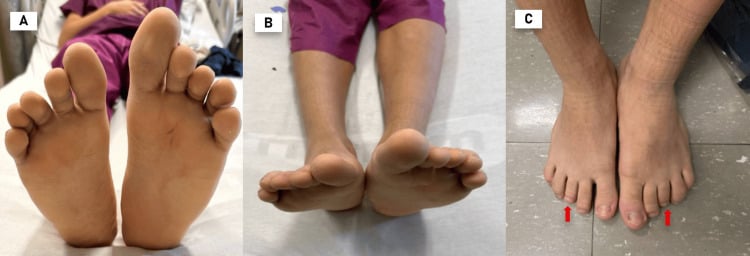
- Hair: fine, sparse, hypopigmented (McKusick)
- Joints: ligamentous laxity (McKusick), and the range of motion
If any of the following are present, reconsider MCD and broaden the differential:
- Epiphyseal involvement (irregular, fragmented epiphyses) = consider SED, multiple epiphyseal dysplasia, or mucopolysaccharidosis
- Spinal abnormalities (platyspondyly, odontoid hypoplasia) = consider SED, mucopolysaccharidosis, or spondylo-metaphyseal dysplasia
- Abnormal biochemistry (low Ca, low PO4, high ALP) = likely rickets, not MCD
- Craniofacial dysmorphism with rhizomelia = consider achondroplasia (large head, trident hand, frontal bossing, FGFR3 mutation)
- Periosteal reaction / multiple fractures = consider non-accidental injury or osteogenesis imperfecta
- Metaphyses
- Irregular, flared, sclerotic
- Epiphyses
- Normal
- Spine
- Normal
- Biochemistry
- Normal
- Key Discriminator
- AD family history, COL10A1, coxa vara
- Metaphyses
- Cupped, frayed, splayed
- Epiphyses
- Normal (late widening)
- Spine
- Normal
- Biochemistry
- Abnormal (low Ca/PO4, high ALP)
- Key Discriminator
- Biochemistry is ALWAYS abnormal
- Metaphyses
- Metaphyseal changes similar to MCD
- Epiphyses
- Normal early
- Spine
- Normal
- Biochemistry
- Low PO4, normal Ca, normal PTH
- Key Discriminator
- Renal phosphate wasting, PHEX mutation (X-linked)
- Metaphyses
- Normal
- Epiphyses
- Delayed, irregular, fragmented
- Spine
- Platyspondyly
- Biochemistry
- Normal
- Key Discriminator
- Epiphyses AND spine involved
- Metaphyses
- Flared, 'ball-in-socket' hip
- Epiphyses
- Normal (may appear delayed)
- Spine
- Lumbar canal stenosis (not platyspondyly)
- Biochemistry
- Normal
- Key Discriminator
- Rhizomelic, macrocephaly, trident hand, FGFR3
- Metaphyses
- May show metaphyseal changes
- Epiphyses
- Normal
- Spine
- Bowing, compression fractures
- Biochemistry
- Normal (except type VI with elevated ALP)
- Key Discriminator
- Blue sclerae, fractures, COL1A1/COL1A2
A metaphyseal abnormality in a young child sits at a medicolegally critical crossroads, and examiners test both directions of error:
- The classic metaphyseal lesion (CML) of abuse: the "corner" or "bucket-handle" fracture is a planar fracture through the primary spongiosa created by shearing/twisting forces. It is focal and often asymmetric, highly specific for non-accidental injury in an infant, and is a different lesion from the symmetric growth-plate-wide change of a dysplasia.
- The discriminator: metaphyseal chondrodysplasia (and rickets) produce symmetric, bilateral, growth-plate-wide metaphyseal irregularity affecting multiple bones in a patterned way - whereas CMLs of abuse are focal corner/bucket-handle lesions at specific sites, often with other features of inflicted injury (rib fractures, differing fracture ages, retinal haemorrhages).
- Both errors harm: missing genuine abuse is catastrophic, but wrongly attributing a dysplasia's or rickets' metaphyseal changes to abuse devastates a family - so the work-up is a full skeletal survey, biochemistry (Ca/PO4/ALP/PTH/vitamin D) and genetic/specialist (paediatric radiology + safeguarding) review before any conclusion.
- Rickets caveat: rickets both mimics CMLs (fraying/cupping) and predisposes to true fractures, compounding the diagnostic difficulty - hence biochemistry is mandatory.
Exam point: a metaphyseal abnormality in an infant requires you to distinguish the symmetric, biochemistry-checked pattern of dysplasia/rickets from the focal corner/bucket-handle lesions of abuse - get a skeletal survey, biochemistry and expert review, because both missing abuse and falsely accusing a dysplasia/rickets child are serious harms.
Investigations
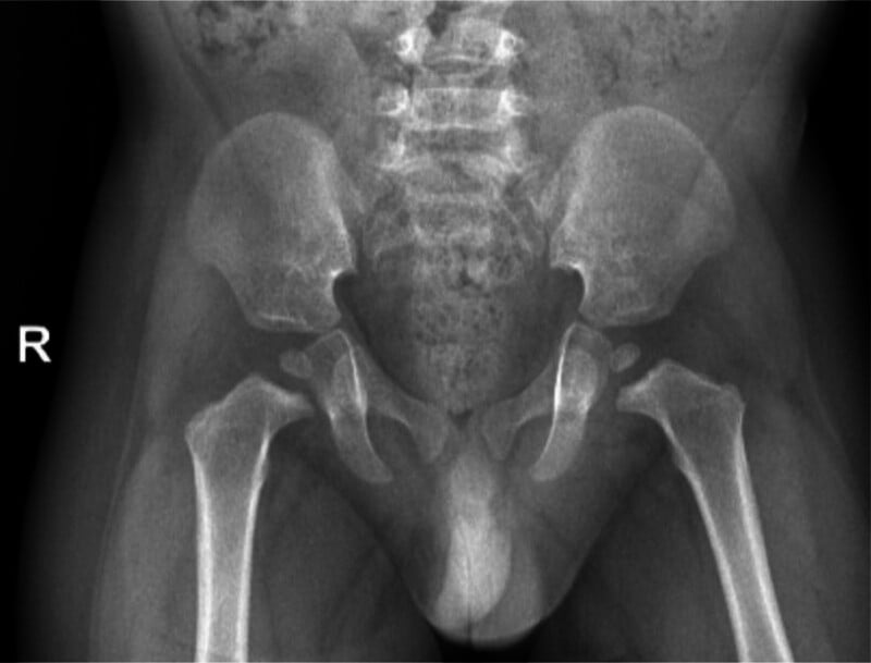
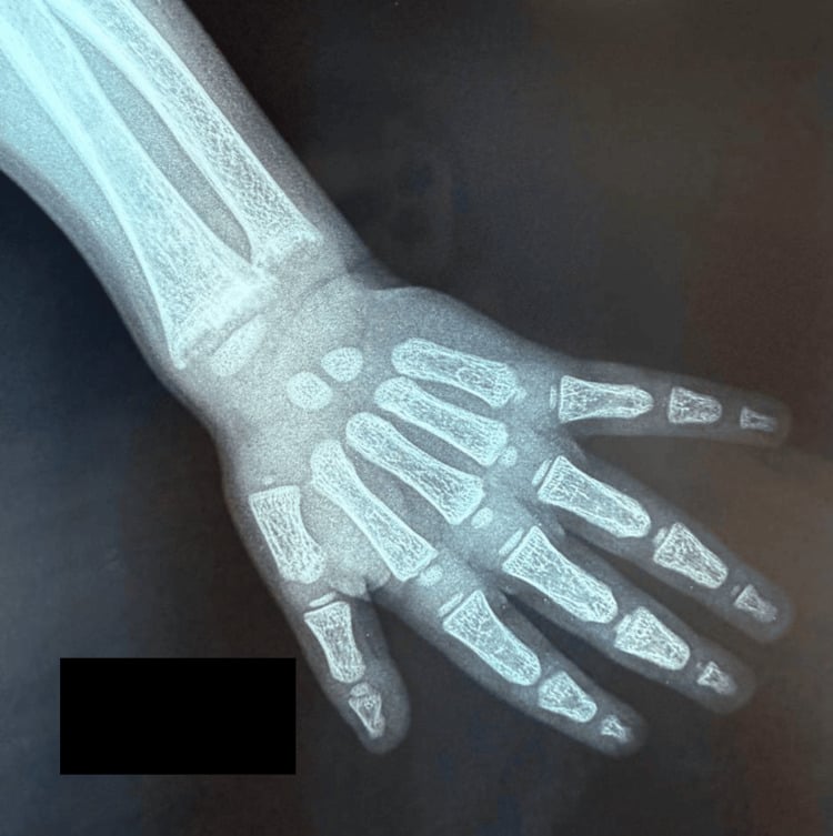
Radiographs first. PA and lateral views of the knees, hips and wrists, a full-length standing lower-limb alignment film and a lateral thoracolumbar spine. Look for metaphyseal irregularity, flaring, sclerosis and cysts, coxa vara, femoral and tibial bowing, and cone-shaped epiphyses in the hands (McKusick). Then critically assess: are the epiphyses truly normal, and is the spine truly normal? If not, reconsider the diagnosis.
The pattern differs by type:
- Schmid: irregular, widened, flared metaphyses with sclerotic and cystic change; growth plates that appear widened, especially at the hips, knees and ankles; coxa vara with horizontal growth plates; anterior bowing of the femora and tibiae; normal epiphyses and spine
- Jansen: severely expanded, irregular, "crumpled" or "fragmented" metaphyses with markedly disorganised growth plates; thin diaphyseal cortices and osteopenia from the hypercalcaemia; a thickened cranial vault with hyperostosis; normal epiphyses and spine. The changes may mimic hyperparathyroidism (subperiosteal resorption)
- McKusick: cupped, flared, scalloped metaphyses with irregular ossification; short tubular bones in the hands with the distinctive cone-shaped epiphyses; anterior flaring of the ribs ("cupped" costochondral junctions); normal spine, and normal epiphyses apart from the cone-shaped ones in the hands; ligamentous laxity that may cause joint subluxations
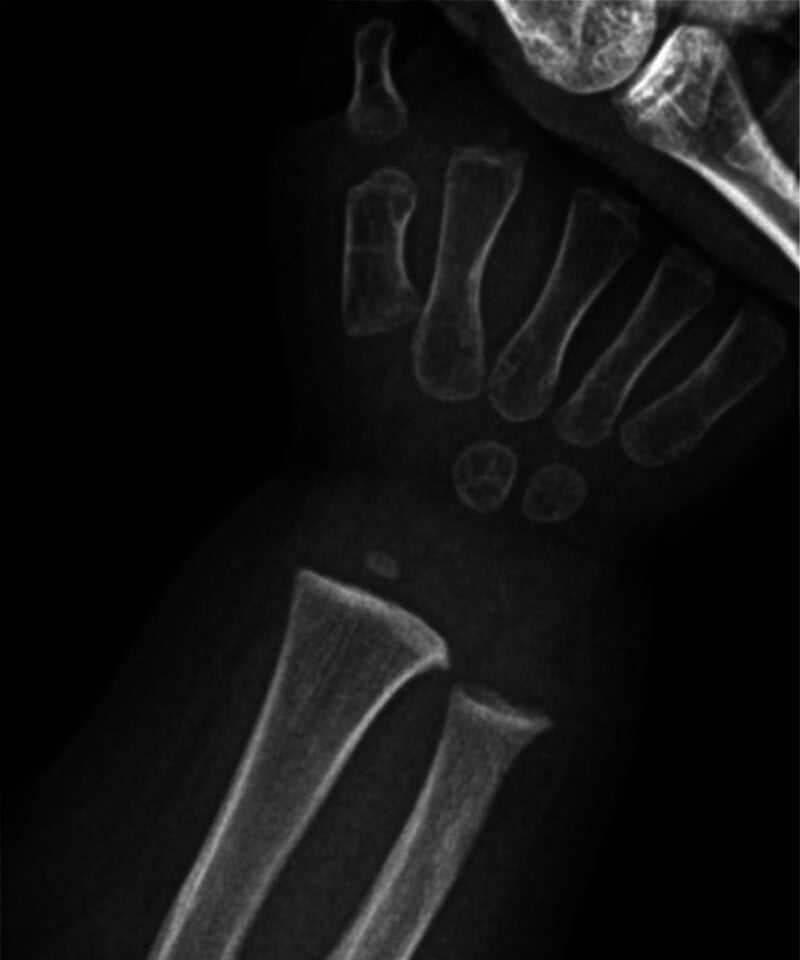
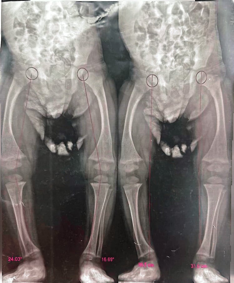
MCD or rickets on the film. Rickets produces metaphyseal cupping, fraying and splaying with Looser zones and generalised osteopenia, and the bone changes are diffuse. MCD produces metaphyseal irregularity that may include sclerosis and cysts rather than only cupping and fraying, with no Looser zones and no osteomalacia, and the changes are isolated to the metaphyses; clinically there is no craniotabes and no widened fontanelle.
Biochemistry. Serum calcium, phosphate, alkaline phosphatase, PTH, 25-hydroxyvitamin D, creatinine and urea. In Jansen type the panel shows hypercalcaemia with suppressed PTH, and there may be hypercalciuria. Any abnormality of calcium, phosphate or ALP means working up rickets, renal osteodystrophy or metabolic bone disease first.
In Schmid and McKusick MCD, calcium, phosphate, alkaline phosphatase, parathyroid hormone and vitamin D are all completely normal. In rickets, at least one parameter is always abnormal. A child with metaphyseal changes on X-ray and normal biochemistry does not have rickets: rickets is excluded, and you should consider MCD.
Genetic testing. Confirms the suspected diagnosis, separates the subtypes and enables genetic counselling and prenatal diagnosis:
- Schmid: targeted COL10A1 sequencing
- Jansen: PTH1R sequencing
- McKusick: RMRP sequencing, including the promoter region
- Unclear phenotype: a skeletal dysplasia gene panel
The McKusick work-up. Lymphocyte subsets (CD4, CD8, CD19, NK cells), immunoglobulin levels and vaccine-response testing characterise the immune deficiency; a full blood count and reticulocyte count look for the macrocytic anaemia; constipation prompts consideration of rectal biopsy to exclude Hirschsprung disease. FBC and immune monitoring then continue annually, for life.
Management Algorithm
Schmid Type Management
The principle is orthopaedic management of deformity. There is no medical treatment to give, because the biochemistry is normal and there is no systemic disease.
At diagnosis. Confirm with normal biochemistry and COL10A1 testing. Counsel the family that inheritance is autosomal dominant, a 50 percent transmission risk, and that intelligence and lifespan are normal. Document the severity of the deformity with full-length standing radiographs and gait analysis as the baseline.
Conservative care. Physiotherapy for gait training, strengthening and joint range of motion; orthotics may help gait instability from coxa vara or severe bowing; no specific restrictions, and encourage normal activity. Review clinically and radiographically every year during growth and follow the coxa vara with the Hilgenreiner-epiphyseal angle.
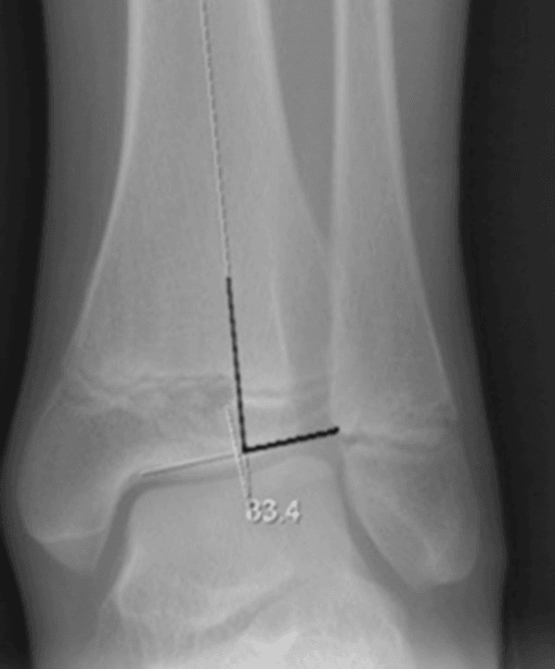
Surgery. Osteotomy during remaining growth carries a significant risk of recurrence, so correction is ideally deferred until the growth plates have closed or the child is near skeletal maturity, unless the deformity is severe or progressive. The options:
- Coxa vara: proximal femoral valgus osteotomy if the Hilgenreiner-epiphyseal angle is greater than 60 degrees or the deformity is progressive
- Genu varum: proximal tibial or distal femoral corrective osteotomy; in the skeletally immature, consider guided growth (hemiepiphysiodesis with plates or staples), which corrects angular deformity gradually without an osteotomy
- Afterwards: protected weight-bearing, physiotherapy and long-term follow-up for recurrence
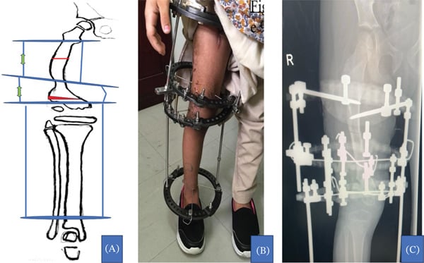
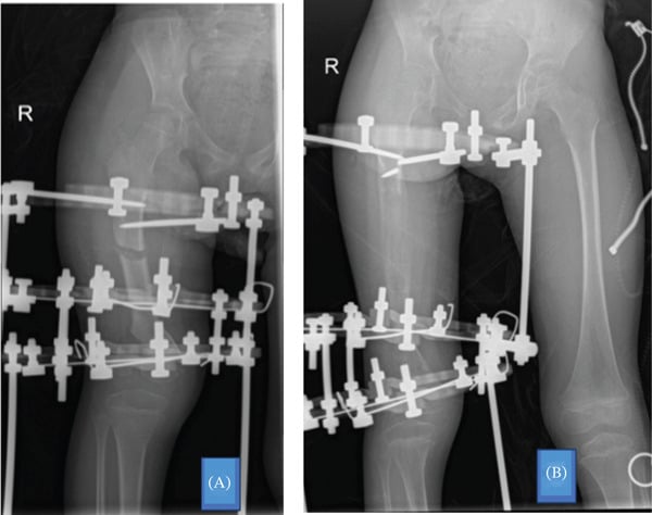
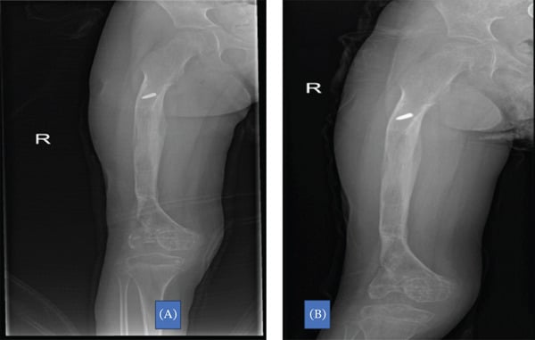
The case that follows shows an Ilizarov frame used for severe femoral deformity, with gradual distraction and then consolidation of the regenerate.
Complications
- MCD Type
- Schmid
- Risk Factors
- Severe Hilgenreiner-epiphyseal angle, continued growth
- Management
- Valgus osteotomy; monitor angle during growth
- MCD Type
- Schmid
- Risk Factors
- Surgery performed too early (active growth plates)
- Management
- Time surgery near skeletal maturity; use guided growth in younger children
- MCD Type
- Jansen
- Risk Factors
- Persistent hypercalcaemia, hypercalciuria
- Management
- Aggressive calcium control, bisphosphonates, renal monitoring
- MCD Type
- McKusick
- Risk Factors
- Profound T-cell deficiency, null mutations in RMRP
- Management
- HSCT referral, infection prophylaxis, immunoglobulin replacement
- MCD Type
- McKusick
- Risk Factors
- Immune surveillance failure, cumulative risk increases with age
- Management
- Annual surveillance, family education, prompt investigation of symptoms
- MCD Type
- McKusick
- Risk Factors
- Macrocytic anaemia in infancy, may be life-threatening
- Management
- Transfusion support, usually self-limiting after early childhood
The most common clinical error is treating MCD patients with vitamin D and calcium supplementation for presumed rickets. In Schmid type, this is unnecessary and may cause iatrogenic hypercalcaemia or hypercalciuria. In Jansen type, additional calcium is actively harmful as the patient is already hypercalcaemic. Always confirm biochemistry before treating metaphyseal changes as rickets.
Outcomes and Prognosis
Schmid has the best prognosis of the three: a normal lifespan and normal intelligence, with the morbidity orthopaedic (coxa vara, bowing). Jansen has the worst: severe dwarfism, the metabolic complications of hypercalcaemia and possible deafness. McKusick is variable; the mortality risk is malignancy (especially lymphoma) and severe immune deficiency, and with modern immune support and malignancy surveillance many patients reach adulthood.
- Final Height
- 135-155 cm (mild-moderate short stature)
- Function
- Good; most achieve independent ambulation and normal activity
- Life Expectancy
- Normal
- Key Determinant
- Severity of coxa vara and bowing
- Final Height
- Often less than 120 cm (severe dwarfism)
- Function
- Variable; limited by severe skeletal dysplasia and metabolic complications
- Life Expectancy
- Reduced (renal complications, hypercalcaemia)
- Key Determinant
- Degree of hypercalcaemia control
- Final Height
- 110-135 cm (moderate-severe short stature)
- Function
- Good with appropriate immune and orthopaedic management
- Life Expectancy
- Reduced (malignancy, severe immune deficiency)
- Key Determinant
- Immune function and malignancy surveillance
Guidelines, Registries & Global Practice
- Schmid type: worldwide distribution; likely under-diagnosed as mild cases may be attributed to physiological bowing or familial short stature
- Jansen type: extremely rare globally; no ethnic or geographic concentration
- McKusick / CHH: founder mutations in Finnish (approximately 1 in 23,000 births) and Old Order Amish (Pennsylvania, USA) populations; cases reported worldwide across all ethnicities
- Overall: MCD accounts for a small proportion of skeletal dysplasias; accurate diagnosis requires genetic confirmation
- High-resource: multidisciplinary care with geneticists, paediatric orthopaedic surgeons, immunologists, and haematologists; skeletal dysplasia gene panels available
- Limited-resource: clinical and radiographic diagnosis with basic biochemistry; genetic testing may require sample shipment to reference laboratories; orthopaedic management prioritised
- Universal principle: biochemistry (Ca, PO4, ALP) is universally available and is the most important first-line test to differentiate MCD from rickets in all settings
- McKusick surveillance: malignancy and immune monitoring requires infrastructure that may be limited in resource-constrained settings
- Diagnosis
- Biochemistry first (exclude rickets); then genetic testing for MCD subtype
- Medical Management
- Jansen: bisphosphonates for hypercalcaemia; McKusick: immune monitoring and prophylaxis
- Orthopaedic Management
- Referral to paediatric orthopaedics for deformity correction
- Diagnosis
- Standing lower limb radiographs + pelvis; classify pattern of metaphyseal involvement
- Medical Management
- No role for vitamin D/calcium supplementation in MCD with normal biochemistry
- Orthopaedic Management
- Corrective osteotomy for progressive coxa vara (Hilgenreiner angle greater than 60 degrees); guided growth for genu varum
- Diagnosis
- Nosology-based classification; genetic confirmation for all MCD subtypes
- Medical Management
- Multidisciplinary management; McKusick requires immunology and oncology input
- Orthopaedic Management
- Individualised surgical planning; recurrence risk high if surgery before skeletal maturity
- Diagnosis
- McKusick CHH is a recognised combined immune deficiency; lymphocyte subset analysis mandatory
- Medical Management
- HSCT for severe combined immune deficiency phenotype; IVIG for antibody deficiency
- Orthopaedic Management
- Coordinate orthopaedic surgery with immunological status; avoid live vaccines if T-cell deficient
There is no dedicated international registry for metaphyseal chondrodysplasias, though the Finnish CHH cohort represents one of the best-characterised single-gene skeletal dysplasia populations worldwide. The ISDS maintains a nosology of skeletal dysplasias that classifies MCD subtypes. Evidence is predominantly from case series, cohort studies, and expert reviews rather than randomised controlled trials, owing to the rarity of these conditions.
In any child with metaphyseal irregularity on radiographs:
- ALWAYS check biochemistry (Ca, PO4, ALP) before prescribing vitamin D or calcium
- If biochemistry is normal, the diagnosis is NOT rickets — do not treat as rickets
- Consider MCD and refer for genetic testing
- In Jansen type, additional calcium is actively dangerous (patient is already hypercalcaemic)
Misdiagnosis of MCD as rickets leads to unnecessary treatment, delayed appropriate management, and potential harm from iatrogenic hypercalcaemia or hypercalciuria.
Controversies & Areas of Uncertainty
The role of GH in MCD remains uncertain. Small series suggest modest benefit in McKusick CHH (improved growth velocity without significant side effects), but there is no evidence of benefit in Schmid or Jansen types. GH does not address the underlying growth plate defect. Most guidelines do not routinely recommend GH for MCD.
The ideal timing of corrective osteotomy in Schmid MCD is debated. Operating during active growth carries significant recurrence risk, but delaying surgery until skeletal maturity may allow prolonged deformity with joint compensation issues. Guided growth techniques offer an intermediate solution but long-term data are limited.
Calcimimetics and PTH1R antagonists have been proposed as targeted therapies for Jansen hypercalcaemia, but clinical data are extremely limited. Current management relies on bisphosphonates and supportive care. A true targeted approach requires development of a functional PTH1R inhibitor, which does not yet exist in clinical practice.
The optimal malignancy surveillance protocol for CHH patients is not standardised. Annual clinical examination and FBC is widely practised, but the role of routine imaging (ultrasound, CT) for lymphoma screening is debated due to radiation risk and low positive predictive value in asymptomatic patients. Current practice relies on clinical vigilance and patient education.
MCQ Practice Points
Q: Which gene is mutated in Schmid metaphyseal chondrodysplasia? A: COL10A1, located on chromosome 6q21. This gene encodes the alpha-1 chain of type X collagen, which is specifically expressed by hypertrophic chondrocytes in the growth plate. Mutations (typically in the NC1 domain) disrupt trimer assembly and impair endochondral ossification at the metaphysis, producing the characteristic metaphyseal irregularity while sparing the epiphyses and spine.
Q: What is the biochemical profile in Schmid metaphyseal chondrodysplasia? A: Completely normal. Calcium, phosphate, alkaline phosphatase, parathyroid hormone, and 25-hydroxyvitamin D are all within normal limits. This is the critical discriminator from rickets, where at least one parameter is always abnormal. Treating a child with Schmid MCD with vitamin D is unnecessary and potentially harmful.
Q: Why is PTH suppressed in Jansen metaphyseal chondrodysplasia despite hypercalcaemia? A: Constitutive activation of the PTH1R receptor. The mutation causes ligand-independent receptor signalling, producing the biological effects of PTH (calcium release from bone, renal calcium reabsorption) without requiring PTH binding. The resulting hypercalcaemia then suppresses parathyroid gland PTH secretion via negative feedback, producing the characteristic pattern of high calcium with low PTH.
Q: How do you differentiate metaphyseal chondrodysplasia from rickets on clinical and biochemical grounds? A: Biochemistry is the key. In rickets, calcium and/or phosphate are low, ALP is elevated, and vitamin D is low (nutritional) or phosphate is low with normal Ca (hypophosphataemic). In MCD (Schmid and McKusick), ALL biochemical parameters are normal. Radiologically, both show metaphyseal changes, but rickets also produces looser zones, craniotabes, and generalized osteopenia, which are absent in MCD.
Q: What extra-skeletal features distinguish McKusick (cartilage-hair hypoplasia) from other MCD types? A: Fine, sparse, hypopigmented hair; combined immune deficiency (T-cell and B-cell); increased malignancy risk (especially non-Hodgkin lymphoma); macrocytic anaemia; and Hirschsprung disease. These systemic features are unique to the McKusick type and reflect the broader role of the RMRP gene in cell proliferation across multiple tissues, not just cartilage.
Q: What are the key imaging features that distinguish MCD from spondyloepiphyseal dysplasia (SED)? A: In MCD, the metaphyses are abnormal but the epiphyses and spine are normal. In SED, the epiphyses are irregular, delayed, or fragmented AND the spine shows platyspondyly or other vertebral anomalies. The pattern of involvement (metaphyses only vs epiphyses plus spine) is the fundamental radiographic discriminator.
Exam Viva Scenarios
Practise clinical reasoning and management decisions out loud
“A 4-year-old boy presents with progressive bilateral genu varum, a waddling gait, and short stature. His parents are unaffected. Biochemistry reveals normal calcium, phosphate, alkaline phosphatase, PTH, and vitamin D. Pelvis and knee radiographs show coxa vara with irregular, flared metaphyses. The epiphyses and spine appear normal. What is the diagnosis and how would you manage this child?”
“An 18-month-old girl of Finnish descent presents with short-limbed short stature, recurrent lower respiratory tract infections, and chronically loose stools. Examination reveals fine, sparse, light-coloured hair, ligamentous laxity, and bilateral genu varum. Hand radiographs show short metacarpals with cone-shaped epiphyses. Knee radiographs show flared metaphyses with normal epiphyses. What is the diagnosis, what investigations are needed, and how would you manage her?”
Key Genetics
- Schmid = COL10A1 (type X collagen), AD, chromosome 6q21 — most common MCD
- Jansen = PTH1R (PTH/PTHrP receptor), AD, chromosome 3p21 — rarest, most severe
- McKusick / CHH = RMRP (RNase MRP RNA component), AR, chromosome 9p13 — Finnish/Amish founder effect
- All three affect the growth plate (physis) but spare epiphyses and spine
Clinical Features
- Schmid: coxa vara, genu varum, bowing, waddling gait, mild-moderate short stature, onset age 2-5
- Jansen: severe dwarfism, hypercalcaemia, prominent forehead, onset neonatal/infancy
- McKusick: fine sparse hair, immune deficiency, short limbs, cone-shaped epiphyses in hands, malignancy risk
- Spine and epiphyses are NORMAL in all MCD types — if abnormal, consider SED
Diagnosis
- Biochemistry is NORMAL in Schmid and McKusick — the critical discriminator from rickets
- Jansen has hypercalcaemia with SUPPRESSED PTH (constitutive receptor activation)
- Radiographs: metaphyseal irregularity, flaring, sclerosis; normal epiphyses and spine
- Genetic testing confirms subtype: COL10A1, PTH1R, or RMRP sequencing
Management
- Schmid: orthopaedic only — corrective osteotomy for coxa vara (angle greater than 60 degrees) or severe genu varum
- Jansen: bisphosphonates for hypercalcaemia, renal monitoring, orthopaedic support
- McKusick: multidisciplinary — immune monitoring, malignancy surveillance, orthopaedic care, genetic counselling
- DO NOT treat with vitamin D or calcium unless biochemistry confirms rickets
Differential Diagnosis
- Rickets: abnormal biochemistry (low Ca, low PO4, high ALP) — MCD has normal labs
- SED: epiphyses AND spine involved — MCD spares both
- Achondroplasia: rhizomelic, macrocephaly, FGFR3 — different gene and pattern
- Hypophosphataemic rickets: low phosphate with renal wasting, PHEX mutation — biochemistry abnormal
Evidence Base and Key Trials
A type X collagen mutation causes Schmid metaphyseal chondrodysplasia
- First identification of a type X collagen (COL10A1) mutation as the cause of AUTOSOMAL DOMINANT Schmid metaphyseal chondrodysplasia
- A 13 base pair deletion in ONE COL10A1 allele segregating with the disease in a single large Mormon kindred, with a lod score of 18.2 at theta = 0
- The deletion causes a frameshift altering the highly conserved C-terminal (NC1) domain of the alpha-1(X) chain and shortening the polypeptide by nine residues
- Proposed mechanism: the mutant chain cannot associate during TRIMER FORMATION, so the effect is a reduced amount of normal protein rather than a toxic product
- Type X collagen expression is restricted to HYPERTROPHIC CHONDROCYTES in regions of endochondral ossification - which is precisely why the disease is metaphyseal and spares the spine and epiphyses
A constitutively active mutant PTH-PTHrP receptor in Jansen-type metaphyseal chondrodysplasia
- A single heterozygous nucleotide exchange in exon M2 of the PTH/PTHrP receptor gene in ONE patient with Jansen MCD, changing a strictly conserved histidine 223 to arginine (H223R) in the receptor's FIRST INTRACELLULAR LOOP
- COS-7 cells expressing the mutant receptor showed constitutive, LIGAND-INDEPENDENT cAMP accumulation; cells expressing wild-type receptor did not
- This explains the severe ligand-independent HYPERCALCAEMIA AND HYPOPHOSPHATAEMIA - note both, as the phosphate is as characteristic as the calcium
- The authors attribute the abnormal endochondral bone formation to the same constitutive signalling, but describe this as the likely rather than demonstrated explanation
Mutations in the RNA component of RNase MRP cause a pleiotropic human disease, cartilage-hair hypoplasia
- Identified numerous mutations in the UNTRANSLATED RMRP gene cosegregating with cartilage-hair hypoplasia (McKusick type MCD), which is autosomal RECESSIVE
- RMRP encodes the RNA component of the endoribonuclease RNase MRP, which has at least two functions: cleaving RNA in mitochondrial DNA synthesis, and nucleolar cleaving of pre-ribosomal RNA
- The mechanistic finding that makes the genotype-phenotype picture coherent: INSERTION MUTATIONS IMMEDIATELY UPSTREAM of the coding sequence SILENCE transcription, whereas mutations WITHIN the transcribed region do not
- Association of the protein subunits with the RNA appeared UNALTERED, so the defect is in the RNA's own function rather than in complex assembly
- The authors conclude only that RMRP mutations disrupt 'a function of RNase MRP RNA that affects multiple organ systems' - which is why the disorder is pleiotropic: short stature, defective cellular immunity and cancer predisposition together