Mechanical Overload | Plantar Plate Pathology | Load Redistribution
- Second metatarsal most commonly affected - longest MT, most fixed at Lisfranc joint
- Plantar plate pathology underlies many cases - drawer test assesses integrity
- Conservative management successful in 80% - metatarsal pad PROXIMAL to MT heads
- Weil osteotomy shortens MT 2-4mm to unload - risks transfer metatarsalgia and floating toe
- First ray insufficiency (HV, hypermobility) is common cause - must be addressed
- “Long second MT (Greek foot/Morton's foot) predisposes to overload
- “Plantar plate tear causes crossover toe (toe drifts medially over hallux)
- “Drawer test: excessive dorsal toe translation indicates plantar plate rupture
- “Weil osteotomy: oblique cut parallel to weightbearing surface, shorten 2-4mm
- “Transfer metatarsalgia prevented by cascade shortening and limiting shortening amount
Overview and Epidemiology
What it is. Metatarsalgia is pain localised to the plantar aspect of the metatarsal heads, typically the lesser metatarsals, second through fifth. It is a symptom complex, not a diagnosis: the end result of abnormal load distribution across the forefoot, with the pain coming from mechanical overload of the lesser metatarsals. Identifying and correcting the underlying mechanical or pathologic cause is the goal of treatment, and the rest of this page is organised around finding it.
Who. It is one of the most common foot complaints in orthopaedic and podiatric practice, and approximately 10-15% of the general population are affected at some point in life.
- Female predominance, 3:1 (high heels, narrow shoes)
- Peak age 40-60 years
- Higher incidence in runners, dancers and sports with repetitive forefoot loading
Why the second metatarsal. The second is the metatarsal most often affected, in 65-70% of cases; the third accounts for 20-25% and the fourth for 5-10%. The second's predisposition has anatomical and biomechanical reasons:
- It is the longest metatarsal in approximately 60% of the population (the Greek or Morton's foot), in whom it is longer than the first
- Its tarsometatarsal joint is the most fixed of the Lisfranc articulations, with no movement, unlike the mobile first and fifth rays
- It sits next to the first ray, so when first ray function is impaired by hallux valgus, hypermobility or prior surgery the load transfers to it
- Its central position bears significant load during the toe-off phase of gait
Pathophysiology
The forefoot. Five metatarsals carry the forefoot. The first is the shortest and thickest; the second is typically the longest and the most fixed at the Lisfranc joint. The first ray, the first metatarsal and medial cuneiform together, normally bears approximately 50% of forefoot load during toe-off, and the lesser metatarsals share the rest:
- First ray: 50%
- Second metatarsal: 15-20%
- Third metatarsal: 10-15%
- Fourth metatarsal: 5-10%
- Fifth metatarsal: 5-10%
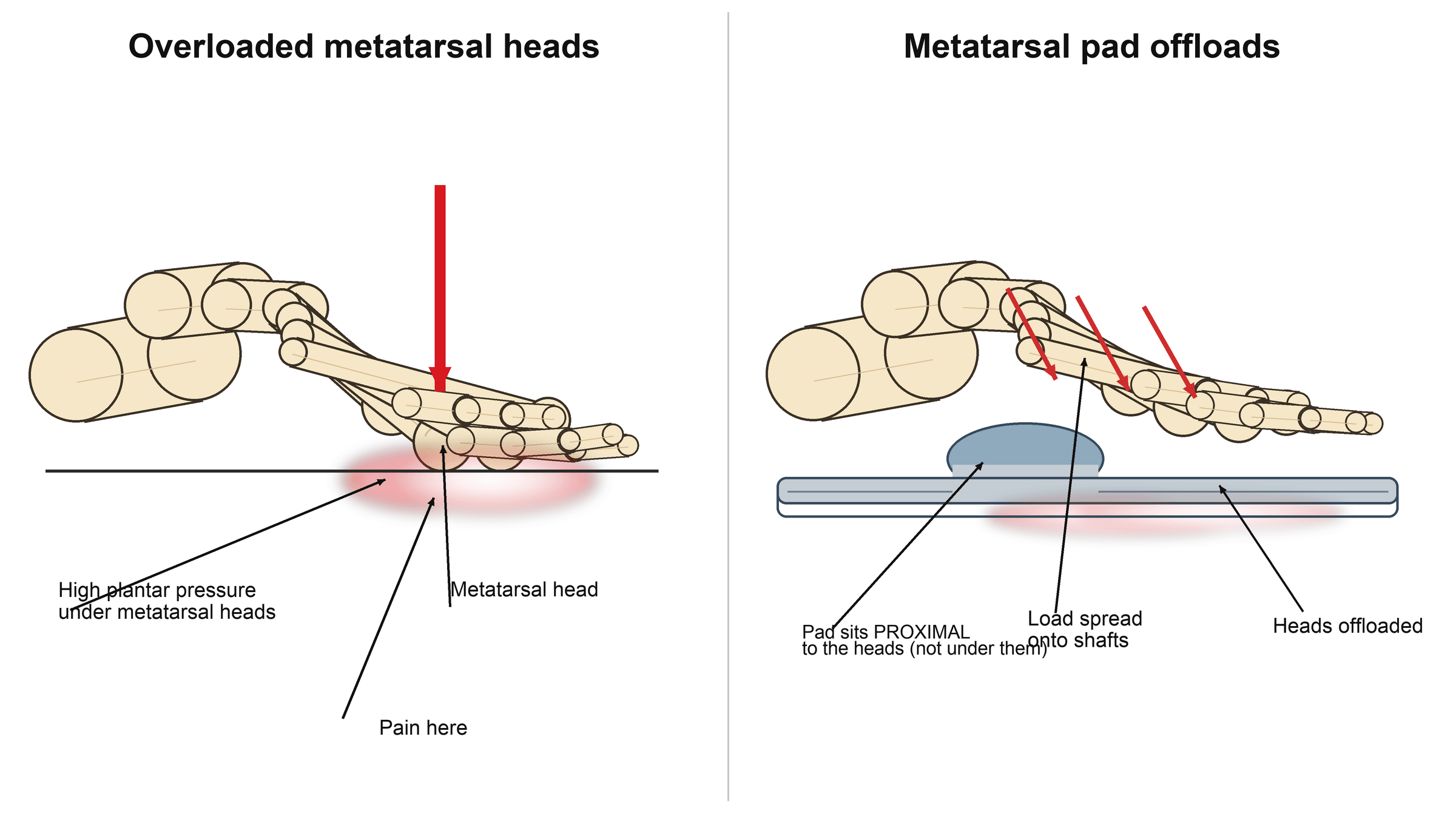
During toe-off the load shifts anteriorly and concentrates on the metatarsal heads, while the plantar plates and the intrinsic muscles hold the metatarsophalangeal joints stable. Anything that overloads the heads, a long or plantarflexed metatarsal, first ray insufficiency shifting load laterally, clawing of the toes, a tight gastrocnemius or high-heeled footwear, produces a focal high-pressure zone and pain directly beneath the head.
The plantar plate. A thick fibrocartilaginous plate on the plantar aspect of each metatarsophalangeal joint, analogous to the meniscus and approximately 1-2mm thick, inserting on the plantar base of the proximal phalanx and on the metatarsal neck. It stabilises the joint in the sagittal and transverse planes, is the attachment site for the plantar fascia, protects the metatarsal head from excessive load and prevents dorsal subluxation of the toe.
How it fails. Chronic repetitive stress breaks down the fibrocartilage. The plate degenerates, then attenuates (thins and weakens), then tears, and a partial tear usually originates at the lateral insertion of the second metatarsophalangeal joint; a complete rupture loses all stabilisation. The consequences follow from what the plate normally does:
- Dorsal subluxation of the toe on the metatarsal head
- Medial deviation, the crossover toe, in which the second toe drifts over the hallux
- Metatarsophalangeal instability
- Pain directly under the head, because the protective cushioning is gone
- A positive drawer test, from the excessive dorsal translation
The mechanisms. Each cause of primary metatarsalgia overloads the lesser heads by a different route, and the treatment follows the route:
- Pathophysiology
- Excessive load on longest MT head
- Clinical Example
- Greek foot (2nd MT longer than 1st)
- Treatment Approach
- Weil osteotomy to shorten and unload
- Pathophysiology
- First ray fails to bear normal 50% load, transfers to 2nd MT
- Clinical Example
- Hallux valgus, first MT hypermobility, prior HV surgery
- Treatment Approach
- Address first ray pathology
- Pathophysiology
- Loss of MTP stabilization, dorsal subluxation, direct MT head overload
- Clinical Example
- Crossover toe, drawer test positive
- Treatment Approach
- Plantar plate repair plus Weil osteotomy
- Pathophysiology
- Increases forefoot loading during gait
- Clinical Example
- Isolated gastrocnemius contracture
- Treatment Approach
- Achilles stretching or gastrocnemius recession
- Pathophysiology
- Fixed forefoot equinus concentrates load on MT heads
- Clinical Example
- High-arched rigid foot
- Treatment Approach
- Cavus correction if severe, orthotics
- Pathophysiology
- Excessive first MT elevation or shortening shifts load to 2nd MT
- Clinical Example
- After aggressive hallux valgus repair
- Treatment Approach
- Revision osteotomy, first MT plantarflexion
Secondary causes. Here the forefoot pain comes from a disease process rather than from load. Freiberg's disease is avascular necrosis of a metatarsal head, typically the second, in adolescent females, with fragmentation among its radiographic changes. Morton's neuroma is an interdigital neuroma, a perineural fibrosis between the heads. Rheumatoid arthritis is a systemic inflammatory arthropathy that produces synovitis, erosions and subluxation at multiple metatarsophalangeal joints bilaterally. A stress fracture is tender over the shaft and most often involves the second or third metatarsal, and sesamoiditis is pain under the first metatarsal head, at the sesamoids, and so distinct from lesser-metatarsal metatarsalgia. How each is told apart is set out under differential diagnosis.
Classification Systems
The etiologic classification is the one that decides the treatment: a primary mechanical cause needs a structural correction, a secondary cause needs the underlying pathology treated, and an iatrogenic cause may need revision surgery to rebalance the forefoot.
- Subcategory
- Structural anatomic abnormality
- Examples
- Long 2nd MT, cavus foot, first ray hypermobility
- Management Principle
- Correct mechanical abnormality
- Subcategory
- Underlying disease process
- Examples
- RA, Freiberg's, Morton's neuroma, sesamoiditis
- Management Principle
- Treat primary pathology
- Subcategory
- Post-surgical load redistribution
- Examples
- After HV repair, first MT osteotomy, excessive MT shortening
- Management Principle
- Revision surgery to rebalance
Clinical Assessment
History. The patient can point to the specific metatarsal head: the pain is on the plantar forefoot under the heads, aching or burning and sharp with weight-bearing, and worst at the end of the day after cumulative loading. What makes it better and worse localises the load:
- Worse with prolonged standing or walking, high heels (which shift load anteriorly), hard surfaces, barefoot walking and thin-soled shoes
- Better with rest, removing the shoes and massaging the forefoot
What the associated symptoms mean. Each points to a cause or away from a simple mechanical one:
- Numbness or burning in a web space that radiates to the toes suggests Morton's neuroma
- A crossover toe indicates plantar plate rupture
- Callus under a specific head confirms the overload pattern
- A toe that feels as if it "gives way" suggests plantar plate insufficiency
- Prominent dorsal metatarsophalangeal swelling suggests synovitis or arthropathy
- Night pain is a red flag for tumour or infection, and is not typical of mechanical metatarsalgia
Inspection. Start standing, because weight-bearing alignment is the point: foot type (cavus, planus or neutral), the first ray (hallux valgus, first metatarsal elevation) and the lesser toes (crossover toe with the second over the hallux, hammer toes, claw toes). Then seated, look at the callus pattern, whose location and severity indicate which metatarsal is overloaded, at dorsal metatarsophalangeal swelling (synovitis, arthropathy) and at the skin for ulceration (diabetes, rheumatoid arthritis) and colour change.
Palpation. Palpate each metatarsal head individually to localise the pain precisely; the second is the most commonly tender, note which heads are symptomatic, and feel for plantar plate prominence (thickening). Squeeze the web spaces between the heads for Morton's neuroma, with Mulder's click elicited by lateral metatarsal compression while pressing the web space; the third web space is the most common site. Palpate the dorsal metatarsophalangeal joints for synovitis, osteophytes and swelling. Palpation is what separates metatarsalgia, tender over the head, from a neuroma, tender in the web space.
The drawer test. This is the single most important special test for metatarsalgia and is performed in every case. Stabilise the metatarsal head firmly with one hand; with the other, grasp the toe and apply a dorsally directed force to translate it on the head. An intact plate resists. A positive test shows excessive translation, more than 2-3mm compared with the adjacent toes and the contralateral foot, reproduces the patient's symptoms, and indicates a plantar plate tear or severe attenuation. Always perform it bilaterally to establish what is normal for that patient. A positive drawer test changes management: plantar plate repair should be considered if surgery is pursued.
Grading the instability. A positive drawer is graded, and the grade drives the operation. The clinical metatarsophalangeal instability grade, from the drawer or "vertical Lachman" test:
- Grade 0: stable
- Grade 1: mild laxity, under half subluxation
- Grade 2: moderate, over half subluxation
- Grade 3: dislocatable joint
- Grade 4: fixed dislocation with rigid deformity
This maps onto the anatomic Nery-Coughlin plantar plate grading seen at surgery or on MRI:
- Grade 0: intact or attenuated
- Grade I: transverse distal tear involving less than half the plate
- Grade II: transverse tear of more than half
- Grade III: extensive tear, often with a longitudinal component
- Grade IV: complete tear or buttonhole with retraction, frequently with collateral involvement
The practical message is that a low grade may settle with a metatarsal pad and offloading, or with a Weil osteotomy that indirectly relaxes the plate, whereas the higher grades, with true subluxation or a crossover toe, need direct plantar plate repair combined with the Weil, and a fixed Grade 4 may need joint salvage rather than repair.
The first ray. Translate the first metatarsal dorsally on the medial cuneiform; excessive mobility, hypermobility, predisposes to transfer metatarsalgia. Look for hallux valgus at the same time.
The Achilles. The Silfverskiold test measures ankle dorsiflexion with the knee extended, and the gastrocnemius is tight if it is limited; repeat with the knee flexed, and if dorsiflexion improves the contracture is an isolated gastrocnemius one.
Range of motion. Assess metatarsophalangeal dorsiflexion and plantarflexion. Limited motion suggests arthropathy or Freiberg's disease.
Gait. Watch for an antalgic gait with a shortened stance phase, early heel-off that avoids toe-off on the painful forefoot, and toe-walking from an Achilles contracture. Toe-off is when load normally shifts to the forefoot, so painful toe-off confirms forefoot pathology.
These features are atypical for simple mechanical metatarsalgia and require a comprehensive workup for an alternative diagnosis:
- Night pain or rest pain: consider tumour, infection, complex regional pain syndrome
- Acute traumatic onset: rule out Lisfranc injury, metatarsal fracture, plantar plate rupture
- Constitutional symptoms (fever, weight loss, malaise) suggest infection or systemic disease
- Rapidly progressive deformity: may indicate inflammatory arthropathy or tumour
- Neurological symptoms, widespread numbness or weakness, suggest peripheral neuropathy or nerve compression
- Vascular insufficiency, with poor pulses, skin changes or ulceration, requires vascular assessment before any intervention
Investigations
Weight-bearing radiographs. The essential first investigation is an AP, lateral and oblique of the foot, and they must be weight-bearing. They are read for:
- Metatarsal length: is the second long (Greek foot)? Measure the relative lengths by the parabola method or by direct measurement
- Metatarsal alignment: elevation, depression or rotation
- Metatarsophalangeal joint space: arthropathy, erosions (rheumatoid arthritis), flattening (Freiberg's)
- First ray position: hallux valgus angle, first metatarsal elevation
- Sesamoid position: lateral displacement with hallux valgus
The second metatarsal is typically 1-2mm shorter than the first or equal to it; a second metatarsal more than 3mm longer than the first predisposes to overload. A calcaneal pitch angle greater than 30 degrees marks a cavus foot.
MRI. The gold standard for soft tissue, and the test to order when the drawer test is positive or the diagnosis is uncertain:
- Positive drawer test, to assess a plantar plate tear
- Suspected neuroma
- To rule out a stress fracture
- Freiberg's disease staging
- Preoperative planning
The protocol is a foot MRI with dedicated coils in sagittal, coronal and axial planes. The plantar plate is the key finding and is best seen on the sagittal and coronal images: a normal plate is a low signal structure on all sequences of uniform 1-2mm thickness; a partial tear shows high signal on T2 or STIR at the insertion, usually lateral, with thinning; a complete rupture shows discontinuity, high signal and metatarsophalangeal subluxation. Beyond the plate, a Morton's neuroma is low signal on T1 and T2 between the heads, a stress fracture shows bone marrow oedema with or without a fracture line, and Freiberg's disease shows head oedema, fragmentation and subchondral change.
Ultrasound. Dynamic, real-time, cheaper than MRI and without radiation, it detects Morton's neuroma as a hypoechoic mass between the heads, can show plantar plate tears (though it is less sensitive than MRI) and guides injections. It is operator-dependent and less detailed than MRI for the plantar plate, so MRI is preferred for a comprehensive assessment.
CT. A limited role, and not routinely required. It is for subtle metatarsal fractures not visible on the radiograph, Lisfranc injury and Freiberg's disease staging.
Laboratory studies. Generally not required for primary mechanical metatarsalgia; order them when a secondary cause is suspected:
- Rheumatoid factor and anti-CCP if there is bilateral metatarsophalangeal synovitis or systemic symptoms
- Uric acid for acute monoarticular metatarsophalangeal pain (gout)
- ESR and CRP if infection or an inflammatory arthropathy is suspected
- HbA1c in diabetic patients with neuropathy or an at-risk foot
Differential Diagnosis
The location of maximal tenderness is the most helpful discriminator: metatarsal head, web space or metatarsal shaft. The drawer test then separates plantar plate pathology from the rest, Mulder's click is pathognomonic for Morton's neuroma, and imaging confirms the diagnosis when the examination is unclear.
- Pain Location
- Plantar MT head (specific MT)
- Key Clinical Features
- Plantar callus, drawer test positive if plate torn
- Diagnostic Test
- Clinical diagnosis, X-ray shows MT length
- Pain Location
- Web space (between MT heads)
- Key Clinical Features
- Burning, radiates to toes, Mulder's click
- Diagnostic Test
- Ultrasound or MRI shows neuroma
- Pain Location
- Dorsal and plantar second MT head
- Key Clinical Features
- Limited MTP motion, adolescent female
- Diagnostic Test
- X-ray shows MT head flattening, sclerosis
- Pain Location
- MT shaft (not plantar head)
- Key Clinical Features
- Acute onset, bony shaft tenderness
- Diagnostic Test
- MRI shows fracture line and edema
- Pain Location
- Multiple MTP joints bilaterally
- Key Clinical Features
- Systemic disease, synovitis, erosions
- Diagnostic Test
- Positive RF/anti-CCP, X-ray erosions
- Pain Location
- Under first MT head (sesamoids)
- Key Clinical Features
- Hallux pain, not lesser MTs
- Diagnostic Test
- X-ray/MRI shows sesamoid pathology
Management
The principle. Treat the mechanical cause, not the symptom alone. The diagnosis is clinical, weight-bearing radiographs establish the metatarsal lengths and alignment, and almost everyone then has a 3-6 month conservative trial. Symptoms that persist despite a compliant trial are imaged with MRI or ultrasound to pin down the structural driver, and surgery is reserved for a function-limiting lesion and matched to the pathology found.
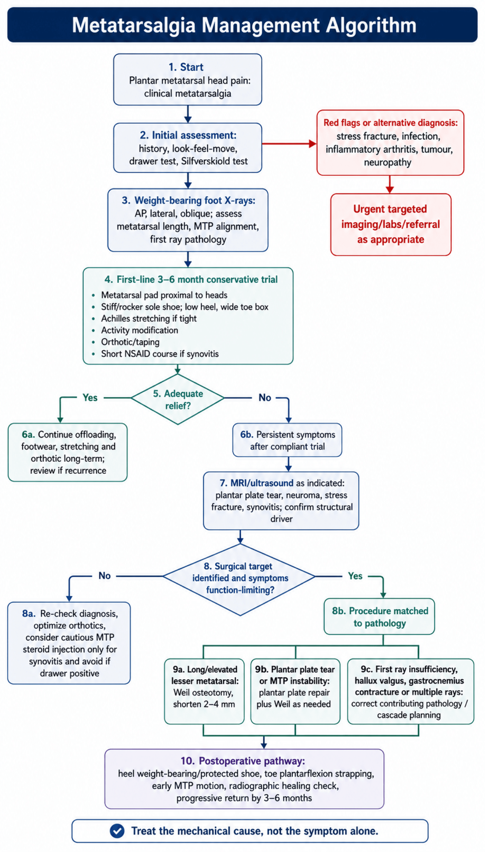
Conservative management succeeds in approximately 80% of cases and should be the first-line approach for all patients with metatarsalgia.
Offloading. A metatarsal pad or dome is the single most effective conservative intervention, and it works only if it is in the right place: proximal to the metatarsal heads, typically 1cm proximal to the point of maximal tenderness, not directly under them. It elevates the metatarsal shafts, transferring load proximally and off the heads, and can be an adhesive gel pad or built into a custom orthotic. The fitting has to be precise, because a pad too far distal, beneath the heads, adds to the pressure on them rather than relieving it, and one too far proximal is uncomfortable.
Footwear. A stiff-soled shoe with a rigid rocker sole reduces metatarsophalangeal dorsiflexion during toe-off and so the load on the heads; options include a Morton's extension or a carbon fibre plate in the shoe. Heels should be low, under 2-3cm, because high heels transfer load anteriorly to the forefoot and low heels keep the distribution more even. A wide toe box accommodates any toe deformity and prevents lateral compression, and a well-cushioned sole absorbs impact.
Activity. Avoid prolonged standing when possible and reduce high-impact activity, running and jumping, during the acute phase, substituting swimming or cycling. This is not complete rest; general fitness is maintained.
Achilles stretching. If the Silfverskiold test is positive, wall stretches with the knee straight (gastrocnemius) and bent (soleus), held for 30 seconds, repeated 3-5 times, two to three times a day. Improving ankle dorsiflexion reduces forefoot loading.
Orthotics and taping. A custom orthotic, usually fabricated by a podiatrist or orthotist, incorporates the pad at the correct position, adds arch support where there is first ray hypermobility or pes planus, and may include a first metatarsal cutout where the first ray is elevated. Plantar plate taping plantarflexes and supports the toe; it is a temporary measure, useful to test whether an orthotic would help.
NSAIDs. Short-term use for an acute flare addresses the synovitis component. They are symptomatic, not curative.
Corticosteroid injection. The indication is metatarsophalangeal synovitis that has not settled with the measures above. Inject into the joint space from a dorsal approach, avoiding the plantar plate, with 1ml of 40mg methylprednisolone and 1ml of local anaesthetic. It may be worth considering when there is a significant inflammatory component, provided the risks are recognised.
Corticosteroid injection for metatarsalgia must be used with caution. Repeated injections can accelerate plantar plate degeneration and precipitate rupture, and while steroid may give short-term relief from metatarsophalangeal synovitis it does not address the underlying mechanical problem and may worsen the structural pathology. Give no more than 2-3 injections per joint, consider other treatments first, and avoid injection altogether if the drawer test is already positive, because the plate is already compromised.
What to expect. About 80% achieve enough symptom control to avoid surgery. Improvement is gradual over 3-6 months, and a patient who is going to respond shows initial improvement within 4-6 weeks. Success is more likely with mild symptoms, recent onset (under 6 months), good compliance with offloading, no structural deformity and a negative drawer test. Failure is more likely with chronic symptoms (over 12 months), a positive drawer test from plantar plate rupture, a significant structural abnormality (a long metatarsal, severe first ray pathology) or a crossover toe deformity.
An adequate conservative trial, a minimum of 3-6 months, is essential before surgery is considered.
PADSConservative Management Steps
Hook:PADS offload the metatarsal heads and allow healing in 80% of cases!
Complications
Shortening one metatarsal reduces the load on it and increases the load on its neighbours; after a second metatarsal osteotomy the third is the one most commonly affected. This page quotes 10-15% after a Weil osteotomy; the pooled review of 1131 Weil osteotomies in the evidence section reported 7%. Prevention is the key: (1) limit shortening to 2-4mm, rarely more than 4mm; (2) consider cascade shortening of the adjacent metatarsals if more than one needs addressing; (3) address first ray insufficiency so that load does not transfer to the lesser metatarsals; (4) plan the relative metatarsal lengths preoperatively on weight-bearing radiographs. Treatment is conservative at first, with metatarsal pads and orthotics; if severe and refractory it may need an adjacent metatarsal osteotomy, at the risk of starting a cascade of transfer problems.
Floating toe. The toe loses contact with the ground during walking. The causes are excessive metatarsal shortening, weak plantar structures and extensor overpull, and it is usually a cosmetic concern rather than a functional problem. The same pooled review that reported 7% transfer reported floating toe as the most common complication, in 36%, against the 15-30% in the table. It is prevented by limiting the shortening, repairing the plantar plate when torn, repairing the capsule at closure and strapping the toe in plantarflexion postoperatively; treatment is toe taping, with revision rarely if it is significantly symptomatic.
Metatarsophalangeal stiffness. A loss of dorsiflexion, typically 10-20 degrees compared with the preoperative range, from capsular adhesions, prolonged immobilisation or an aggressive osteotomy. Early range of motion, physiotherapy and minimal soft tissue disruption prevent it. It is usually well tolerated; severe stiffness gets aggressive physiotherapy, and manipulation under anaesthesia is rarely needed.
Recurrent pain. Inadequate shortening, a plantar plate that was not addressed, the wrong diagnosis or progression of an arthropathy. Review the imaging, look for an alternative diagnosis, and check fixation and union. Management is conservative first, with revision rarely, and only if a clear structural issue is identified.
Intraoperative complications. Neurovascular injury, to the digital nerve or vessel during dissection, is prevented by careful dissection and identifying the structures. Malposition of the osteotomy at the wrong angle causes a dorsal or plantar step-off, and is prevented by cutting parallel to the weight-bearing surface. Excessive shortening beyond 4mm raises the transfer risk, and is prevented by measuring and planning; inadequate fixation, a stripped screw or a bent K-wire, by proper technique and appropriately sized implants.
Early postoperative complications, within 6 weeks:
- Infection in under 2%, typically superficial, managed with antibiotics and wound care
- Haematoma, a collection causing painful swelling, which usually resolves and is aspirated if large
- Wound dehiscence from poor healing (diabetes, smoking), managed with local wound care and rarely revision
- Fixation failure, screw loosening or displacement, which may need revision fixation if symptomatic
Late complications, after 6 weeks:
- Delayed union, visible on the radiograph at 3-6 months, usually asymptomatic and progressing to union
- Nonunion, a persistent gap with no bridging callus, which may need revision if painful
- Malunion, healing in an abnormal position, which can cause persistent pain or deformity
- Hardware prominence, a palpable screw head, which may need removal if symptomatic
- Complex regional pain syndrome, rare: disproportionate pain, swelling and vasomotor changes
The complications of any foot operation. Digital paraesthesia, temporary or permanent, affects under 10%, usually the medial or lateral aspect of the toe; most resolve over 3-6 months, and desensitisation techniques help if it is permanent. Scars can be hypertrophic or adherent, and keloid is rare; massage, silicone gel and rarely revision. Hammer toe and claw toe can develop or worsen and may need soft tissue balancing, and first ray hypermobility or hallux valgus that was not addressed may progress and cause recurrent symptoms. Some patients are dissatisfied despite a technically successful operation, because of stiffness, residual pain or unrealistic expectations, which is why the preoperative counselling matters.
Guidelines, Registries & Global Practice
Metatarsalgia is a worldwide forefoot complaint with no single dedicated national-society guideline; practice is driven by foot-and-ankle literature and the closely related conditions of hallux valgus and lesser-toe instability. The principles below are consistent across UK (BOFAS/BOA), US (AAOS/AOFAS), European (EFAS/EFORT) and AO Foundation teaching.
Global Epidemiology
- Evidence
- Hallux valgus - a major driver of first-ray insufficiency and lesser-MT overload - has a pooled prevalence of 23% in adults aged 18-65 and 36% in adults over 65 (systematic review/meta-analysis)
- Source
- Nix 2010 (J Foot Ankle Res), PMID 20868524
- Evidence
- Hallux valgus and related forefoot deformity are markedly more common in women than men, consistent with the female predominance seen in metatarsalgia and crossover toe
- Source
- Nix 2010, PMID 20868524
- Evidence
- The second MTP joint is the most commonly affected by plantar plate failure (63% in a prospective surgical series)
- Source
- Nery 2012 (Foot Ankle Int), PMID 22735202
Guidance and Consensus, Side by Side
- Position
- Identify and treat the specific cause; staged grade-matched plantar plate protocol (shrinkage / direct repair / tendon transfer) combined with Weil osteotomy
- Evidence basis
- Level IV-V (expert review and prospective series): Espinosa 2008, Nery 2014
- Position
- Exhaust structured non-operative care (offloading orthoses, rocker/stiff sole, footwear advice) before surgery; reserve osteotomy for refractory mechanical overload
- Evidence basis
- Consensus / Level V - aligned with 80% non-operative success in reviews
- Position
- Distal oblique (Weil) osteotomy is the best-evaluated lesser-MT osteotomy; limit shortening and consider cascade balancing to minimise transfer metatarsalgia and floating toe
- Evidence basis
- Level III: Trnka 1999 (Weil superior to Helal); Highlander 2011 (complication profile)
Registry & Pooled Evidence
No national joint registry captures lesser-metatarsal osteotomy (registries focus on arthroplasty). The best population-level evidence is therefore pooled review data: across 1131 Weil osteotomies, floating toe occurred in 36%, recurrence in 15%, transfer metatarsalgia in 7%, and delayed/non-/malunion in 3% (Highlander 2011, PMID 21490179). These figures should frame consent worldwide.
Practice Variation
First-line conservative management (metatarsal pad placed proximal to the heads, stiff/rocker sole, footwear modification, Achilles stretching) is universal, succeeding in around 80% of cases. Threshold for surgery varies by health system and access to podiatry/orthotics rather than by any difference in the underlying evidence.
The Weil osteotomy is the global default for refractory mechanical overload. Minimally invasive distal metatarsal metaphyseal osteotomy (DMMO) is increasingly used in some European centres but has a recognised learning curve. Choice is largely surgeon- and resource-dependent, not guideline-mandated.
Consent and Documentation (Universal Principles)
- Document an adequate conservative trial (3-6 months of specific offloading and footwear measures) before offering surgery - inadequate trial is a recurrent source of dissatisfaction and litigation.
- Quantify complication risk at consent: floating toe (reported up to 36% in pooled data), transfer metatarsalgia (7-15%), expected MTP stiffness (10-20 degrees), and the possibility of further surgery.
- Record the planned and achieved shortening (target 2-4mm); excessive shortening is the principal avoidable cause of transfer metatarsalgia.
- Assess and document first ray pathology (hallux valgus, hypermobility) and whether it was addressed - failure to do so is a common cause of recurrence.
MCQ Practice Points
Q: What is the most common cause of metatarsalgia related to hallux valgus correction?
A: Transfer metatarsalgia - excessive shortening or elevation of first ray transfers load to lesser metatarsals. Occurs after over-aggressive first MT shortening, dorsal malunion, or first MTP fusion in excessive dorsiflexion. Prevention: Maintain first ray length and plantar position. Treatment: Metatarsal osteotomy to offload affected rays.
Q: What is the Weil osteotomy and its indication?
A: Oblique distal metatarsal head osteotomy cut roughly parallel to the weightbearing surface (from dorsal-distal to plantar-proximal) for metatarsalgia with a relatively long metatarsal. The head fragment slides proximally to shorten the metatarsal with inherent stability. Fix with 1-2 screws. Indicated for intractable metatarsalgia, subluxed/dislocated MTP joint, and crossover toe deformity.
Q: What is the clinical significance of plantar plate tears in metatarsalgia?
A: Plantar plate attenuation/tear causes MTP joint instability leading to hammer toe, crossover toe, and metatarsalgia. Drawer test positive (greater than 2mm dorsal translation or greater than 50% compared to contralateral). MRI shows plantar plate disruption. Treatment: Direct plantar plate repair through dorsal approach + Weil osteotomy. Grade II-III tears require repair.
Q: How do you differentiate Morton's neuroma from primary metatarsalgia?
A: Morton's neuroma: Burning, electrical pain in 3rd webspace (sometimes 2nd), Mulder's click positive, sensory changes in adjacent toes, pain relieved by removing shoes. Primary metatarsalgia: Aching pain under metatarsal heads, callus formation, worsened by weightbearing. Morton's: Webspace injection diagnostic and therapeutic; metatarsalgia: Offloading insoles first-line.
Q: What is the relative metatarsal length formula and its clinical relevance?
A: Ideal formula: first metatarsal longest, then decreasing 2, 3, 4, 5 (or first equals second). Abnormally long 2nd metatarsal predisposes to metatarsalgia under 2nd MT head. Index-minus foot (short first MT) transfers stress to lesser metatarsals. Relative overlength guides which metatarsals require shortening osteotomy.
Exam Viva Scenarios
Practise clinical reasoning and management decisions out loud
“A 55-year-old woman presents with 6 months of right forefoot pain under the ball of her foot, worse with prolonged standing and wearing heels. She points to the area under her second metatarsal head. Examination shows plantar tenderness under the second MT head, and you perform a drawer test which shows some increased translation compared to the other toes. How do you approach this patient?”
“A 60-year-old woman presents with progressive second toe deformity - the toe is now drifting medially over her hallux. She has pain under the second metatarsal head. On examination, the second toe is deviated medially and when you perform a drawer test, the toe subluxates dorsally very easily. How do you manage this patient?”
“You see a 52-year-old patient in clinic who underwent Weil osteotomy of the second metatarsal 6 months ago for metatarsalgia. The second MT pain has resolved, but she now has significant pain under the third metatarsal head that wasn't present before surgery. Weight-bearing X-rays show the second MT has been shortened by approximately 6mm. What has happened and how do you manage this complication?”
Key Anatomy
- Second MT: Longest (60% population), most fixed at Lisfranc joint, most commonly affected (65-70%)
- Plantar plate: Fibrocartilaginous structure 1-2mm thick, stabilizes MTP joint, prevents dorsal subluxation
- First ray: Should bear 50% of forefoot load - if insufficient, transfers to second MT
- Load distribution: 1st ray 50%, 2nd MT 15-20%, 3rd MT 10-15%, 4th and 5th MT 5-10% each
Clinical Diagnosis
- Pain: Plantar MT head (patient can point to specific MT), worse with standing/walking
- Drawer test: Dorsally translate toe on MT head - positive if excessive translation (plantar plate tear)
- Crossover toe: Second toe drifts medially over hallux - indicates plantar plate rupture
- First ray exam: Check for hallux valgus, hypermobility, prior surgery (causes second MT overload)
- Achilles tightness: Silfverskiold test - increases forefoot loading if positive
Causes
- Plantar plate pathology (tear causes crossover toe, positive drawer)
- Long second MT (Greek/Morton's foot - mechanical overload)
- Achilles tightness (increases forefoot loading)
- Neuroma Morton's (web space pain - differential)
- Transfer lesion (after HV surgery, first MT osteotomy)
- Arthritis (RA, Freiberg's disease - secondary causes)
- Rigid cavus foot (fixed forefoot equinus)
Conservative Management (PADS) - 80% Success
- P - Pad placement PROXIMAL to MT heads (1cm proximal to pain point)
- A - Activity modification (avoid standing, walking, heels)
- D - Dense-soled (stiff) shoes reduce MTP bending
- S - Stretching Achilles tendon if tight
- Trial 3-6 months before considering surgery
Weil Osteotomy Technique
- Indication: Failed 3-6 months conservative, structural abnormality
- Approach: Dorsal longitudinal incision between extensor tendons
- Osteotomy: Oblique cut parallel to weightbearing surface (25-30 degrees to shaft)
- Shortening: 2-4mm (6mm or more is excessive - high transfer risk)
- Fixation: 2.0-2.4mm screw dorsal distal to plantar proximal
- Concurrent: Plantar plate repair if drawer positive, address first ray if HV/hypermobility
Complications
- Transfer metatarsalgia: 10-15% (load shifts to adjacent MT from excessive shortening)
- Floating toe: 15-30% (toe loses ground contact - usually not functionally limiting)
- MTP stiffness: 20-40% (10-20 degree dorsiflexion loss - expected)
- Recurrent pain: 5-10% (inadequate shortening, plate not addressed)
- Prevention: Limit shortening 2-4mm, cascade planning, address first ray, plate repair if torn
Evidence Base and Key Trials
Current Concept Review: Metatarsalgia
- Metatarsalgia is a symptom, not a diagnosis - the underlying cause must be identified
- Primary metatarsalgia arises from intrinsic anatomic factors (long lesser metatarsal, plantarflexed metatarsal, first ray insufficiency)
- Secondary metatarsalgia is driven by systemic or local pathology (RA, Freiberg's disease, interdigital neuroma)
- Iatrogenic (transfer) metatarsalgia follows first ray surgery that alters load distribution
- Treatment must be directed at the specific mechanical or pathologic cause, not just the symptom
Complications of the Weil Osteotomy
- Literature review pooling 1131 Weil osteotomies from 17 studies
- Floating toe was the most common complication - reported in 233 cases, overall occurrence 36%
- Recurrence of deformity/symptoms reported in 15% of cases
- Transfer metatarsalgia reported in 7% of cases
- Delayed union, non-union and malunion collectively reported in 3% of cases
Lesser MTP Instability: Prospective Evaluation and Repair of Plantar Plate and Capsular Insufficiency
- 22 patients (40 MTP joints) treated by direct plantar plate repair through a dorsal approach combined with Weil osteotomy
- The second MTP joint was the most commonly affected (63%); Grade III tears were the most frequent type
- Mean AOFAS score improved from 52 preoperatively to 92 postoperatively
- Direct anatomic repair corrected medial/dorsal/dorsomedial toe deviation and restored MTP alignment
- Plain radiographs, MRI and MTP arthroscopy were correlated with the anatomic grading of the tear
Comparison of Weil and Helal Osteotomies for Metatarsalgia Secondary to Lesser MTP Dislocation
- 30 patients (47 metatarsals) compared - 15 Weil osteotomy vs 15 Helal osteotomy
- Transfer lesions: 0% after Weil vs 41% after Helal (significant)
- Recurrent metatarsalgia: 0% after Weil vs 27% after Helal
- Radiographic reduction maintained in 84% of Weil vs 36% of Helal joints
- No malunion or pseudarthrosis in the Weil group; 5 malunions and 3 pseudarthroses with Helal
Prospective Evaluation of a Protocol for Surgical Treatment of Lesser MTP Plantar Plate Tears
- 68 patients (100 MTP joints) graded by the anatomic plantar plate tear classification and treated by a grade-matched protocol
- Grades 0-I: thermal shrinkage; Grades II-III: direct reinsertion; Grade IV: flexor-to-extensor transfer - all combined with a Weil osteotomy
- All grades improved significantly in VAS and AOFAS scores after surgery (P less than .0001)
- Grade IV tears had the poorest outcome (fair AOFAS average of 72 points)
- Grades I, III and IV had less stable joints and poorer toe purchase/ground touch than grades 0 and II