Degenerative arthritis of the midcarpal joint, commonly from SLAC/SNAC patterns
- Differentiate midcarpal from radiocarpal arthritis - different surgical solutions
- SLAC wrist progresses in a fixed order (Stage I radial styloid, II scaphoid fossa, III midcarpal, IV pancarpal)
- Four-corner fusion preserves 60% wrist motion with 80% grip strength
- Proximal row carpectomy requires intact lunate fossa cartilage
- Total wrist fusion position: 10-15 degrees extension, neutral to 5-10 degrees ulnar deviation
- “Know the Watson SLAC staging system (SLAC staging is Watson's; Lichtman staging is for Kienbock) and which stages are amenable to motion-preserving surgery
- “Describe physical examination findings: midcarpal shift test, Watson test
- “Explain why scaphoid excision necessary in four-corner fusion
- “Discuss fixation options for four-corner fusion: headless screws vs circular plate vs Spider plate
- “Know contraindications to PRC: lunate fossa arthritis, capitate chondromalacia, inflammatory arthritis
Overview and Epidemiology
Midcarpal arthritis affects the articulation between the proximal carpal row (scaphoid, lunate, triquetrum) and the distal row (trapezium, trapezoid, capitate, hamate). Radiocarpal arthritis affects the radius-carpal interface instead. Midcarpal arthritis frequently results from chronic carpal instability, particularly scapholunate advanced collapse (SLAC) and scaphoid nonunion advanced collapse (SNAC).
Surgical management has evolved over the past three decades. Motion-preserving procedures, including four-corner fusion and proximal row carpectomy, offer alternatives to total wrist arthrodesis in appropriately selected patients, and choosing well depends on the pattern of progression, the patient and the technical details of each operation.
Who. SLAC wrist is the most common degenerative wrist pattern. Prevalence increases with age, men predominate 3:1, and involvement is often bilateral (30%). Scaphoid nonunion is present in 50% of trauma cases.
The pathology. Normal carpal kinematics are lost and articular cartilage degenerates progressively, with subchondral sclerosis, cyst formation and osteophytes. Synovial inflammation is variable, and capsular contracture develops.
Natural history. SLAC progresses over 10-20 years, and pain typically precedes the radiographic changes. Function declines with advancing stage, grip strength is typically reduced by 40-60%, motion limitation is variable, and spontaneous fusion is rare.
Surgical Options Overview
Several procedures address midcarpal arthritis. Selection depends on the extent of arthritis, patient factors and surgeon experience.
- indication
- SLAC/SNAC Stage I only
- motionPreservation
- 100% preserved
- gripStrength
- 90-95% preserved
- advantages
- Simple, preserves anatomy, low morbidity
- disadvantages
- Only for Stage I, progression possible
- indication
- SLAC/SNAC Stage II-III
- motionPreservation
- 50-60% arc
- gripStrength
- 75-80% normal
- advantages
- Reliable pain relief, preserves motion, proven outcomes
- disadvantages
- Technically demanding, nonunion 5-10%, scaphoid excision required
- indication
- SLAC/SNAC Stage II-III, low demand
- motionPreservation
- 50-60% arc
- gripStrength
- 70-75% normal
- advantages
- Single-stage, no implants, no nonunion risk
- disadvantages
- Requires intact lunate fossa, progression possible, lower strength
- indication
- Stage IV, salvage, high demand
- motionPreservation
- 0% (complete fusion)
- gripStrength
- 85-90% normal
- advantages
- Definitive, reliable pain relief, maximum strength
- disadvantages
- Loss of all motion, impacts ADLs, adjacent joint stress
- indication
- Pan-arthritis low demand
- motionPreservation
- 60-80% arc
- gripStrength
- 60-70% normal
- advantages
- Preserves motion, bilateral procedures possible
- disadvantages
- Implant failure risk, infection, loosening, revision difficult
Four-Corner Fusion
The workhorse for SLAC/SNAC Stage II-III. Excising the scaphoid eliminates the radioscaphoid arthritis, and fusing the capitate, hamate, lunate and triquetrum removes the midcarpal joint while preserving the radiolunate articulation.
The ideal candidate has SLAC or SNAC Stage II or III with preserved radiolunate cartilage, is aged 30-70, has moderate to high functional demands, has failed a minimum of 6 months of conservative treatment, and is willing to accept limited postoperative immobilisation. It suits the manual labourer who needs grip strength, the active patient who wants motion preserved, and the younger patient who wants to avoid a complete wrist fusion.
Contraindications. Four-corner fusion is not for:
- Stage IV, with radiolunate arthritis
- DRUJ arthritis (consider total wrist fusion)
- Active infection
- Avascular necrosis of the lunate (Kienböck)
- Severe osteoporosis, or bone too poor to hold fixation
Relative contraindications are inflammatory arthritis (higher nonunion risk), smoking (counsel cessation for a minimum of 6 weeks), workers' compensation (controversial) and unrealistic expectations.
The procedure achieves pain relief in 80-90% of patients while preserving functional wrist motion and grip strength.
Proximal Row Carpectomy
The alternative motion-preserving procedure. Removing the scaphoid, lunate and triquetrum creates a radiocapitate articulation, the capitate head sitting in the lunate fossa of the radius. It is simpler than four-corner fusion, but the capitate head and the lunate fossa cartilage must both be intact.
PRC contraindications. Lunate fossa chondromalacia, capitate cartilage degeneration and inflammatory arthritis contraindicate PRC, and high demand is a relative contraindication. Intraoperative assessment of cartilage quality is essential: if the lunate fossa shows Grade III-IV changes, convert to four-corner fusion or total wrist arthrodesis. Do not proceed with PRC if the cartilage is inadequate.
The ideal candidate has SLAC or SNAC Stage II, intact lunate fossa and capitate articular cartilage, low to moderate functional demands and a wish to preserve motion, is typically older than 50, and has failed conservative treatment. The procedure works best in older, lower-demand patients whose well-preserved cartilage is confirmed intraoperatively.
Against four-corner fusion. PRC is a single-stage procedure with no fusion to wait for, no implants and so no hardware complications, and no nonunion risk. It is technically simpler, shorter and cheaper. The price is strict cartilage criteria, a higher risk of progressive arthritis, less predictable long-term outcomes and unsuitability for inflammatory arthritis. Grip strength is quoted as lower (70-75% against 75-80%), but the comparative data below qualify that.
Absolute contraindications.
- Lunate fossa chondromalacia (Grade III-IV)
- Capitate head cartilage degeneration, or avascular necrosis of the capitate
- Inflammatory arthritis (rheumatoid, psoriatic)
- Kienböck disease involving the capitate
- SLAC Stage IV
Relative contraindications are the high-demand manual labourer, the young patient (younger than 30), tobacco use and workers' compensation (controversial).
Before surgery. CT evaluates the lunate fossa, and wrist arthroscopy is worth considering to confirm cartilage quality. The decision made in theatre is paramount, so have a back-up plan (four-corner fusion or total wrist fusion).
Total Wrist Arthrodesis
The definitive salvage procedure, providing reliable pain relief and maximum grip strength at the cost of all wrist motion. It is the gold standard when motion preservation is not possible or has failed.
Indications.
- SLAC/SNAC Stage IV (pancarpal arthritis)
- Failed four-corner fusion or failed PRC
- Severe inflammatory arthritis
- Post-traumatic pancarpal arthritis
- The high-demand patient who needs maximum strength
- Salvage after infection or osteonecrosis
The patient it suits. The high-demand manual labourer, the patient who wants maximum pain relief and strength and understands the functional limitations, and the patient whose motion-preserving procedures have failed. In bilateral disease the dominant wrist can be fused and motion preserved on the other side.
What it costs. Wrist motion is lost completely and irreversibly, with an impact on daily activities (hygiene, typing, driving), stress on adjacent joints (CMC, elbow) and a psychosocial impact. In return it is a single definitive procedure that allows heavy labour and impact activities.
Total Wrist Arthroplasty
A motion-preserving alternative for pancarpal arthritis in selected low-demand patients. High complication rates limit its role, but it is valuable in bilateral disease and specific situations; total wrist fusion is more reliable, and in midcarpal arthritis arthroplasty is reserved for exceptional cases.
Indications. Bilateral wrist arthritis, preserving motion on one side when the other is fused; the low-demand elderly patient; inflammatory arthritis with good bone stock; and the patient unable to tolerate the limitations of a fusion.
Contraindications. High-demand activities or manual labour, a young patient, poor bone quality, active infection and an inadequate soft-tissue envelope.
Implants and results. Designs include the Universal 2 (commonly used), the modular Maestro and the third-generation ReMotion, and the technology is evolving. Expect a 60-80° flexion-extension arc and 60-70% of normal grip, with revision in 15-25% at 10 years for loosening, subsidence, dislocation or infection.
Salvaging the Radiocarpal Pattern: Radioscapholunate Fusion
The indication. Isolated radiocarpal arthritis with a healthy midcarpal joint, classically radiocarpal post-traumatic osteoarthritis after an intra-articular distal radius fracture, or in some inflammatory and instability patterns. The pain generator is the articulation of the radius with the scaphoid and lunate, not the midcarpal joint.
The procedure. A radioscapholunate (RSL) fusion joins the radius to the scaphoid and lunate. It abolishes the painful radiocarpal articulation while preserving midcarpal motion, since the capitolunate and STT joints continue to move: the mirror image of a four-corner fusion, which sacrifices the midcarpal joint to preserve the radiolunate.
The refinements. Adding a distal scaphoid pole excision removes the scaphotrapeziotrapezoid impingement that otherwise limits radial deviation and reduces the fusion surfaces; some also excise the triquetrum to further unload and increase midcarpal motion. These adjuncts improve range of motion and are thought to lower the historically high RSL nonunion rate.
The fallback. If the midcarpal joint is also involved (pancarpal disease), RSL is not appropriate and the reliable salvage reverts to total wrist arthrodesis, the same endpoint as a failed four-corner fusion or PRC.
Aetiology and Classification
SLAC wrist. Chronic scapholunate dissociation leads to a predictable pattern of cartilage loss, staged by Watson. SLAC staging is Watson's; do not confuse it with Lichtman's, which stages Kienböck disease.
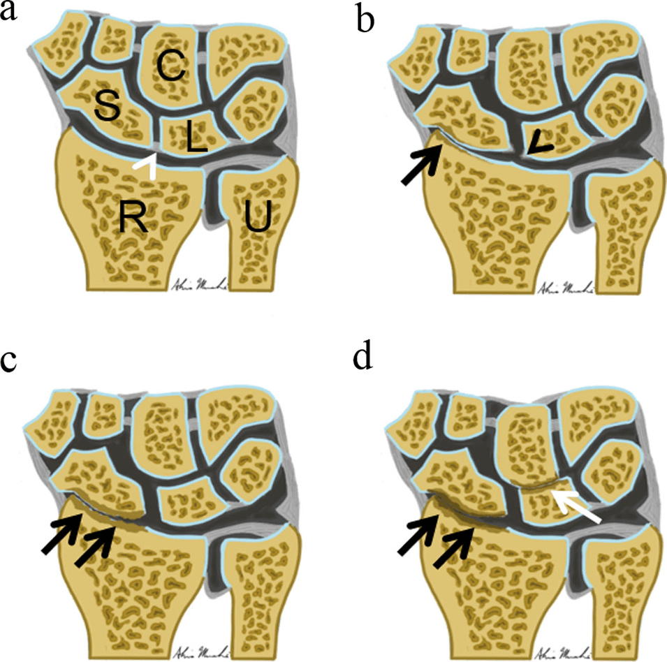
- Arthritis
- Scaphoid and radial styloid
- Features
- Radial styloid beaking; scaphoid fossa preserved
- Treatment
- Radial styloidectomy
- Arthritis
- Entire scaphoid fossa (radioscaphoid joint)
- Features
- Scaphoid fossa cartilage loss; capitate migrates proximally
- Treatment
- Four-corner fusion vs PRC
- Arthritis
- Midcarpal (capitate-lunate)
- Features
- Radiolunate joint preserved (the key feature); hamate-triquetrum involvement
- Treatment
- Four-corner fusion vs PRC
- Arthritis
- Pancarpal, including the radiolunate joint
- Features
- DRUJ arthritis common; motion preservation not possible
- Treatment
- Total wrist arthrodesis
Why the stage matters. The radiolunate joint remains preserved until late disease, and that is why scaphoid excision works. PRC in Stage III depends on the capitate head as well, because capitate cartilage degeneration contraindicates it; it is possible only if the capitate head is intact.
SNAC wrist. Scaphoid nonunion advanced collapse follows an identical progression but starts from a scaphoid fracture nonunion rather than a ligament injury. There is a history of scaphoid fracture; the proximal fragment remains attached to the lunate while the distal fragment rotates into flexion. The same radiographic staging and the same surgical algorithm apply.
Other causes.
- Post-traumatic - sequelae of perilunate dislocation, trans-scaphoid perilunate fracture-dislocation, axial loading injuries, distal radius malunion
- Inflammatory - rheumatoid arthritis (the wrist is involved in 70%), psoriatic arthritis, systemic lupus erythematosus, crystal arthropathy (gout, pseudogout)
- Degenerative - primary osteoarthritis (rare in the wrist), Kienböck disease (avascular necrosis of the lunate), Preiser disease (avascular necrosis of the scaphoid)
Clinical Assessment
History. Patients typically present with chronic wrist pain, stiffness and functional limitation. Distinguishing midcarpal from radiocarpal pathology clinically guides imaging and treatment planning, so ask about:
- Pain location - dorsal midcarpal or radial styloid
- Onset - acute injury or insidious progression
- Mechanical symptoms - catching, clicking, giving way
- Functional limitation - grip, keyboard use, weight-bearing
- Prior treatment - injections, splinting, therapy
- Occupational demands - labour or sedentary
Inspection and palpation. Look for a dorsal prominence from carpal collapse, muscle atrophy (a late finding), the condition of the skin for planning incisions, and prior surgical scars. Feel for point tenderness over the scapholunate interval, midcarpal joint-line tenderness, radial styloid tenderness (Stage I SLAC), and DRUJ stability and tenderness.
Movement. Measure and document a baseline: the flexion-extension arc against the contralateral wrist, radial-ulnar deviation and forearm rotation.
Special tests.
- Watson test (scaphoid shift) - positive with scapholunate instability
- Midcarpal shift test - a clunk with ulnar-to-radial deviation (technique below)
- Shear test - pain with axial loading
Where the tenderness points. Radioscaphoid and radial-styloid disease gives dorsoradial pain and tenderness over the radioscaphoid joint and radial styloid, with a positive Watson test. Midcarpal disease gives central dorsal pain and a positive midcarpal shift, a painful clunk on ulnar-to-radial translation of the distal row. The pattern of tenderness helps, but overlap is common.
Midcarpal shift test. Stabilise the proximal carpal row dorsally with one hand. With the other, grasp the metacarpals and translate the distal row palmarly; a palpable clunk indicates midcarpal instability. Perform it in neutral, then in ulnar and radial deviation. A positive test suggests capitolunate instability or midcarpal pathology.
Investigations
Radiographs are essential: a standard wrist series of PA, lateral and bilateral clenched-fist views.
- PA - scapholunate interval widening (greater than 3mm), scaphoid ring sign (flexed scaphoid), radial styloid beaking, joint-space narrowing, subchondral sclerosis and cysts, osteophytes
- Lateral - scapholunate angle (normal 30-60°, DISI greater than 70°), radiolunate angle (normal 0±10°), carpal height ratio (less than 0.5 abnormal), proximal migration of the capitate relative to the radius
- Clenched-fist PA - accentuates the scapholunate gap for dynamic instability, compared with the relaxed PA view
CT gives a detailed assessment of the extent of arthritis and the quality of the subchondral bone, evaluates the lunate fossa cartilage when PRC is being considered, and plans a four-corner fusion. 3D reconstruction helps in complex cases.
MRI is rarely needed for arthritis. It assesses the soft tissues and ligament integrity in early disease, rules out avascular necrosis and looks for concurrent pathology.
Wrist arthroscopy is the gold standard for cartilage assessment. It confirms the stage before definitive surgery, identifies salvageable joints and allows limited debridement.
Localising the Arthritis: Radiocarpal versus Midcarpal
The radiograph is the decisive step. The SLAC/SNAC pattern spares the radiolunate joint: narrowing progresses radioscaphoid → capitolunate (midcarpal) while the radiolunate space stays normal. This is the "midcarpal-salvageable" pattern that permits four-corner fusion or PRC.
The pattern that closes those doors. Radiolunate or radiocarpal narrowing and sclerosis, for example after an intra-articular distal radius fracture or in inflammatory disease, contraindicates both four-corner fusion and PRC. Four-corner fusion relies on a healthy radiolunate surface and PRC on a healthy lunate fossa, so this pattern needs total wrist arthrodesis or a radiocarpal-based fusion.
When the films are equivocal. A selective image-guided diagnostic injection, first of the radiocarpal and then of the midcarpal joint on separate occasions, localises the pain generator. Arthroscopy then grades the radiolunate/lunate-fossa and capitate-head cartilage directly, and that cartilage is the true determinant of PRC versus four-corner fusion versus total fusion, which is why arthroscopy is the gold standard for staging before committing to a salvage.
Differential Diagnosis
Dorsal and radial wrist pain has several causes that mimic or coexist with midcarpal arthritis. Distinguishing them prevents inappropriate salvage surgery.
- keyFeature
- Chronic pain, prior SL injury or scaphoid fracture
- radiology
- Radioscaphoid then capitolunate narrowing; radiolunate spared until late
- distinguisher
- Watson/scaphoid-shift positive; staged collapse pattern
- keyFeature
- Pain over proximal dorsal wrist
- radiology
- Radiolunate joint narrowing and sclerosis
- distinguisher
- Radiolunate involvement contraindicates motion-preserving salvage
- keyFeature
- Central dorsal wrist pain, no clear injury
- radiology
- Lunate sclerosis, fragmentation, collapse (Lichtman)
- distinguisher
- Lunate is the primary problem; PRC contraindicated if capitate involved
- keyFeature
- Pain at thumb base or distal scaphoid
- radiology
- STT joint narrowing, distal and radial
- distinguisher
- Tenderness distal to anatomical snuffbox, not dorsal midcarpal
- keyFeature
- Pain with pinch and grip at thumb base
- radiology
- Trapeziometacarpal narrowing and subluxation
- distinguisher
- Grind test positive; pain volar-radial, not dorsal carpal
- keyFeature
- Dorsal swelling or activity-related ache
- radiology
- Normal radiographs; cyst on ultrasound or MRI
- distinguisher
- No joint-space loss; transilluminates when visible
- keyFeature
- Bilateral, morning stiffness, systemic features
- radiology
- Periarticular erosions, uniform joint-space loss, osteopenia
- distinguisher
- Serology and synovitis; contraindicates PRC, alters fixation expectations
Non-Operative Management
Initial treatment aims at symptom control and function, and many patients achieve acceptable outcomes without surgery.
Splintage. A wrist immobilisation splint for acute flares, a custom thermoplastic orthosis for activity and night splinting for pain control, typically as a 4-6 week trial.
Medication. NSAIDs are first-line for pain, with paracetamol for mild symptoms and topical diclofenac gel. Avoid long-term opioid use.
Corticosteroid injection. Into the midcarpal joint under ultrasound or fluoroscopic guidance, triamcinolone 40mg mixed with local anaesthetic. Relief typically lasts 3-6 months; limit it to 3 injections a year. The injection can also help confirm the pain generator.
Therapy and activity. Gentle range-of-motion exercises, strengthening when pain permits, activity modification training and ergonomic optimisation, although therapy has limited efficacy in advanced arthritis. Avoid heavy lifting and impact loading, attend to keyboard ergonomics, modify the job when possible and use adaptive equipment for daily activities.
When to operate. Surgery is considered when conservative measures fail to give acceptable symptom control or function.
- Absolute - severe pain limiting daily activities despite 6 months of conservative therapy; progressive deformity; functional impairment affecting occupation or quality of life
- Relative - moderate pain with imaging showing advanced arthritis; patient preference after understanding the options; a young patient with high functional demands
Timing. Delay surgery until conservative measures are exhausted. Weigh the patient's age and occupation, their expectations and likely compliance, and coordinate with other medical conditions.
Guidelines, Registries & Global Practice
Global Epidemiology
- SLAC and SNAC are the most common patterns of degenerative wrist arthritis worldwide; primary midcarpal osteoarthritis is rare.
- Untreated scaphoid nonunion carries a high arthritic burden: radioscaphoid change in roughly three-quarters of patients by 4 years and midcarpal change in around 60% by 9 years (Vender/Watson sequence).
- Scaphoid fractures peak in young men (15-35 years); delayed or missed diagnosis is the dominant driver of the SNAC population globally and is over-represented in manual-labour and resource-limited settings.
- Scapholunate ligament injury underlies the SLAC population and is frequently under-recognised at initial trauma presentation.
Society Guidance: No Single Disease-Specific Guideline
There is no dedicated AAOS, BOA/BOAST, AO or EFORT guideline for midcarpal arthritis; management derives from comparative outcome literature and hand-surgery society consensus. Practice differs mainly in procedure preference rather than indication.
- Common ground (AAOS / BOA / AO / EFORT-aligned practice)
- Conservative trial (NSAIDs, splinting, activity modification, image-guided corticosteroid) before surgery
- Where practice varies
- Threshold/duration of trial (commonly 3-6 months)
- Common ground (AAOS / BOA / AO / EFORT-aligned practice)
- Watson SLAC / SNAC staging directs surgical choice
- Where practice varies
- Use of arthroscopy vs CT to confirm capitate-head cartilage
- Common ground (AAOS / BOA / AO / EFORT-aligned practice)
- Motion-preserving surgery (PRC or scaphoid excision + 4CF) preferred over total fusion
- Where practice varies
- PRC-first (motion, lower morbidity) vs 4CF-first (grip, demand)
- Common ground (AAOS / BOA / AO / EFORT-aligned practice)
- Total wrist arthrodesis is the reliable salvage
- Where practice varies
- Selective total wrist arthroplasty in low-demand or bilateral disease
Registry and High-Quality Trial Evidence
- No carpal-specific salvage registry exists; the evidence base is comparative cohorts, systematic reviews and meta-analysis (PRC vs 4CF), now being tested prospectively.
- The multicentre double-blind PROOF randomised non-inferiority trial (NCT04260165) is directly comparing PRC and 4CF with PRWE as the primary endpoint - the highest-level data anticipated in this field.
- Total wrist arthroplasty implant survivorship is captured in national joint registries (e.g. national registries reporting implant outcomes) and remains inferior to arthrodesis durability, supporting fusion as the default salvage in higher-demand patients.
High- vs Limited-Resource Practice Variation
- High-resource settings: routine CT and/or wrist arthroscopy for cartilage assessment, headless-screw or low-profile plate fixation for 4CF, and certified hand-therapy rehabilitation pathways.
- Limited-resource settings: greater reliance on plain radiographs and intraoperative cartilage assessment; PRC is often favoured for its single-stage, implant-free nature, shorter operative time and lower cost (direct surgical costs for 4CF are several-fold higher than PRC, driven mainly by implants).
- Across all settings, late presentation of scaphoid nonunion increases the proportion requiring salvage rather than reconstruction.
Rehabilitation Principles (Universal)
- Post-PRC: early protected mobilisation, typically from 2-3 weeks.
- Post-4CF: immobilisation usually 8-12 weeks until radiographic union, then progressive strengthening.
- Post-total fusion: splint weaning with strengthening once fixation is secure; counsel on compensatory elbow/shoulder/finger motion.
- Return to sedentary work generally precedes return to heavy manual work by several weeks to months in every health system.
Controversies and Areas of Uncertainty
PRC or four-corner fusion as the default. The largest meta-analysis favours PRC for motion and lower complications with comparable grip, yet many surgeons still default to four-corner fusion in younger, higher-demand patients. True equipoise persists pending the multicentre double-blind PROOF randomised trial (NCT04260165), the first head-to-head RCT.
The age threshold for PRC. PRC is traditionally reserved for patients over 35-50 years because of radiocapitate contact stress, but long-term series with durable results to 15 years despite universal radiographic change challenge a strict age cut-off. The decisive factor is capitate-head and lunate-fossa cartilage quality, not age alone.
Fixation for four-corner fusion. Headless compression screws, dorsal circular plates, staples and K-wires all achieve union, but circular plates carry higher hardware-related morbidity and impingement, while K-wires show higher partial-union rates. No construct is clearly superior; reported four-corner nonunion remains around 9%.
Capsular interposition and resurfacing PRC. Dorsal capsular interposition, or a resurfacing graft when the capitate head is marginal, is proposed to extend PRC indications, but evidence is limited and it is not yet standard.
Radial styloidectomy with salvage. Routine adjunctive radial styloidectomy to prevent impingement is debated. Over-resection risks radiocarpal instability, so most surgeons limit it to a few millimetres and only when impingement is demonstrated.
The role of total wrist arthroplasty. Improving implant designs have renewed interest in motion-preserving arthroplasty for low-demand or bilateral disease, but registry-level survivorship still trails arthrodesis, keeping fusion the reliable salvage in active patients.
Related pages: SLAC Wrist and SNAC Wrist for the two collapse patterns in full, with Scapholunate Dissociation and Scaphoid Nonunion for the upstream injuries that produce them; Proximal Row Carpectomy and Scaphoid Excision and Four-Corner Fusion for the two operations compared here, step by step; Kienböck's Disease for the lunate-driven route to the same salvage decision; and Wrist Arthroplasty for the motion-preserving alternative to arthrodesis.
MCQ Practice Points
Q: What is the typical progression pattern of SLAC (Scapholunate Advanced Collapse) wrist?
A: SLAC progression: Stage I - radial styloid to scaphoid. Stage II - entire radioscaphoid joint. Stage III - capitolunate joint. The radiolunate joint is spared due to preserved spherical congruency. This pattern guides surgical decision-making: PRC possible if capitate head intact.
Q: What distinguishes SNAC from SLAC wrist arthritis?
A: SNAC (Scaphoid Nonunion Advanced Collapse) results from scaphoid nonunion. SLAC results from scapholunate ligament injury. Both progress similarly through radioscaphoid then capitolunate joints. Key difference: SNAC shows scaphoid nonunion with humpback deformity and DISI pattern. Both spare radiolunate joint.
Q: What are the surgical options for midcarpal arthritis with intact radiolunate joint?
A: Options include: Proximal row carpectomy (PRC) - requires intact capitate head and radiolunate joint; Four-corner fusion (scaphoid excision + capitate-lunate-hamate-triquetrum fusion) - preserves more motion than total wrist fusion. PRC simpler with faster recovery; four-corner preserves more grip strength.
Q: What radiographic measurement indicates scapholunate dissociation leading to SLAC wrist?
A: Scapholunate angle greater than 70° (normal 30-60°) indicates dissociation. Terry Thomas sign - scapholunate gap greater than 3mm (or greater than 2mm difference from contralateral). Scaphoid ring sign on PA view indicates flexed scaphoid. DISI pattern (dorsal intercalated segment instability) shows lunate extended dorsally.
Q: Why is the radiolunate joint preserved in SLAC and SNAC wrist arthritis?
A: The radiolunate joint maintains spherical congruency even with carpal malalignment, distributing load evenly. The radioscaphoid joint has elliptical articulation, making it susceptible to point-loading with scaphoid malposition. This preservation makes PRC and four-corner fusion viable options.
Summary
Midcarpal arthritis represents a complex spectrum of degenerative wrist conditions requiring thorough understanding of carpal biomechanics, staging systems, and surgical options. SLAC and SNAC wrist patterns progress through predictable stages, with radiolunate joint preservation until late disease enabling motion-preserving surgery.
Four-corner fusion and proximal row carpectomy both achieve approximately 50-60% motion preservation with good pain relief in appropriately selected patients. Four-corner fusion provides superior grip strength and more predictable long-term outcomes but requires fusion healing and carries nonunion risk. Proximal row carpectomy offers simpler surgery with faster recovery but demands pristine lunate fossa cartilage and shows higher late progression rates.
Total wrist arthrodesis remains the gold standard for definitive treatment when motion preservation fails or is not feasible, providing maximum grip strength and reliable pain relief despite complete motion loss. Patient selection, understanding individual functional demands, and mastery of multiple surgical techniques are essential for optimal outcomes in this challenging condition.
Clinical Decision Scenarios
Practise clinical reasoning and management decisions out loud
“A 55-year-old carpenter presents with chronic dorsal wrist pain following a scaphoid fracture 10 years ago. Radiographs show scaphoid nonunion with arthritis between the proximal scaphoid fragment and radius, as well as between the capitate and lunate. The radiolunate joint appears well preserved. How would you classify and manage this?”
“You perform a dorsal approach for four-corner fusion in a 62-year-old woman with Stage III SLAC wrist. After excising the scaphoid and exposing the midcarpal joint, you inspect the lunate fossa cartilage and find it pristine with no evidence of degeneration. The capitate head also shows excellent cartilage. Your surgical plan was four-corner fusion but the findings suggest PRC might be possible. How do you proceed?”
“A 48-year-old labourer presents 18 months after a four-corner fusion done elsewhere with persistent dorsal wrist pain. Radiographs show a lucent line across the capitolunate interval and CT confirms a fibrous nonunion. The radiolunate joint remains well preserved. How do you assess and manage this failed four-corner fusion?”
SLAC Staging
- Radial styloid arthritis (Stage I) - radial styloidectomy
- Scaphoid fossa (Stage II) - 4CF or PRC
- Midcarpal capitolunate arthritis (Stage III) - 4CF or PRC
- Stage IV adds pancarpal + DRUJ - total wrist fusion
- Radiolunate preserved until Stage IV (key concept)
- SNAC follows identical staging from scaphoid nonunion
Four-Corner Fusion Essentials
- Scaphoid excision + fuse capitate-hamate-lunate-triquetrum
- Preserves radiolunate joint (motion source)
- Outcomes: 75-80% grip, 50-60% motion, 85-90% pain relief
- Union rate 90-95% with rigid fixation
- Immobilize 8-12 weeks until fusion
- Complications: nonunion 5-10%, hardware prominence 10-20%
PRC vs 4CF Decision
- PRC requires pristine lunate fossa cartilage (Grade I-II)
- PRC: faster recovery, no nonunion, 70-75% grip
- 4CF: better grip (75-80%), more predictable long-term
- PRC contraindications: inflammatory arthritis, poor cartilage, high demand
- Intraoperative cartilage assessment definitive
- Both achieve similar 50-60% motion preservation
Total Wrist Arthrodesis
- Indications: Stage IV SLAC, failed 4CF/PRC, high demand
- Position: 10-15° extension, neutral to 5-10° ulnar deviation
- Outcomes: 85-90% grip (best), 90-95% pain relief, 95% union
- Complete motion loss impacts ADLs significantly
- Plate fixation most common, allows early mobilization
- Consider bilateral disease (fuse one, preserve other)
PRC Contraindications
- Lunate fossa chondromalacia (Grade III-IV)
- Capitate articular cartilage degeneration
- Inflammatory arthritis (RA, psoriatic)
- Kienbock disease involving the capitate (AVN lunate)
- High-demand manual laborer
- Young patient (relative, consider 4CF instead)
Physical Examination
- Watson test: scaphoid shift with SL instability
- Midcarpal shift test: clunk ulnar to radial deviation
- Point tenderness scapholunate interval (dorsal)
- Grip strength typically 40-60% reduced
- ROM measurement and comparison to contralateral
- DRUJ assessment (Stage IV consideration)
Imaging Assessment
- PA wrist: SL gap (greater than 3mm), scaphoid ring sign
- Lateral: SL angle (normal 30-60°, DISI greater than 70°)
- Clenched fist: Dynamic SL instability
- CT scan: Assess arthritis extent, plan fixation, evaluate lunate fossa
- MRI: Limited role in arthritis (ligament assessment early)
- Arthroscopy: Gold standard cartilage evaluation
Surgical Pearls
- 4CF: Complete scaphoid excision prevents impingement
- 4CF: Thorough cartilage removal to bleeding bone essential
- PRC: Don't proceed if cartilage inadequate (convert to 4CF)
- Total fusion: Position critical (cannot easily revise)
- All: Preserve soft tissue envelope and blood supply
- All: Rigid fixation improves union rates
Evidence Base and Outcomes Studies
4CF vs PRC for SLAC/SNAC: Systematic Review & Meta-Analysis
- 61 studies, 3,174 wrists (54% PRC, 46% 4CF); weighted mean follow-up 61 months
- PRC had significantly greater postoperative extension and ulnar deviation
- No significant difference in grip strength between procedures
- Conversion to arthrodesis: 5.2% (PRC) vs 11% (4CF)
- 4CF nonunion rate 8.9% (57/640); hardware removal 2.2% after 4CF
PRC vs 4CF for Post-Traumatic Arthropathy: Systematic Review
- 7 studies (Levels I-III), 240 patients, 242 wrists
- Flexion-extension arc better after PRC: 75 degrees vs 62 degrees for 4CF
- Grip strength better after 4CF: 74% vs 67% of contralateral
- Radial deviation better after 4CF
- Overall complication rate 29% (4CF) vs 14% (PRC); nonunion 7% after 4CF
SLAC Wrist Salvage: Scaphoid Excision and Limited Fusion (Landmark)
- 100 cases of scaphoid excision plus limited (four-corner) fusion; mean follow-up 44 months
- Flexion-extension averaged 72 degrees (53% of normal contralateral)
- Grip strength 80% of the opposite side; pain relief good to excellent in most
- Radiolunate joint preserved in 98/100 despite long follow-up
- Nonunion in 3 cases; dorsal capitate-radius impingement in 12% (technique-related)
Degenerative Sequence of Scaphoid Nonunion (SNAC, Landmark)
- 64 untreated symptomatic scaphoid nonunions analysed radiographically
- Radioscaphoid changes in 75% by 4 years; midcarpal changes in 60% by 9 years
- Pattern follows SLAC (now termed SNAC) from rotary subluxation of the distal fragment
- Radius-proximal fragment and radiolunate joints consistently spared
- Instability progression associated with earlier arthritis onset