Two or More Ligaments | Vascular Assessment Mandatory | Surgical Emergency
SCHENCK CLASSIFICATION
Critical Must-Knows
- Vascular injury is the life/limb-threatening priority - assess immediately
- ABI less than 0.9 mandates angiography or surgical exploration
- Spontaneous reduction occurs in up to 50% - high index of suspicion
- Timing: Surgery at 1-3 weeks optimal (soft tissue settles, before scarring)
- PCL is key to knee stability - must be addressed in reconstruction
Clinical Pearls
- "Knee dislocation = MLKI until proven otherwise
- "Popliteal artery is tethered at adductor hiatus and popliteal arch
- "Common peroneal nerve injury in 25-40% of lateral dislocations
- "Posterolateral corner must not be missed - chronic PLC deficiency = failure
Critical MLKI Exam Points
Vascular Assessment First
Life before limb, limb before function. Popliteal artery injury occurs in up to 32% of knee dislocations. Ischemia greater than 6-8 hours leads to 86% amputation rate. ABI less than 0.9 requires angiography.
High Index of Suspicion
50% spontaneously reduce before imaging. Low-energy mechanisms in obese patients are often missed. Any multiligament laxity after knee trauma = assume dislocation occurred.
Posterolateral Corner
The PLC is the most commonly missed structure. FCL, popliteus, popliteofibular ligament must be assessed. Chronic PLC deficiency causes failure of ACL/PCL reconstructions.
Surgical Timing
1-3 weeks is optimal. Allows soft tissue swelling to settle, avoids arthrofibrosis from early surgery, prevents scarring of late reconstruction.
Multi-Ligament Knee Injury Decision Algorithm
| Priority | Assessment | Finding | Action |
|---|---|---|---|
| 1 - IMMEDIATE | Vascular status | Absent pulse or ABI less than 0.9 | Urgent angiography or surgical exploration |
| 2 - IMMEDIATE | Reduction status | Dislocated | Reduce urgently, splint, reassess pulses |
| 3 - URGENT | Nerve function | Common peroneal palsy | Document, may need exploration if open |
| 4 - EARLY | Soft tissue | Open injury, severe swelling | Washout if open, spanning ex-fix if unstable |
| 5 - DEFINITIVE | Ligament pattern | MRI defines injury pattern | Plan reconstruction at 1-3 weeks |
MLKI - IMLKI - Initial Assessment
| M | Mechanism and energy High or low energy, direction of force |
| L | Limb vascularity Pulses, ABI, capillary refill |
| K | Keep reduced Reduce if dislocated, splint in extension |
| I | Imaging urgently X-rays, angiography if indicated, MRI when stable |
| M | Mechanism and energy High or low energy, direction of force | K | Keep reduced Reduce if dislocated, splint in extension |
| L | Limb vascularity Pulses, ABI, capillary refill | I | Imaging urgently X-rays, angiography if indicated, MRI when stable |
Hook:MLKI reminds you the M is for limb-threatening vascular assessment first
SCHENCK CSCHENCK Classification
| I | One cruciate intact ACL or PCL torn, other cruciates intact |
| II | Both cruciates ACL and PCL torn, collaterals intact |
| III | Bicruciate + one collateral III-M (medial) or III-L (lateral) |
| IV | All four major ligaments Complete disruption |
| V | Periarticular fracture Any pattern with associated fracture |
| I | One cruciate intact ACL or PCL torn, other cruciates intact | IV | All four major ligaments Complete disruption |
| II | Both cruciates ACL and PCL torn, collaterals intact | V | Periarticular fracture Any pattern with associated fracture |
| III | Bicruciate + one collateral III-M (medial) or III-L (lateral) |
Hook:I-II-III-IV-V: Progressive severity from one cruciate to fracture-dislocation
PLC - PPLC - Posterolateral Corner Components
| P | Popliteus tendon Primary static restraint to external rotation |
| L | Lateral (fibular) collateral ligament Primary varus restraint |
| C | Capsule and popliteofibular ligament Secondary stabilizers |
| P | Popliteus tendon Primary static restraint to external rotation |
| L | Lateral (fibular) collateral ligament Primary varus restraint |
| C | Capsule and popliteofibular ligament Secondary stabilizers |
Hook:PLC = Popliteus, LCL, popliteofibular ligament - all must be addressed
VANS - VVANS - Vascular Assessment
| V | Vascular exam and ABI ABI less than 0.9 = abnormal |
| A | Angiography if abnormal CT-angio or conventional |
| N | Nerve exam Common peroneal especially |
| S | Serial exams Re-examine every 6 hours for 48 hours |
| V | Vascular exam and ABI ABI less than 0.9 = abnormal | N | Nerve exam Common peroneal especially |
| A | Angiography if abnormal CT-angio or conventional | S | Serial exams Re-examine every 6 hours for 48 hours |
Hook:VANS takes you to the hospital - vascular assessment is the priority
SPORTSSPORTS - Athletic MLKI Assessment
| S | Sports mechanism Cutting, pivoting, contact, hyperextension |
| P | Pulses and ABI Vascular assessment (lower risk but still check) |
| O | Orthopaedic exam Ligament testing, PLC assessment critical |
| R | Return-to-sport 60-80% overall, better for KD-I/II |
| T | Timing Surgery 1-3 weeks optimal |
| S | Schenck classification KD-I through KD-IV, guides treatment |
| S | Sports mechanism Cutting, pivoting, contact, hyperextension | O | Orthopaedic exam Ligament testing, PLC assessment critical | T | Timing Surgery 1-3 weeks optimal |
| P | Pulses and ABI Vascular assessment (lower risk but still check) | R | Return-to-sport 60-80% overall, better for KD-I/II | S | Schenck classification KD-I through KD-IV, guides treatment |
Hook:SPORTS MLKI needs SPORTS assessment - Sports mechanism, Pulses, Orthopaedic exam, Return-to-sport, Timing, Schenck
RTSRTS - Return-to-Sport Factors
| R | Reconstruction quality PCL and PLC critical for success |
| T | Time to surgery 1-3 weeks optimal, avoid delay |
| S | Schenck type KD-I/II better (70-80%), KD-IV guarded (40-60%) |
| R | Reconstruction quality PCL and PLC critical for success |
| T | Time to surgery 1-3 weeks optimal, avoid delay |
| S | Schenck type KD-I/II better (70-80%), KD-IV guarded (40-60%) |
Hook:RTS depends on Reconstruction quality, Timing, Schenck type
Overview and Epidemiology
Multi-ligament knee injury (MLKI) refers to injury to two or more of the four major knee ligaments (ACL, PCL, MCL, LCL/PLC). Knee dislocation is defined as complete loss of tibiofemoral contact.
Mechanism of injury:
- High-energy trauma: Motor vehicle accidents, pedestrian vs car, motorcycle
- Low-energy trauma: Sports injuries, falls in obese patients (ultra-low velocity)
- Direction of force: Anterior, posterior, lateral, medial, rotational
Ultra-Low Velocity Dislocations
Obese patients (BMI greater than 40) can dislocate their knee with minimal trauma. These are often missed because the mechanism seems trivial. Have a high index of suspicion in obese patients with multiligament laxity.
Epidemiology:
- Male predominance (3:1)
- Peak age 20-40 years
- Motor vehicle accidents most common (55%)
- Sports injuries (25%)
- Falls and industrial accidents (20%)
Associated injuries:
- Popliteal artery injury: 5-32%
- Common peroneal nerve palsy: 25-40%
- Meniscal tears: 40-50%
- Periarticular fractures (Schenck KD-V): 15-20%
- Tibial plateau or femoral condyle involvement
Anatomy and Biomechanics
Knee stability structures:
The knee relies on four major ligaments working in concert:
| Structure | Primary Function | Failure Leads To |
|---|---|---|
| ACL | Anterior tibial translation, rotational stability | Giving way, pivot shift |
| PCL | Posterior tibial translation | Posterior sag, quadriceps dysfunction |
| MCL | Valgus stability | Medial opening |
| LCL/PLC | Varus stability, external rotation control | Varus thrust, external rotation instability |
PCL as the Central Pillar
The PCL is the central pillar of knee stability. It is stronger than the ACL and provides the primary restraint to posterior translation. In MLKI, PCL reconstruction quality correlates strongly with outcomes.
Posterolateral Corner (PLC):
The PLC is a complex of structures providing varus and rotational stability:
- Fibular collateral ligament (FCL/LCL): Primary varus restraint
- Popliteus tendon: Primary restraint to external rotation at 30 degrees flexion
- Popliteofibular ligament: Links popliteus to fibular head
- Lateral capsule: Secondary stabilizer
- Arcuate ligament: Variable importance
PLC Must Not Be Missed
Chronic PLC deficiency causes failure of ACL and PCL reconstructions due to increased stress on the grafts. The PLC must be assessed clinically (dial test, external rotation recurvatum) and addressed surgically.
Popliteal artery anatomy:
The popliteal artery is at risk because:
- Tethered at two points: Adductor hiatus (proximal) and soleal arch (distal)
- Travels close to the posterior capsule
- Cannot move away from displacing tibia
- Intimal tears may not be immediately evident
Common peroneal nerve:
- Wraps around fibular neck
- At risk in lateral dislocations and PLC injuries
- Injury in 25-40% of MLKI
- Recovery variable (50% partial recovery)
Classification Systems
Schenck Classification (anatomic, most commonly used)
| Type | Pattern | Description |
|---|---|---|
| KD-I | ACL or PCL intact | Single cruciate + collateral(s) |
| KD-II | Bicruciate only | ACL and PCL torn, collaterals intact |
| KD-III-M | Bicruciate + MCL | Medial-sided injury |
| KD-III-L | Bicruciate + LCL/PLC | Lateral-sided injury (worse prognosis) |
| KD-IV | All four ligaments | Complete disruption |
| KD-V | Any + fracture | Fracture-dislocation |
Suffix C = vascular injury requiring repair
KD-III-L Prognosis
KD-III-L (lateral pattern) has the worst prognosis due to PLC involvement and high rate of common peroneal nerve injury (up to 40%). These require meticulous PLC reconstruction.
Clinical Presentation and Assessment
History:
- Mechanism of injury (crucial for understanding pattern)
- Direction of force if witnessed
- Any sensation of knee "going out" or "snapping back"
- Presence of gross deformity initially
- Time since injury
Physical examination priorities:
Initial Examination Priorities
| Finding | Significance | Action |
|---|---|---|
| Absent pulses | Popliteal artery injury | Immediate vascular surgery consult |
| ABI less than 0.9 | Arterial injury likely | Angiography or surgical exploration |
| Gross deformity | Unreduced dislocation | Reduce urgently under sedation |
| Foot drop | Common peroneal nerve injury | Document, observe, may need exploration |
| Tense swelling | Possible compartment syndrome | Compartment pressure measurement |
| Open wound | Open dislocation | Urgent washout, antibiotics |
Vascular assessment protocol:
- Palpate pulses: Popliteal, dorsalis pedis, posterior tibial
- Calculate ABI: Ankle-Brachial Index (normal more than 0.9)
- If ABI less than 0.9: Angiography (CT-angio or conventional)
- If pulses absent: Immediate vascular surgery consult
- Serial exams: Every 6 hours for 48 hours (intimal tears can progress)
ABI Threshold
ABI less than 0.9 is abnormal and requires further investigation. An ABI of 0.9 or above has 95% negative predictive value for significant vascular injury. However, intimal tears can progress, so serial exams are mandatory.
Ligament examination (once vascular status confirmed):
| Test | Assesses | Positive Finding |
|---|---|---|
| Lachman | ACL | Soft endpoint, increased translation |
| Posterior drawer | PCL | Posterior tibial translation |
| Valgus stress 30 degrees | MCL | Medial opening |
| Varus stress 30 degrees | LCL | Lateral opening |
| Dial test (30 and 90 degrees) | PLC | External rotation asymmetry more than 10 degrees |
| External rotation recurvatum | PLC | Hyperextension with external rotation |
| Posterolateral drawer | PLC | Posterolateral subluxation |
Nerve examination:
- Common peroneal: Ankle dorsiflexion, toe extension, lateral leg sensation
- Tibial nerve: Ankle plantarflexion, toe flexion, plantar sensation
- Saphenous: Medial leg and ankle sensation
Differential diagnosis:
The acutely unstable or grossly swollen knee after trauma has several mimics. The cardinal point is that a multiligament injury or spontaneously reduced dislocation must be actively excluded, because the others do not carry the same limb-threatening vascular risk.
Differential Diagnosis of the Acutely Unstable / Swollen Knee
| Condition | Discriminating features | Vascular risk | Key investigation |
|---|---|---|---|
| Multiligament injury / reduced dislocation | Laxity in two or more planes, high-energy or ultra-low velocity mechanism, possible foot drop | High - assess immediately | ABI then MRI; CT angiography if ABI less than 0.9 |
| Isolated ACL rupture | Single-plane anterior laxity, positive Lachman, haemarthrosis, pivot shift | Negligible | MRI |
| Isolated PCL rupture | Posterior sag and posterior drawer only, dashboard mechanism | Low (unless high-energy) | MRI, posterior stress radiographs |
| Patellar dislocation | Lateral patellar displacement, apprehension, medial tenderness, no tibiofemoral laxity | Negligible | Skyline radiograph, MRI |
| Tibial plateau fracture | Bony tenderness, lipohaemarthrosis, deformity, fracture on radiograph | Moderate (Schatzker patterns) | Radiograph then CT |
| Extensor mechanism rupture (patellar/quad tendon) | Inability to straight-leg raise, palpable gap, patella alta or baja | Negligible | Lateral radiograph, ultrasound or MRI |
| Septic arthritis / haemarthrosis (atraumatic) | Fever, no clear mechanism, hot effusion | Negligible | Joint aspiration, inflammatory markers |
Investigations
Radiographic assessment:
Initial X-rays:
- AP and lateral knee (may show reduced or dislocated position)
- Look for periarticular fractures
- Avulsion fragments (fibular head = PLC, Segond = ACL/PLC, PCL avulsion)
- Calculate tibial subluxation
CT imaging:
- If reduction maintained, CT is helpful for fracture assessment
- CT angiography is study of choice for vascular assessment
- Sensitivity greater than 95% for significant arterial injury
CT Angiography
CT angiography has largely replaced conventional angiography for initial assessment. It is non-invasive, rapid, and highly sensitive. Reserve conventional angiography for planned intervention.
MRI imaging:
MRI is essential for surgical planning:
- Defines which ligaments are injured
- Identifies repairable vs reconstructable injuries
- Assesses meniscal tears (common)
- Evaluates cartilage injury
- Determines PLC involvement
Timing of MRI:
- Once patient stable and vascular status confirmed
- Usually within first week
- May be limited by swelling initially
Key MRI findings:
| Structure | MRI Finding |
|---|---|
| ACL rupture | Discontinuity, abnormal signal, edema |
| PCL rupture | Discontinuity, posterior tibial sag on sagittal |
| MCL tear | Medial soft tissue edema, ligament discontinuity |
| PLC injury | FCL disruption, popliteus abnormality, arcuate sign |
| Bone bruise | Kissing contusions indicate mechanism |
Vascular assessment algorithm:
Vascular Assessment Pathway
| Finding | Action | Timeframe |
|---|---|---|
| Hard signs (absent pulse, expanding hematoma) | Immediate surgical exploration | Minutes |
| ABI less than 0.9 | CT angiography or conventional angio | Within 2 hours |
| ABI 0.9 or more with good pulses | Serial exams every 6 hours for 48 hours | Ongoing |
| Deteriorating pulses/ABI on serial exam | Urgent angiography | Immediate |
Management
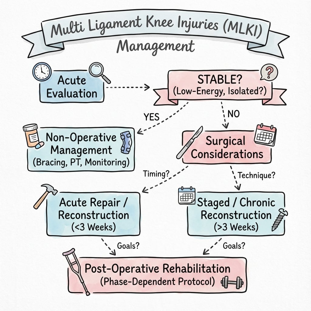
Priority 1: Vascular Assessment
- Assess pulses and calculate ABI
- If hard signs of vascular injury: immediate surgery
- If ABI less than 0.9: angiography
Priority 2: Reduction
- If still dislocated: reduce urgently under sedation
- Apply longitudinal traction
- Reverse the deformity
- Post-reduction X-ray and pulse check
Priority 3: Stabilization
- Splint in slight flexion (20-30 degrees)
- Avoid hyperextension (stretches vascular repair)
- Consider spanning external fixator if grossly unstable
Post-reduction protocol:
- Serial vascular exams every 6 hours for 48 hours
- Ice, elevation, DVT prophylaxis
- MRI when swelling allows
- Surgical planning
Post-Reduction Vascular Check
After reduction, pulses may appear normal but intimal injury can progress. Mandatory serial vascular examinations every 6 hours for at least 48 hours. Any deterioration requires urgent investigation.
Surgical Technique
MRI review:
- Identify all injured structures
- Assess repairability (avulsions vs mid-substance tears)
- Plan graft requirements
- Note meniscal and cartilage injury
Graft planning:
- Multiple grafts usually needed
- Autograft: hamstrings, BTB, quadriceps
- Allograft: Achilles, tibialis anterior, posterior tibial
- Consider graft availability and patient factors
Patient positioning:
- Supine with leg holder
- Lateral post for valgus stress
- Ability to flex knee fully
Careful preoperative planning prevents intraoperative surprises and ensures all necessary grafts and equipment are available.
Complications
Complications of MLKI and Reconstruction
| Complication | Incidence | Prevention/Management |
|---|---|---|
| Vascular injury | 5-32% | Immediate recognition, vascular surgery, serial exams |
| Peroneal nerve palsy | 25-40% | Recognize early, exploration if open, observe most |
| Arthrofibrosis/stiffness | 20-40% | Optimal timing, early motion, may need MUA/arthrolysis |
| Persistent instability | 10-20% | Address all structures, especially PLC |
| Graft failure | 5-15% | Adequate graft, protect rehabilitation, address alignment |
| Post-traumatic OA | 20-50% | Anatomic restoration, treat associated injuries |
| DVT/PE | Increased risk | Chemoprophylaxis, early mobilization |
Vascular complications:
- Amputation rate 86% if ischemia greater than 6-8 hours
- Missed injury due to intimal tear - serial exams critical
- Compartment syndrome may follow reperfusion
- Long-term claudication if vessel stenosis
Nerve complications:
- Common peroneal nerve palsy in 25-40%
- Only 50% make meaningful recovery
- Consider tendon transfers if no recovery by 12 months
- Posterior tibial nerve injury less common but more devastating
Stiffness Prevention
Arthrofibrosis is the most common complication affecting function. Prevention includes optimal surgical timing (1-3 weeks), early ROM, and sometimes hinged knee brace allowing protected motion. If more than 90 degrees lost, consider MUA at 6-12 weeks.
Persistent instability:
- Usually due to missed PLC injury
- Or inadequate PCL reconstruction
- Revision surgery required
- Address all structures and consider alignment
Postoperative Care and Rehabilitation
Postoperative protocol:
- Knee immobilizer or hinged brace locked in extension
- Touch-down weight bearing (10-15 kg) with crutches
- Quad sets, straight leg raises
- Wound care, DVT prophylaxis
- Ice and elevation
- Progressive ROM (goal 90 degrees by 4 weeks, 120 by 6 weeks)
- Hinged brace unlocked for ROM exercises
- Continue partial weight bearing
- Avoid active hamstrings if PCL reconstructed (prevents posterior tibial sag)
- Prone hangs for extension
- Progress to full weight bearing
- Continue ROM progression (goal full ROM)
- Begin closed chain strengthening
- Stationary bike, pool exercises
- Proprioception training
- Progressive strengthening
- Full ROM expected
- Jogging at 4-6 months if PCL stable
- Sport-specific training begins
- Brace wean as strength improves
- Return to sport typically 9-12 months
- Functional testing before clearance
- Ongoing strengthening and proprioception
- Some patients require longer before high-demand activities
Key rehabilitation principles:
- Slower progression than isolated ACL
- Protect PCL graft (no active hamstrings early)
- Balance motion restoration with stability
- Patient-specific based on tissue quality and compliance
- Return to sport later than isolated ligament injuries
Active Hamstrings
Avoid active hamstrings in the early postoperative period after PCL reconstruction. The hamstrings cause posterior tibial translation which stresses the PCL graft. Use quad-dominant exercises and protect the graft.
Outcomes and Prognosis
Outcomes by pattern:
| Pattern | Outcome | Key Factor |
|---|---|---|
| KD-II (bicruciate only) | Best outcomes | No collateral injury |
| KD-III-M | Good outcomes | MCL often heals |
| KD-III-L | Worst outcomes | PLC complexity, nerve injury |
| KD-IV | Variable | Depends on surgical quality |
| KD-V | Worse | Fracture complicates healing |
Prognostic factors:
- Positive: Early surgery, complete reconstruction, good compliance
- Negative: Vascular injury, nerve injury, delayed surgery, missed PLC
PLC Critical
Missed or inadequately treated PLC is the most common cause of failure. PLC deficiency leads to increased stress on ACL and PCL grafts. Always assess and address the posterolateral corner.
Long-term concerns:
- Post-traumatic osteoarthritis (20-50%)
- Residual laxity (often acceptable if functional)
- Stiffness (10-20% require intervention)
- Chronic pain (10-15%)
- Return to high-level sport is often not achieved
Evidence Base
- Prospective series of 38 knee dislocations. An ABI less than 0.90 was present in 11 patients (29%) and all 11 had a surgically confirmed arterial injury. Sensitivity, specificity and positive predictive value of ABI less than 0.90 were all 100%, and the negative predictive value of an ABI of 0.90 or higher was 100%.
- Across 99,688 knee dislocations in a national all-payer database, vascular injury occurred in 1066 (1.1%), of which 262 (24.6%) required repair. Obesity, male sex, alcohol misuse and high comorbidity burden were independent risk factors.
- French national prospective series of 67 knees (mean age 37, 82% male). Popliteal artery lesion in 9 knees, and crucially only 1 of these 9 had a discernible distal pulse, underscoring that palpable pulses do not exclude arterial injury. Isolated common peroneal nerve injury occurred in 12 knees (18%).
- Prospective series of 30 acute grade-III posterolateral injuries treated by anatomic repair of avulsions, reconstruction of mid-substance tears and concurrent cruciate reconstruction. Side-to-side lateral compartment gap improved from 6.2 mm to 0.1 mm and IKDC subjective scores rose from 29 to 82.
- Surgical treatment gave more excellent/good IKDC results than non-operative care (58% vs 20%). Early surgery (3 weeks or less) outperformed delayed surgery (Lysholm 90 vs 82). Repair of the posterolateral corner failed far more often than reconstruction (37% vs 9%).
- Prospective comparison of 43 knee dislocations. Overall ligament failure was 7% with a hinged external fixator versus 29% with bracing. Posterolateral corner repair failed in 54% versus PLC reconstruction failure of 25%, again favouring reconstruction over repair.
- Long-term (2 to 18 year) experience with combined PCL/ACL and collateral reconstruction. Found no conclusive superiority of double-bundle over single-bundle PCL reconstruction, supported delayed surgery of about 2 to 3 weeks to reduce arthrofibrosis, and emphasised addressing all components of the instability with allograft.
Clinical Decision Scenarios
Use these scenarios to practise clinical reasoning and management decisions
Scenario 1: High-Energy Knee Dislocation
"A 35-year-old motorcyclist is brought to ED after a crash. His right knee is grossly deformed. There is no open wound. The foot is pale and cool. What is your immediate management?"
Scenario 2: Surgical Planning for MLKI
"A 28-year-old footballer sustained a knee dislocation that spontaneously reduced. Vascular exam is normal. MRI shows complete ACL and PCL tears, plus posterolateral corner injury with popliteus and FCL disruption. How do you plan surgical management?"
Scenario 3: Failed Multi-Ligament Reconstruction
"A patient presents 1 year after multi-ligament knee reconstruction (ACL and PCL) at another hospital. They have persistent instability with varus thrust and external rotation instability during gait. Examination shows positive dial test and external rotation recurvatum. What is your assessment?"
MCQ Practice Points
Definition Question
Q: What defines a multi-ligament knee injury? A: Injury to two or more of the four major knee ligaments (ACL, PCL, MCL, LCL/PLC). Knee dislocation is complete loss of tibiofemoral contact.
Vascular Question
Q: What ABI threshold requires further vascular investigation in knee dislocation? A: ABI less than 0.9 requires angiography (CT or conventional). An ABI of 0.9 or more has 95% negative predictive value for significant vascular injury.
Classification Question
Q: What is a Schenck KD-III-L injury? A: Bicruciate injury (ACL + PCL) plus lateral-sided/posterolateral corner injury. The "L" denotes lateral involvement. This pattern has the worst prognosis.
PLC Question
Q: What are the three main components of the posterolateral corner? A: Fibular collateral ligament (FCL), popliteus tendon, and popliteofibular ligament. These provide varus stability and external rotation control.
Timing Question
Q: What is the optimal timing for multi-ligament knee reconstruction? A: 1-3 weeks after injury. This allows soft tissue swelling to settle while tissues remain identifiable for repair/reconstruction, and avoids scarring that complicates delayed surgery.
Guidelines, Registries & Global Practice
Global epidemiology. Knee dislocation is rare. In a US national all-payer dataset of 99,688 coded knee dislocations (2010-2022), associated vascular injury occurred in only 1.1%, of which one quarter required repair (Dubin et al, J Orthop 2024). This contrasts sharply with the historical 30% figure derived from high-energy true dislocations, and the gap reflects case mix rather than contradiction. Prospective national series confirm a young, male-predominant population (mean age around 37, roughly 80% male) with common peroneal nerve injury in the order of 18% (Lustig et al, Orthop Traumatol Surg Res 2009). Obesity, including ultra-low velocity dislocation, is an independent risk factor for vascular injury and for missed diagnosis worldwide.
Major guidance, side by side. No single high-level society guideline governs the multiligament knee; practice is driven by expert consensus and systematic-review evidence, which is remarkably consistent across regions:
| Body / region | Position on key issues | Evidence basis |
|---|---|---|
| AAOS / US sports-medicine consensus | ABI-based vascular triage (ABI 0.90 cut-off); operative reconstruction preferred over non-operative care; reconstruct (not repair alone) the PLC | Prospective and systematic-review (Level I-IV) |
| BOA / BOAST (UK, open & vascular limb injury principles) | Combined orthoplastic and vascular pathway, urgent senior vascular review for any perfusion deficit, time-critical revascularisation | Consensus standard of care |
| AO Foundation | Reduce and assess perfusion first; spanning external fixation for the grossly unstable, open, or vascular-repair knee; staged ligament surgery | Consensus / case series |
| EFORT / European consensus | Early single-stage anatomic reconstruction where soft tissues allow; address every injured structure | Systematic review (Level IV) |
The areas of genuine international agreement are: vascular assessment first with ABI 0.90 as the action threshold; early surgery (within roughly 3 weeks) beats delayed surgery; and the posterolateral corner should be reconstructed rather than repaired in isolation (Levy et al, Arthroscopy 2009).
Registry evidence. There is no dedicated multiligament knee registry; this is a ligament-reconstruction rather than an implant-arthroplasty problem, so the major joint registries (NJR, AJRR, AOANJRR, Swedish/SHAR, Norwegian, NZJR) do not capture it. The relevant population-level data come instead from national administrative datasets and prospective multicentre cohorts (e.g. the French Society of Orthopaedic Surgery series and US national database studies cited above). Where end-stage post-traumatic arthritis later requires arthroplasty, those joint registries become relevant for implant selection and survivorship.
Global practice variation.
- High-resource settings favour MRI-planned, single-stage anatomic reconstruction of all torn ligaments at 1-3 weeks, often with allograft.
- Limited-resource settings more often rely on staged surgery, autograft (allograft and graft banks may be unavailable), bracing of selected collateral injuries, and acceptance of non-operative management in older or comorbid patients.
- Allograft availability is the single largest driver of technique differences worldwide; autograft-only strategies (hamstrings, quadriceps, contralateral grafts) are well described where banks are limited.
Keep It Global and Clinical
This is a worldwide resource. Management is framed around the limb-salvage decision pathway rather than any single country's health system. Examiners test clinical decision-making, so the focus stays on vascular triage, classification and reconstruction strategy.
Exam Context
For any board worldwide, be ready to: run the vascular assessment algorithm with the ABI 0.90 threshold, apply the Schenck classification to a described pattern, and articulate a systematic surgical plan addressing every injured structure early. The single highest-yield concept is that a missed posterolateral corner causes reconstruction failure.
MULTI-LIGAMENT KNEE INJURIES
Clinical summary
IMMEDIATE PRIORITIES
- •Vascular assessment first - limb-threatening emergency
- •Pulse check and ABI (less than 0.9 = angiography)
- •Reduce if dislocated
- •Serial vascular exams every 6 hours for 48 hours
SCHENCK CLASSIFICATION
- •KD-I: Single cruciate + collateral(s)
- •KD-II: Bicruciate only (collaterals intact)
- •KD-III-M or III-L: Bicruciate + medial or lateral
- •KD-IV: All four ligaments
- •KD-V: Any pattern + periarticular fracture
SURGICAL TIMING AND APPROACH
- •Optimal: 1-3 weeks (swelling settled, before scarring)
- •Address ALL injured structures
- •PCL is priority - central pillar of stability
- •PLC must not be missed (causes failure)
PLC RECONSTRUCTION
- •Modified Larson (fibular-based) technique
- •Recreates FCL and popliteofibular ligament
- •Uses allograft (Achilles or tibialis anterior)
- •Protect peroneal nerve
KEY COMPLICATIONS
- •Vascular injury (86% amputation if missed)
- •Peroneal nerve palsy (25-40%)
- •Arthrofibrosis (avoid with proper timing, early ROM)
- •Persistent instability (usually missed PLC)
TRAPS AND PEARLS
- •50% spontaneously reduce - high suspicion needed
- •Ultra-low velocity in obese patients still needs vascular assessment
- •Intimal tears can progress - serial exams essential
- •PLC deficiency most common cause of reconstruction failure
- •Avoid active hamstrings early after PCL reconstruction