Bacterial Bone Infection | Biofilm Formation | Chronic Sequestration
- Staphylococcus aureus causes 80% of all osteomyelitis cases
- Biofilm formation on necrotic bone prevents antibiotic penetration
- Sequestrum = necrotic bone, Involucrum = new bone shell around infection
- Chronic osteomyelitis requires surgical debridement - antibiotics alone insufficient
- An antistaphylococcal penicillin (flucloxacillin/nafcillin) or cefazolin is first-line for MSSA bone infection worldwide
- “Cierny-Mader combines anatomic location with host physiologic status
- “CRP more sensitive than ESR for monitoring treatment response
- “MRI has 90% sensitivity for osteomyelitis detection
- “Approximately 6 weeks of pathogen-directed antibiotic therapy; paediatric RCTs support around 3 weeks
Overview
Osteomyelitis is infection of bone, caused by bacteria, fungi or mycobacteria. Acute and chronic osteomyelitis differ significantly in clinical presentation, microbiology and management, and understanding the pathophysiology, particularly biofilm formation on necrotic bone, is fundamental to effective treatment.
Host and pathogen. The outcome of a bone infection depends on the balance between bacterial virulence and the host immune response. Local factors (vascular supply, foreign material) and systemic factors (diabetes, immunosuppression) determine whether the infection resolves, becomes chronic or disseminates.
Pathophysiology
Haematogenous spread. Bacteraemia seeds bone during transient episodes, and S. aureus is the most common organism. Different bones have unique vascular patterns that influence their susceptibility:
- Metaphysis of long bones in children - slow sinusoidal blood flow with hairpin loops creates stagnant zones for bacterial seeding
- Vertebral bodies in adults - Batson's valveless venous plexus allows retrograde seeding from pelvic and abdominal infections
Contiguous spread. Infection spreads into bone from a neighbouring focus: after surgery, around hardware and fracture fixation, or from a diabetic foot ulcer. It often involves soft tissue and bone together, and it is polymicrobial in trauma and diabetic cases.
Direct inoculation. The organism is carried in at the time of injury or operation:
- Open fractures - contamination at injury
- Combat injuries - high-energy trauma
- Puncture wounds - a nail through the shoe (Pseudomonas)
- Implant surgery - low-virulence organisms such as Staph epidermidis
From acute to chronic. The infection progresses through three stages, and the sequestrum forms in the second.
Progression from Acute to Chronic
Bacteria invade and an inflammatory response begins. Vascular congestion raises the intraosseous pressure, pus forms under the periosteum as a subperiosteal abscess, and purulent material tracks along the Volkmann canals.
Vascular compromise develops as the raised intraosseous pressure compresses vessels and causes ischaemia. Necrotic bone forms the sequestrum, and the body attempts to wall off the infection with a new bone shell, the involucrum.
Biofilm forms on the sequestrum. Sinus tracts form to the skin, and the infection flares repeatedly despite antibiotics.
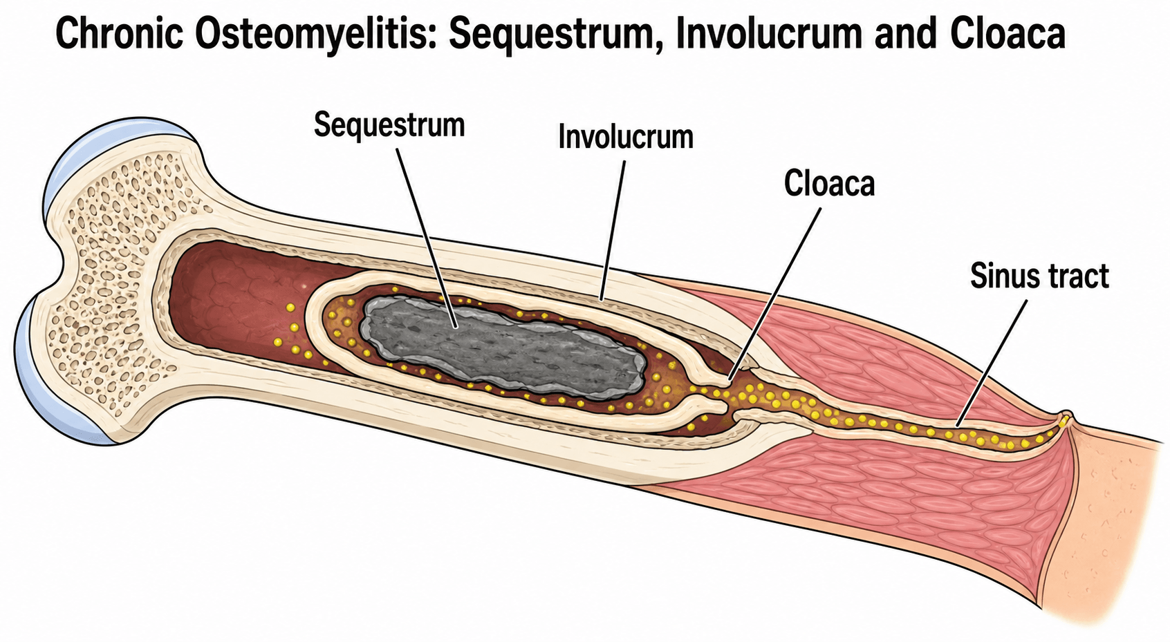
Dead bone and its shell. As infection thromboses the local blood supply, a fragment of bone dies and separates. Cortical bone is supplied by periosteal vessels, so damage to them leads to avascular sequestrum formation. Chronic osteomyelitis is described by the structures that follow:
- Sequestrum - a segment of necrotic, avascular bone, dead and sclerotic, that becomes colonised by bacteria and serves as the nidus for chronic infection
- Involucrum - reactive new bone laid down by the periosteum around the sequestrum as the body attempts to wall off the infection
- Cloaca - an opening in the involucrum through which pus and dead bone escape, often leading to a sinus tract through the soft tissues
Biofilm and Bacterial Persistence
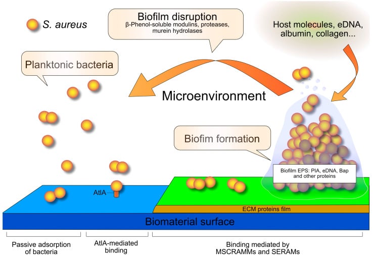
Biofilm. Biofilm is a structured community of bacteria enclosed in a self-produced polysaccharide matrix, the glycocalyx, and it is the key to chronicity. The bacteria adhere to necrotic bone inside it, and the matrix:
- is a physical barrier against antibiotics
- prevents neutrophil phagocytosis
- allows the bacteria to enter a dormant, metabolically inactive state
- enables quorum sensing between bacteria
Bacteria within biofilm are 1000 times more resistant to antibiotics than planktonic, free-floating bacteria. Initial surface adhesion is followed by matrix-mediated maturation, and dispersion seeds recurrent infection elsewhere.
Antibiotics alone cannot cure chronic osteomyelitis. The biofilm-colonised sequestrum is unreachable by antibiotics and has to be removed surgically: debridement, not antibiotics, is the cornerstone of treatment.
Beyond biofilm. Biofilm is only one of several reasons bacteria survive in bone. Chronic osteomyelitis is notorious for relapsing months or even years after apparently successful treatment, and the mechanisms below are why "cure" is better described as long-term remission.
- What Happens
- Bacteria adhere to sequestrum/implant in a self-produced polysaccharide matrix and enter a dormant state
- Why It Drives Chronicity / Relapse
- Tolerant to antibiotics and shielded from phagocytes - must be physically removed by surgery
- What Happens
- A slow-growing subpopulation with defective electron transport (often haemin/menadione auxotrophs) forming small, non-pigmented colonies
- Why It Drives Chronicity / Relapse
- Low metabolic activity reduces susceptibility (especially to aminoglycosides, which need membrane potential); they persist intracellularly and can revert to virulent wild-type, causing late relapse
- What Happens
- S. aureus is internalised by osteoblasts and osteocytes and survives within the osteocyte lacuno-canalicular network
- Why It Drives Chronicity / Relapse
- Creates a protected reservoir hidden from extracellular antibiotics and the immune system, and induces osteoblast apoptosis - a source of recurrence after therapy stops
- What Happens
- Phenotypically dormant (non-mutant) cells that tolerate rather than resist antibiotics
- Why It Drives Chronicity / Relapse
- Survive a treatment course and regrow once antibiotics are withdrawn, even with a fully susceptible organism
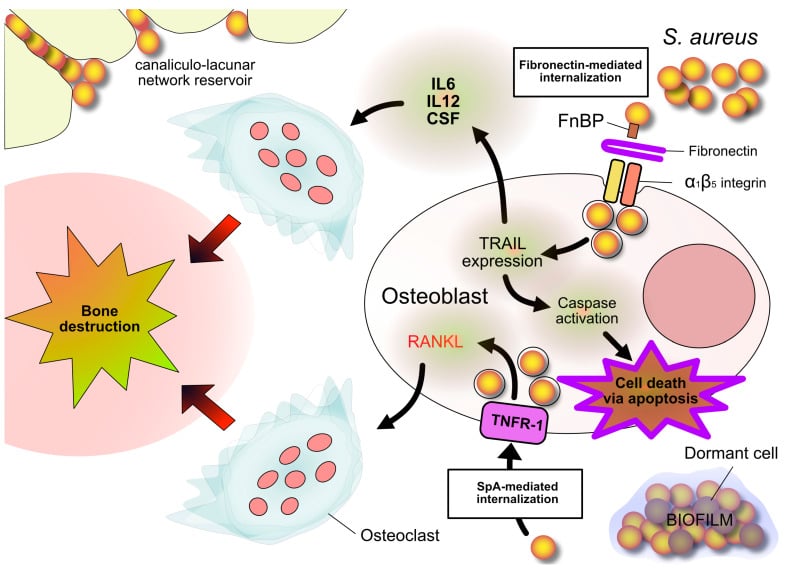
Antibiotic sensitivity testing measures the susceptibility of fast-growing planktonic bacteria. It does not capture biofilm tolerance, small-colony variants, intracellular reservoirs or persister cells, which is why chronic osteomyelitis can recur despite "appropriate" antibiotics. Two principles follow: surgical source control removes the biofilm and sequestrum reservoir, and agents with intracellular and biofilm activity (rifampicin in combination, clindamycin, fluoroquinolones) target the persisting populations. Counsel patients in terms of remission and recurrence risk rather than guaranteed cure.
Microbiology
Staphylococcus aureus causes 80% of osteomyelitis and is the most common organism in all age groups. Beyond it, the clinical scenario predicts the organism.
- Site
- Long bone metaphysis
- Organism
- Group B Streptococcus, E. coli
- Key Feature
- Crosses physis - can cause septic arthritis
- Site
- Metaphysis of long bones
- Organism
- S. aureus, Kingella kingae
- Key Feature
- Physis acts as barrier after 4 months
- Site
- Metaphysis
- Organism
- S. aureus 90%
- Key Feature
- Physis acts as barrier
- Site
- Vertebral bodies
- Organism
- S. aureus, E. coli, Mycobacterium TB
- Key Feature
- Batson's plexus spread
- Site
- Metatarsals, calcaneus
- Organism
- Polymicrobial, anaerobes, MRSA
- Key Feature
- Contiguous spread from ulcer
- Site
- Diaphysis, vertebrae
- Organism
- Salmonella more common than S. aureus
- Key Feature
- Only scenario where Salmonella predominates
- Site
- Calcaneus
- Organism
- Pseudomonas aeruginosa
- Key Feature
- Direct inoculation
- Site
- Vertebrae, long bones
- Organism
- S. aureus, Pseudomonas, Candida
- Key Feature
- Haematogenous seeding
- Site
- Fracture site
- Organism
- S. aureus, S. epidermidis, Propionibacterium
- Key Feature
- Low virulence biofilm formers
Cierny-Mader Classification
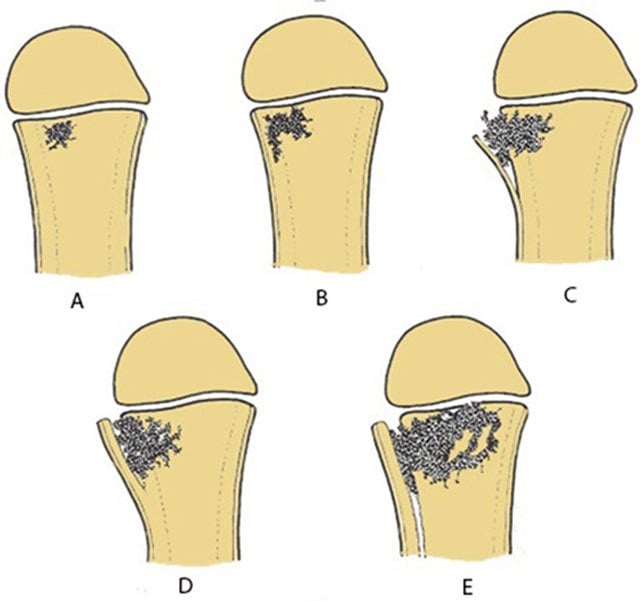
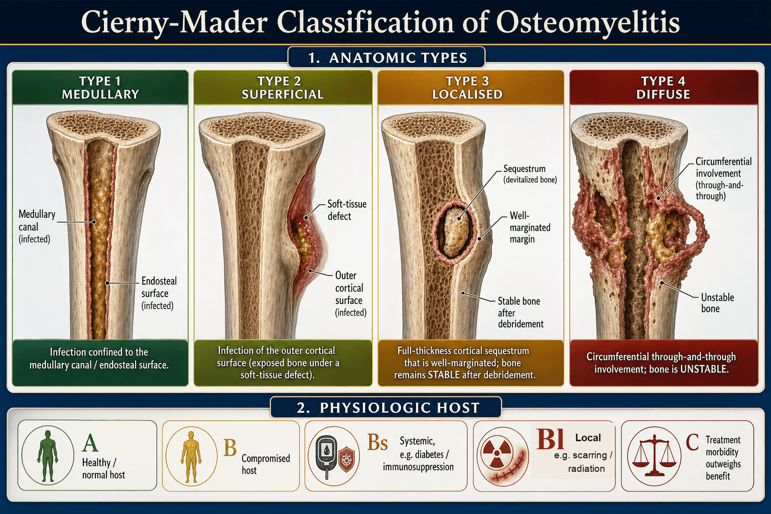
Cierny-Mader is the gold standard. It combines the anatomic type of the infection (I-IV) with the physiologic status of the host (A-C).
- Location
- Intramedullary canal
- Characteristics
- Endosteal surface, haematogenous
- Treatment
- Reaming, antibiotics, nail
- Location
- Surface of bone
- Characteristics
- Exposed cortex, soft tissue loss
- Treatment
- Debridement, flap coverage
- Location
- Full thickness, stable
- Characteristics
- Cortical sequestrum, stable bone
- Treatment
- Debride, maintain stability
- Location
- Permeative, unstable
- Characteristics
- Loss of structural integrity
- Treatment
- Resection, bone transport, arthrodesis
Type III leaves the bone stable after debridement, so the localised infected segment can simply be excised. Type IV becomes unstable after debridement and needs reconstruction with bone transport, free fibula or arthrodesis. The distinction changes the whole surgical plan.
Diagnosis
Confirming it. The diagnosis is confirmed clinically (pain, a draining sinus), on imaging (MRI) and with inflammatory markers (CRP).
Inflammatory markers. CRP is the most sensitive. It rises in 6-12 hours and falls with treatment, and it is better than ESR for monitoring the response. ESR is slower to rise and to fall. The white cell count is often normal in chronic cases and is less useful.
Blood cultures are positive in 50% of acute haematogenous cases and usually negative in chronic osteomyelitis.
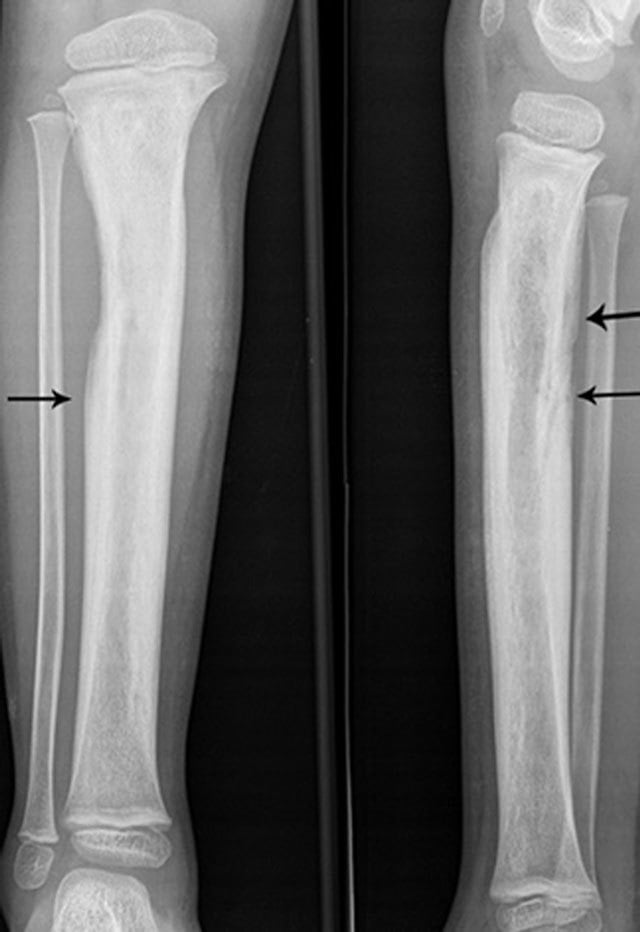
Radiographs are first line, and late: changes take 10-14 days to appear, and 30-50% of the bone must be lost before lysis shows. Look for periosteal reaction, lytic lesions, sequestrum and involucrum.
MRI is the investigation of choice, 90% sensitive and 80% specific. It shows marrow oedema (T2 hyperintense), abscess and soft-tissue extension, and it differentiates abscess from cellulitis; gadolinium enhances inflamed tissue.
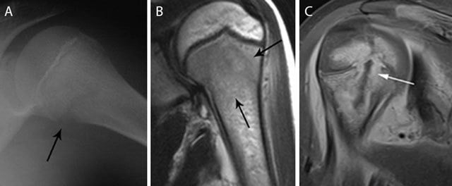
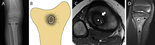
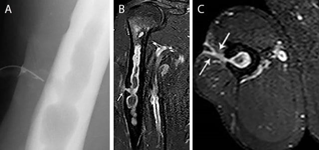
Nuclear medicine. A bone scan is sensitive but not specific; a labelled white cell scan is more specific for infection.
Microbiology. Tissue culture is the gold standard.
- Take multiple deep samples, not from the sinus tract; a minimum of 3 increases the yield
- Send them for aerobic, anaerobic, fungal and TB culture, and send a specimen for histology
- Hold antibiotics until the cultures are obtained; the exception is the septic patient, who needs empiric therapy
Cultures from sinus tract openings have only 50% concordance with deep bone cultures. They grow skin commensals and colonisers, not the true pathogen, so do not base antibiotic choice on a superficial swab. Always obtain intraoperative bone and tissue cultures before starting definitive antibiotics.
Differential Diagnosis
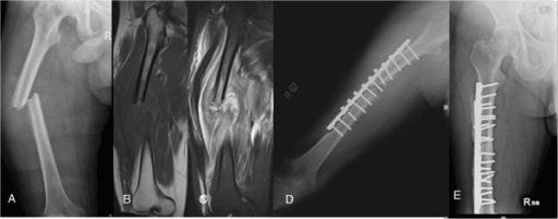
Osteomyelitis is a notorious mimic, and is mimicked by several aggressive lesions. The cardinal rule is that a lytic, painful bone lesion with periosteal reaction must be biopsied before infection is assumed, because Ewing sarcoma and osteosarcoma can look identical.
- Typical Patient
- Child/adolescent
- Discriminating Features
- Onion-skin periosteal reaction, fever and raised inflammatory markers can mimic infection, diaphyseal
- Resolving Step
- Biopsy (small round blue cells, EWSR1 translocation)
- Typical Patient
- Adolescent/young adult
- Discriminating Features
- Sunburst/Codman triangle, metaphyseal, soft-tissue mass with ossification
- Resolving Step
- Biopsy and staging MRI/CT
- Typical Patient
- Child
- Discriminating Features
- Well-defined lytic lesion, vertebra plana, may be painful
- Resolving Step
- Biopsy
- Typical Patient
- Diabetic, neuropathic foot
- Discriminating Features
- Midfoot, joint-centred destruction and deformity, less focal cortical erosion
- Resolving Step
- MRI pattern plus probe-to-bone; bone biopsy if uncertain
- Typical Patient
- Sickle cell disease
- Discriminating Features
- Serpentine sclerosis, no soft-tissue abscess; vs Salmonella osteomyelitis
- Resolving Step
- MRI, aspiration/culture
- Typical Patient
- Variable
- Discriminating Features
- Joint-centred rather than intraosseous; crystals or pus on aspiration
- Resolving Step
- Joint aspiration and culture
A permeative, painful bone lesion with periosteal reaction in a child is Ewing sarcoma until proven otherwise. Fever, raised CRP/ESR and a lytic diaphyseal lesion overlap completely with osteomyelitis; never debride such a lesion as "infection" without a diagnostic biopsy first.
Management
The principle. Surgery and antibiotics are synergistic. Debride all necrotic tissue, obtain cultures and manage the dead space, then give approximately 6 weeks of pathogen-directed therapy.
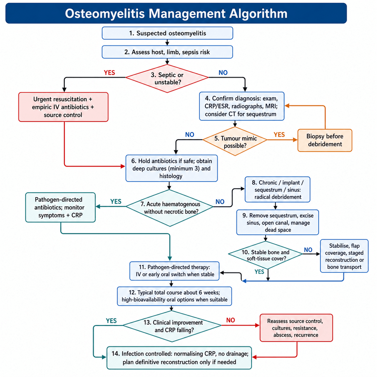
Chronic osteomyelitis, week by week. The sequence below runs from diagnosis to reconstruction; the deep cultures and histology described under Diagnosis are taken at the first operation.
Treatment Algorithm for Chronic Osteomyelitis
Confirm the osteomyelitis clinically, on MRI and with CRP, as set out under Diagnosis. Hold antibiotics pending cultures and plan the surgical intervention.
Radically debride all necrotic tissue: remove every sequestrum, open the medullary canal and excise the sinus tracts. Manage the dead space with antibiotic beads, a muscle flap or bone graft.
Give organism-specific therapy guided by the intraoperative cultures and adjusted to the sensitivities (agents in the table below). Monitor CRP weekly; it should fall by roughly 50% at 2 weeks.
Switch to oral antibiotics once the patient is clinically improving and the CRP is falling: the landmark OVIVA RCT (N=1054) established that oral antibiotics are non-inferior to intravenous therapy once the patient is clinically stable. High-bioavailability agents are the fluoroquinolones (often with rifampicin for staphylococcal biofilm), clindamycin and linezolid. Continue until the CRP normalises and the patient is asymptomatic.
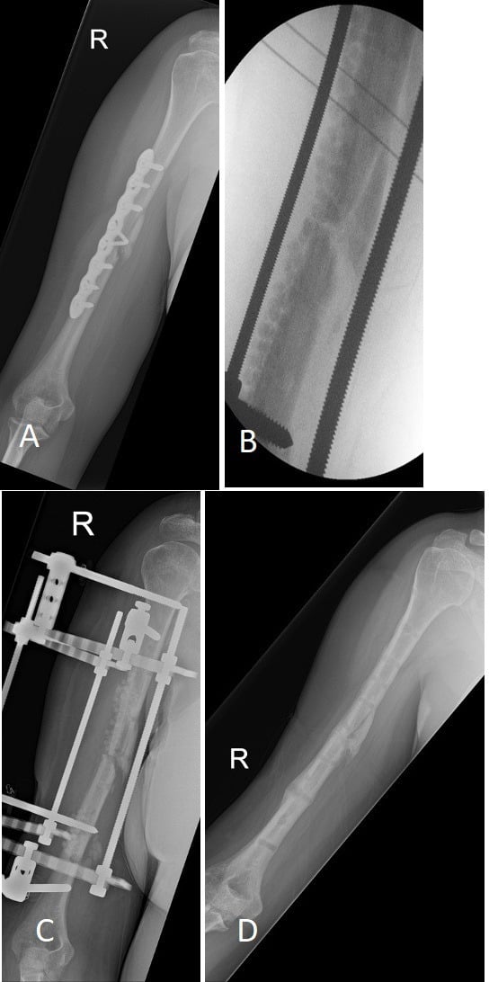
Reconstruct definitively if needed, and only once the infection is controlled (normal CRP, no drainage): bone transport for segmental defects, free fibula for massive defects, arthrodesis for joint involvement.
Choosing the antibiotic. The agents below reflect convergent IDSA, BOA-BOAST, AAOS and EFORT/European practice. The exact agent and dose vary with local resistance patterns and renal function; the principle, narrow, high-bioavailability and pathogen-directed, is universal.
- First-Line Parenteral
- Flucloxacillin/nafcillin 2g 6-hourly or cefazolin 2g 8-hourly
- Oral Step-Down
- Flucloxacillin 1g 6-hourly; add rifampicin for biofilm
- Duration
- Approximately 6 weeks
- First-Line Parenteral
- Vancomycin 15-20 mg/kg 12-hourly or daptomycin
- Oral Step-Down
- Linezolid 600mg 12-hourly or doxycycline
- Duration
- Approximately 6 weeks
- First-Line Parenteral
- Benzylpenicillin 1.8g 4-hourly
- Oral Step-Down
- Amoxicillin 1g 8-hourly
- Duration
- Approximately 6 weeks
- First-Line Parenteral
- Ceftriaxone 2g daily
- Oral Step-Down
- Ciprofloxacin 750mg 12-hourly
- Duration
- Approximately 6 weeks
- First-Line Parenteral
- Piperacillin-tazobactam 4.5g 6-hourly or ceftazidime
- Oral Step-Down
- Ciprofloxacin 750mg 12-hourly
- Duration
- 6-8 weeks
Complications of Chronic Osteomyelitis
Beyond persistent infection, chronic osteomyelitis carries structural and systemic complications that are high-yield in the viva. The pathological femoral fracture in the tumour-mimic figure above is one of them.
- Mechanism
- Infected and post-debridement bone loses structural integrity (cortical destruction, large defects)
- Key Point
- May require stabilisation and reconstruction (bone transport, free fibula); diffuse Type IV disease is the classic setting
- Mechanism
- Malignant transformation in a long-standing (years to decades) chronically draining sinus tract
- Key Point
- Usually well-differentiated SCC but can metastasise
- Mechanism
- Sustained inflammation raises serum amyloid A, which deposits in kidneys and other organs
- Key Point
- Rare in the antibiotic era but a classic cause of renal failure in neglected chronic osteomyelitis
- Mechanism
- Physeal involvement or damage from infection/surgery
- Key Point
- Limb-length discrepancy or angular deformity - mandates skeletal follow-up after paediatric osteomyelitis
- Mechanism
- Spread from an intra-articular metaphysis or direct joint extension
- Key Point
- Particularly hip, shoulder and elbow where the metaphysis is intracapsular; threatens the articular cartilage
- Mechanism
- Persisting biofilm/sequestrum and bacterial persistence reservoirs
- Key Point
- Recurrence roughly 10-30 percent; persistent drainage, repeated debridements and amputation in refractory diffuse disease
Any change in a long-standing osteomyelitis sinus - new pain, increased or bloody discharge, or an everted, fungating, non-healing edge - is squamous cell carcinoma until biopsy proves otherwise. The same transformation can occur in chronic scar. A changing chronic sinus is biopsied, not simply re-dressed.
Guidelines, Registries & Global Practice
Global Epidemiology
- Acute haematogenous osteomyelitis is predominantly a paediatric disease; reported incidence is roughly 1 in 5,000 children per year in high-income settings, higher in low- and middle-income countries where presentation is often delayed.
- Chronic and contiguous osteomyelitis is increasingly an adult disease driven by open fractures, internal fixation, diabetic foot disease and prosthetic joints. Diabetic foot osteomyelitis is rising globally in parallel with the diabetes pandemic.
- Staphylococcus aureus remains the dominant pathogen worldwide across all settings; MRSA prevalence varies markedly by region (high in parts of the Americas and Asia, lower in much of Scandinavia and the Netherlands due to search-and-destroy policies).
- In regions with high tuberculosis burden, skeletal TB (including spinal Pott disease) is an essential differential for subacute/chronic bone infection.
Side-by-Side Society Guidance
- Emphasis
- Vertebral osteomyelitis and diabetic foot guidelines
- Distinctive Position
- Image-guided/open biopsy before antibiotics; culture-directed therapy approximately 6 weeks
- Emphasis
- Combined ortho-plastic single-stage management
- Distinctive Position
- Specialist-centre, ortho-plastic teams; radical debridement plus immediate soft-tissue cover
- Emphasis
- Musculoskeletal infection principles
- Distinctive Position
- Surgical source control plus pathogen-directed therapy; MDT model
- Emphasis
- Fracture-related infection (FRI) consensus
- Distinctive Position
- Standardised FRI definition (confirmatory vs suggestive criteria); debridement, stabilise, soft-tissue cover
- Emphasis
- Peri-implant and bone-joint infection definitions
- Distinctive Position
- Biofilm-focused; structured diagnostic criteria; rifampicin combinations for staphylococci
- Emphasis
- Diabetic foot osteomyelitis
- Distinctive Position
- Probe-to-bone test; conservative (medical) management viable for selected forefoot disease
Practice Variation: High- vs Limited-Resource Settings
- MRI, image-guided biopsy and molecular diagnostics readily available
- Single-stage ortho-plastic reconstruction with free flaps and local antibiotic carriers (calcium sulfate/Cerament)
- Early oral switch supported by OVIVA-level evidence
- OPAT services enable outpatient parenteral therapy
- Later presentation; chronic sequestrum and large involucrum more common
- Reliance on plain radiographs and clinical diagnosis
- Sequestrectomy plus local bone-cement beads and staged management
- Higher TB-osteomyelitis burden mandates routine mycobacterial work-up
Applicable in every health system:
- Obtain deep tissue cultures before definitive antibiotics (unless the patient is septic)
- Justify and document antibiotic choice, dose and duration; review against culture at 48-72 hours under antimicrobial stewardship
- Informed consent for surgery must discuss recurrence risk (chronic osteomyelitis recurrence approximately 10-30%)
- Multidisciplinary input (infectious diseases, microbiology, plastic surgery for soft-tissue cover)
Common pitfalls and litigation themes: starting antibiotics before cultures, inadequate debridement, and relying on sinus-tract swabs instead of deep tissue.
Controversies & Areas of Uncertainty
Antibiotic duration. The traditional 6 weeks is dogma more than data. OVIVA standardised 6 weeks but did not test shorter courses, and paediatric RCTs support around 3 weeks. The optimal duration in adults, and whether it differs by Cierny-Mader type, remains unresolved.
Oral or intravenous. OVIVA shifted practice toward early oral switch, but uptake varies, and concerns persist for poorly bioavailable agents, non-adherent patients and certain organisms. Route is now individualised rather than reflexively intravenous.
Single- or two-stage surgery. Single-stage debridement with immediate soft-tissue cover and local antibiotic carriers (e.g. calcium sulphate) challenges the classic staged Lautenbach/bead approach. The outcome data are promising but largely observational.
Diabetic foot osteomyelitis. This is where the surgical rule is contested. Whether forefoot diabetic osteomyelitis needs surgery or can be cured medically is debated; IWGDF accepts antibiotic-only management in selected cases, but recurrence and amputation risk remain high.
MCQ Practice Points
Q: What is the most common causative organism in all age groups for osteomyelitis? A: Staphylococcus aureus - causes 80-90% of all osteomyelitis cases regardless of age. The exception is sickle cell disease where Salmonella is more common than S. aureus.
Q: Bacteria within biofilm are how many times more resistant to antibiotics than planktonic bacteria? A: 1000 times more resistant - this is why surgical debridement is mandatory for chronic osteomyelitis. Antibiotics cannot penetrate the glycocalyx matrix.
Q: What is the sensitivity of MRI for diagnosing osteomyelitis? A: 90% sensitive - MRI is the gold standard imaging modality, showing marrow oedema and soft tissue extension. Plain radiographs take 10-14 days to show changes.
Q: For complex bone and joint infection, is oral antibiotic therapy inferior to intravenous therapy? A: No - the OVIVA RCT (Li et al, NEJM 2019, N=1054) showed oral antibiotics are non-inferior to IV for the first 6 weeks once the patient is clinically stable, with fewer line complications. First-line MSSA agent remains an antistaphylococcal penicillin (flucloxacillin/nafcillin) or cefazolin; MRSA needs vancomycin or daptomycin.
Exam Viva Scenarios
Practise clinical reasoning and management decisions out loud
“A 45-year-old male presents 18 months after open tibial fracture ORIF. He has persistent drainage from the anterior shin. MRI shows intramedullary signal abnormality with cortical sequestrum but the tibia appears structurally intact. CRP 45. How would you classify and manage this?”
“Explain why acute haematogenous osteomyelitis in children typically affects the metaphysis of long bones, and why it can lead to septic arthritis in some cases but not others.”
“A 58-year-old man has had radical debridement and a local muscle flap for chronic tibial osteomyelitis. Intraoperative cultures grow methicillin-susceptible Staphylococcus aureus. The medical team plans 6 weeks of intravenous flucloxacillin via a PICC line. The patient is systemically well and keen to go home. How would you advise on antibiotic route and duration, and what evidence supports your view?”
Key Pathophysiology
- Biofilm on sequestrum = 1000x antibiotic resistance
- Metaphyseal sinusoidal flow = seeding site in children
- Sequestrum = dead bone, Involucrum = new bone shell
- Elevated intraosseous pressure causes vascular compromise
Cierny-Mader Classification
- Type I = Medullary, Type II = Superficial
- Type III = Localized (stable after debridement)
- Type IV = Diffuse (unstable after debridement)
- A = Normal host, B = Compromised, C = Treatment worse than disease
Microbiology
- S. aureus = 80% all cases
- Pseudomonas = puncture wound through shoe
- Salmonella = sickle cell disease
- Polymicrobial = diabetic foot, trauma
Diagnosis
- MRI = 90% sensitive - gold standard
- CRP better than ESR for monitoring treatment
- Deep tissue culture (NOT sinus tract swab)
- Plain XR needs 10-14 days to show changes
Treatment
- Surgery + antibiotics synergistic
- Debride all necrotic tissue and sequestra
- Approximately 6 weeks antibiotics; OVIVA = oral non-inferior to IV when stable
- MSSA: flucloxacillin/cefazolin; MRSA: vancomycin/daptomycin
Evidence Base
Short vs Long Antibiotic Course - Childhood AHOM (Peltola RCT)
- Prospective RCT of 131 culture-positive acute haematogenous osteomyelitis cases in children
- 20 days vs 30 days of clindamycin or first-generation cephalosporin (IV only first 2-4 days)
- S. aureus caused 89% of cases; all strains methicillin-susceptible
- Except for 1 mild sequela per arm, all patients fully recovered - no difference between arms
- 24% needed no surgery; most had only diagnostic aspiration or drilling
Osteomyelitis and the Role of Biofilms in Chronic Infection
- Reviews biofilm attachment, development and maturation using S. aureus as the model organism
- Self-produced glycocalyx matrix shields bacteria from antibiotics and host immune cells
- Biofilm-embedded bacteria are orders of magnitude more tolerant of antibiotics than planktonic forms
- Explains the persistence of chronic bone infection and rationale for surgical source control
Surgical Management of Chronic Osteomyelitis
- Review of surgical techniques for chronic osteomyelitis (most commonly post-traumatic tibial)
- Cure requires pathogen isolation, radical debridement of all necrotic/infected tissue, then dead-space and soft-tissue reconstruction
- Discusses debridement, dead space management, Ilizarov bone transport and vascularised reconstruction
- Host morbidity strongly influences propagation and outcome - individualise treatment
A Clinical Staging System for Adult Osteomyelitis (Cierny-Mader)
- Seminal paper formalising the Cierny-Mader staging system for adult osteomyelitis
- Combines anatomic type (I medullary, II superficial, III localised, IV diffuse) with host physiologic class (A, B-local/systemic, C)
- Stage directs both the extent of surgery and the prognosis
- Type IV in a class-C host carries the worst outlook and may favour amputation over reconstruction
Oral versus IV Antibiotics for Bone and Joint Infection (OVIVA)
- Multicentre non-inferiority RCT across 26 UK centres, 1054 adults with bone or joint infection
- Oral vs intravenous antibiotics for the first 6 weeks after surgery or start of treatment
- Oral therapy was non-inferior to IV for definitive treatment failure at 1 year (margin 7.5 percentage points)
- Oral arm had fewer intravenous-catheter complications and shorter hospital stay
Osteomyelitis (Lew & Waldvogel Seminal Review)
- Authoritative review establishing the contiguous-focus / vascular-insufficiency / haematogenous framework
- Bone biopsy for microbiology and histology is key to targeted, long-lasting therapy
- Chronic osteomyelitis with sequestrum requires surgical debridement for cure; acute disease may respond to antibiotics alone
- Multidisciplinary care (orthopaedics, infectious diseases, plastic and vascular surgery) underpins success