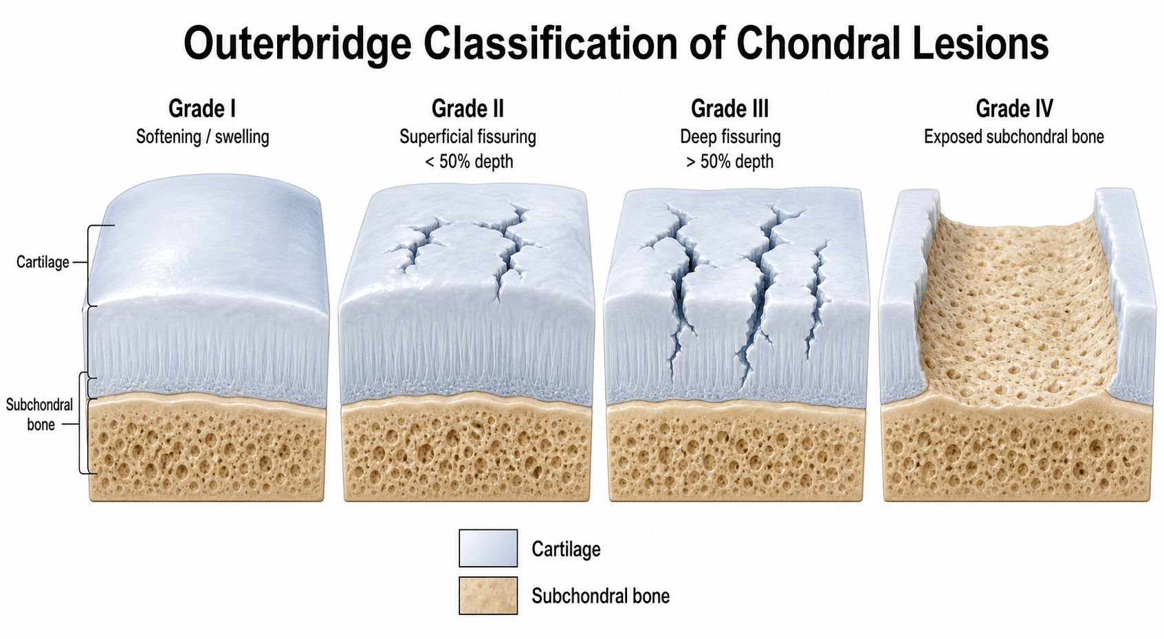
Outerbridge Classification of Chondral Lesions
A chondral lesion of grade IV (full-thickness, bone exposed) is the surgical threshold — reparative or restorative cartilage surgery is generally indicated for symptomatic grade IV lesions. Grades I and II are typically managed with debridement and physiotherapy, and grade III lesions sit in a grey zone where the diameter (greater than or less than 1.3 cm), location, and patient factors guide treatment.
The classic basic-science viva that explains the grade-IV threshold:
- Articular cartilage is hyaline cartilage — an extracellular matrix of type II collagen and proteoglycan (aggrecan) holding a high water content (~70–80%), maintained by sparse chondrocytes (its only cell type) with very low metabolic turnover.
- Zonal architecture — a superficial (tangential) zone (flat cells, collagen parallel to the surface, resisting shear), a middle/transitional zone, a deep/radial zone (collagen perpendicular, highest proteoglycan, resisting compression), and calcified cartilage anchoring to bone, separated from the deep zone by the tidemark.
- It is avascular, aneural and alymphatic, nourished by diffusion from synovial fluid, so it mounts no inflammatory/reparative response. Partial-thickness lesions (Outerbridge I–III, not reaching bone) do NOT heal; a full-thickness lesion (grade IV) reaching the subchondral marrow gains access to mesenchymal stem cells and heals with fibrocartilage (type I collagen) — mechanically inferior to hyaline. This is exactly why grade IV is the surgical threshold and why marrow stimulation (microfracture) deliberately breaches the subchondral plate.
The Outerbridge Grading System (0 to IV)
The Outerbridge system was originally described in 1961 for chondromalacia of the patella, and is now applied to any articular cartilage surface visualised at arthroscopy. Grading is performed after probing the lesion, ideally with the joint distended and the cartilage surface viewed tangentially.
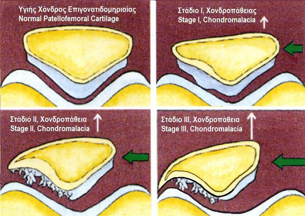
- Macroscopic Appearance
- Normal cartilage
- Depth
- None
- Probe Finding
- Firm, smooth, no softening
- Macroscopic Appearance
- Softening or swelling of an otherwise intact surface
- Depth
- Superficial
- Probe Finding
- Soft on probing; no visible break or fissure
- Macroscopic Appearance
- Fissuring and fragmentation under 1.3 cm in diameter
- Depth
- Partial-thickness (under 50 percent depth)
- Probe Finding
- Rough surface; cartilage still covers subchondral bone
- Macroscopic Appearance
- Fissuring and fragmentation over 1.3 cm in diameter
- Depth
- Partial-thickness (over 50 percent depth, not full-thickness)
- Probe Finding
- Crab-meat fibrillation; bone not yet exposed
- Macroscopic Appearance
- Erosion of cartilage with exposed subchondral bone
- Depth
- Full-thickness
- Probe Finding
- Bone visible; crater edges may be eburnated
Normal · Soft · Small crack · Big crack · BoneThe five grades in order
Hook:The only number to recite is the original half-inch (~1.3 cm) diameter cutoff separating grade II from III.
Grade I is the most subjective grade — it is defined by softening or swelling of an intact surface, and whether the surface is "soft" depends on the surgeon's perception at arthroscopy. Modern classifications therefore collapse grade 0 and grade I together, treating grade I as essentially normal. Examiners may still expect you to know the original five-grade system.
Modified and Related Grading Systems
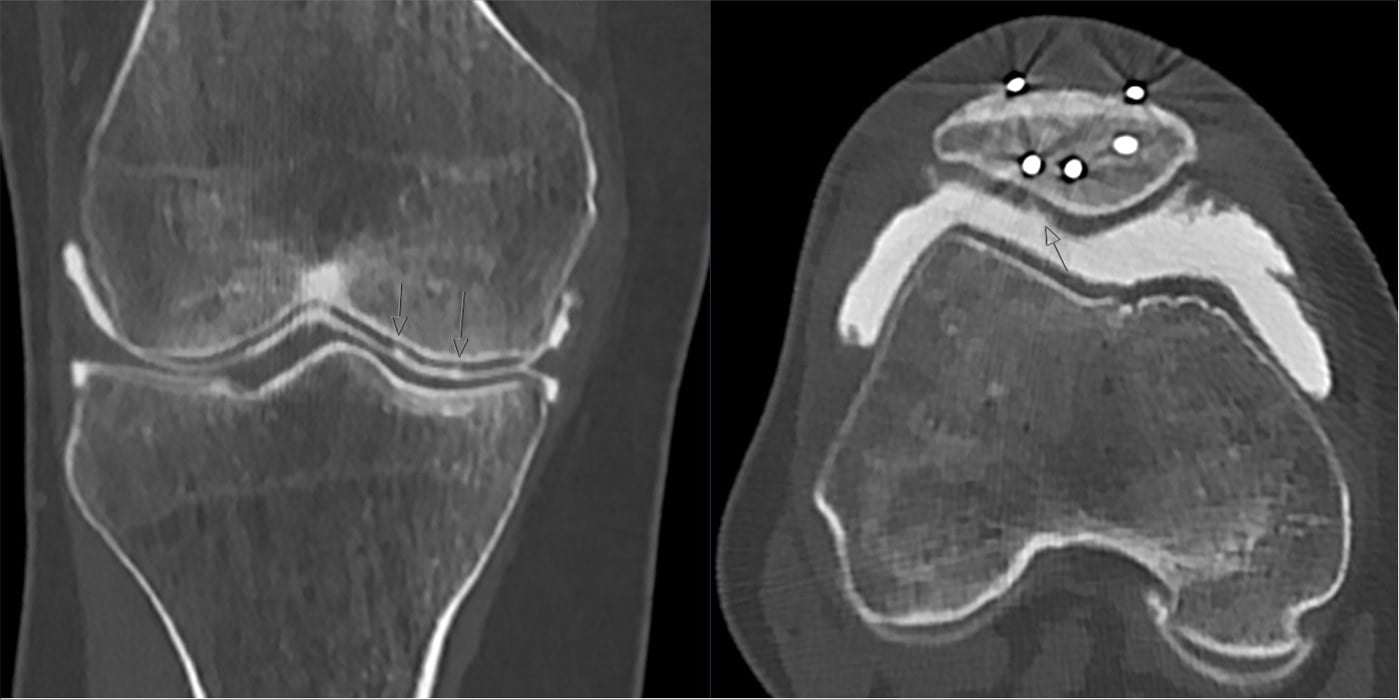
The original Outerbridge system has been modified because the 1.3 cm diameter cutoff and the lack of a depth criterion limit its precision. The most widely used contemporary alternative is the ICRS (International Cartilage Repair Society) classification, which is depth-based and used in cartilage repair trials and registries.
- Grades
- 0, I, II, III, IV
- Key Distinguishing Feature
- 1.3 cm diameter cutoff separates grade II from grade III
- Common Use
- Most widely quoted in clinical practice and the exam
- Grades
- I, II, III, IV
- Key Distinguishing Feature
- Drops grade 0; emphasises softening and fibrillation
- Common Use
- Patellofemoral chondrosis
- Grades
- 0, 1a, 1b, 2, 3a, 3b, 3c, 4
- Key Distinguishing Feature
- Depth-based, with subgrades for blistering and deep fissuring
- Common Use
- Cartilage repair research and registries
- Grades
- 1 to 4
- Key Distinguishing Feature
- Includes depth and bone involvement separately
- Common Use
- Sports knee research
- Grades
- 1 to 4
- Key Distinguishing Feature
- Adds chondral fracture line and osteochondral fracture
- Common Use
- Acute trauma assessment
0=0 · I=1 · II=2 · III=3 · IV=4How Outerbridge maps to ICRS
Hook:ICRS is the research standard (depth-based); Outerbridge is the clinical and exam standard.
The ICRS system is the standard for cartilage repair trials and registries. The Outerbridge system remains the most quoted in viva and clinical practice because it is simple, has five grades, and the 1.3 cm threshold is easy to remember. If asked to grade a lesion in a viva, give the Outerbridge grade first and then translate to ICRS if the question is about cartilage repair.
Limitations and Modern Context
- Inter-observer agreement is moderate at best. The grade depends on the surgeon's assessment of "softness" and the size estimate at arthroscopy. In the Cameron cadaver study, overall accuracy versus arthrotomy was 68% with an inter-observer kappa of 0.52, and the arthroscopic grade tended to be HIGHER than the true (arthrotomy) grade; agreement was better among more experienced surgeons.
- The 1.3 cm cutoff is arbitrary. It was based on Outerbridge's original patellar series and was never validated against clinical outcome. Modern systems use area (in square centimetres) or depth (in millimetres) instead of diameter.
- MRI grading is approximate. Standard MRI is reliable for grade IV (bone exposure) but unreliable for separating grade I from grade II. High-field 3-Tesla MRI with dedicated cartilage sequences (3D-DESS, T2 mapping) improves accuracy.
- The grade is a morphological description, not a treatment algorithm in isolation. A small grade IV lesion in a young athlete with a normal meniscus is treated very differently from a large grade IV lesion in an older patient with malalignment and meniscal deficiency. Use the grade together with age, alignment, meniscal status, ligamentous stability, and lesion area.
- Patient-reported outcome does not correlate strongly with grade. Many patients with grade II to III lesions are asymptomatic, and conversely a small grade IV lesion in a high-demand athlete can be disabling. The grade describes what is seen at arthroscopy, not what the patient feels.
Management Implications by Grade
The grade is one of several factors that guide cartilage management at arthroscopy. Location (weight-bearing versus non-weight-bearing), lesion size, patient age, alignment, and meniscal status all matter — but the grade sets the basic surgical threshold.
- Typical Surgical Treatment
- No surgical treatment; physiotherapy, activity modification, address alignment
- Rationale
- Intact surface; no mechanical symptom to debride
- Typical Surgical Treatment
- Mechanical debridement of unstable flaps; chondroplasty
- Rationale
- Removes mechanical catching while preserving subchondral bone
- Typical Surgical Treatment
- Debridement versus microfracture; decision based on size and patient factors
- Rationale
- Borderline partial-thickness; can convert to full-thickness microfracture if small
- Typical Surgical Treatment
- Microfracture, nanofracture, or subchondral drilling
- Rationale
- Marrow stimulation recruits mesenchymal stem cells to form fibrocartilage
- Typical Surgical Treatment
- Osteochondral autograft (OATS or mosaicplasty) or autologous chondrocyte implantation (ACI or MACI)
- Rationale
- Restores hyaline cartilage for larger lesions; microfracture alone is less durable
Microfracture · OATS · ACITreatment escalator for grade IV
Hook:The grade IV diagnosis is the entry point; lesion AREA (plus age, alignment, meniscal status) then selects the technique.
Microfracture works best for small, contained, full-thickness lesions in young, low-demand patients. The fibrocartilage fill is mechanically inferior to hyaline cartilage and deteriorates over five to seven years in higher-demand patients. For lesions larger than about 2 sq cm, or in athletes, plan a restorative procedure such as OATS or ACI rather than defaulting to microfracture.
The grade and area pick a technique, but the examiner wants the principles, the trade-offs and the prerequisites:
- Optimise the joint environment first ("treat the whole knee") — cartilage repair fails if co-pathology is ignored: correct malalignment (a realignment osteotomy — high tibial osteotomy for varus with a medial-compartment lesion, or tibial-tubercle anteromedialisation for a patellofemoral lesion), meniscal deficiency (meniscal allograft transplant) and ligamentous instability (e.g. ACL reconstruction).
- Microfracture / marrow stimulation — subchondral perforations recruit marrow mesenchymal stem cells → fibrocartilage fill; cheap, single-stage; best for small (~under 2–2.5 cm²), contained lesions in younger/lower-demand patients; deteriorates over ~5–7 years (nanofracture/subchondral drilling are variants).
- Osteochondral autograft (OATS / mosaicplasty) — hyaline-capped plugs from a low-load donor (lateral trochlea/notch) → true hyaline restoration in a single stage; limited by donor availability (up to ~4 cm²) and donor-site morbidity, and the need to match surface contour.
- Autologous chondrocyte implantation (ACI / MACI) — a two-stage cell-based technique (harvest → culture → reimplant under a periosteal patch or a collagen membrane) giving hyaline-like cartilage over larger or less-contained lesions; downsides are two operations, cost, slow maturation and graft hypertrophy (periosteal).
- Osteochondral allograft (OCA) — a fresh donor osteochondral graft for large or uncontained lesions or those with subchondral bone loss, restoring bone plus hyaline in one stage with no donor-site morbidity; limited by graft availability/cost, time-sensitive chondrocyte viability and a small immunogenicity/disease-transmission concern.
Guidelines, Registries and Global Practice
- AAOS (US) Appropriate Use Criteria for knee arthroscopy recommend chondroplasty (debridement) for symptomatic focal chondral lesions and support microfracture as a first-line reparative option for small full-thickness lesions. Larger lesions or athletic patients warrant referral for restorative procedures.
- NICE (UK) guidance does not endorse knee arthroscopy for degenerative chondral lesions in the setting of generalised osteoarthritis; focal traumatic chondral lesions in younger patients remain an accepted indication for cartilage repair surgery.
- ESSKA (European Society for Sports Traumatology, Knee Surgery and Arthroscopy) consensus recommends the ICRS classification for clinical research and registries, while recognising that the Outerbridge system remains the most widely used in routine surgical practice across Europe.
- ISAKOS (International Society of Arthroscopy, Knee Surgery and Orthopaedic Sports Medicine) global initiatives collect both Outerbridge and ICRS grades to allow cross-comparison between centres. Both grading systems are reported in major cartilage repair registries such as the German Cartilage Registry and Swedish Knee Registry.
- Global variation: In resource-limited settings, microfracture remains the dominant reparative technique because it is cheap, requires no specialist equipment, and uses a single-stage arthroscopic approach. OATS, ACI, and MACI are concentrated in tertiary cartilage centres in high-income settings. The grading system used (Outerbridge versus ICRS) varies by region, but the underlying surgical principles are universal.
Viva practice
Exam Viva
Practise clinical reasoning and management decisions out loud
“A 24-year-old footballer has persistent pain and clicking in his knee after a twisting injury. MRI shows a focal chondral defect on the medial femoral condyle. At arthroscopy you find a 1 cm diameter lesion with fissuring and fragmentation but no exposed bone. How do you grade it and what do you do?”
“A 32-year-old runner has ongoing pain and mechanical catching two years after an ACL reconstruction. MRI shows a 2.5 sq cm full-thickness chondral defect on the medial femoral condyle, intact menisci, neutral alignment, and a stable knee. At arthroscopy you confirm exposed subchondral bone. What is the grade and what surgical options would you discuss?”
Exam cheat sheet
The five grades (0 to IV)
- Grade 0: normal cartilage
- Grade I: softening or swelling of an intact surface (no visible break)
- Grade II: partial-thickness fissuring under 1.3 cm in diameter
- Grade III: partial-thickness fissuring over 1.3 cm in diameter (no exposed bone)
- Grade IV: full-thickness loss with exposed subchondral bone
Management by grade
- Grades 0 to I: no surgical treatment; physiotherapy and activity modification
- Grade II: arthroscopic debridement (chondroplasty) of unstable flaps
- Grade III: debridement versus microfracture — depends on area and patient factors
- Grade IV under 2 sq cm: microfracture or nanofracture
- Grade IV over 2 sq cm: OATS (single-stage) or ACI or MACI (two-stage)
How it maps to other systems
- Outerbridge 0 equals ICRS 0 (normal)
- Outerbridge I equals ICRS 1a (fibrillation) or 1b (blister)
- Outerbridge II equals ICRS 2 (under 50 percent depth)
- Outerbridge III equals ICRS 3a, 3b, or 3c (over 50 percent depth, no bone)
- Outerbridge IV equals ICRS 4 (full-thickness, bone exposed)
Key pearls and pitfalls
- The 1.3 cm cutoff is the only number examiners need
- Grade I is the most subjective grade — it depends on probing
- Inter-observer agreement is only moderate (kappa ~0.5) and improves with surgeon experience; arthroscopy tends to over-grade
- Grade guides but does not dictate treatment — age, alignment, meniscus, and area all matter
- Outerbridge is the exam and clinical standard; ICRS is the research standard
Evidence
The etiology of chondromalacia patellae
- Original four-grade (I–IV) macroscopic classification of patellar chondromalacia, with a HALF-INCH (≈1.3 cm) diameter cutoff separating grade II from grade III.
- Distinguished softening of an intact surface (grade I) from surface fissuring/fragmentation (grade II onward) and full erosion to bone (grade IV).
- Subsequently extrapolated beyond the patella to all articular surfaces seen at arthroscopy.
Cartilage injuries: a review of 31,516 knee arthroscopies
- 31,516 knee arthroscopies; 53,569 hyaline cartilage lesions documented in 19,827 patients.
- Grade III lesions of the PATELLA were the most common; grade IV lesions were predominantly on the medial femoral condyle.
- Patients under 40 with a grade IV lesion accounted for 5% of all arthroscopies (most with a single chondral lesion).
Articular cartilage defects: study of 25,124 knee arthroscopies
- Retrospective study of 25,124 knee arthroscopies; chondral lesions were found in 60% of patients, graded by the Outerbridge classification.
- Outerbridge grade II was the most frequent grade (42%); the patellar surface (36%) and medial femoral condyle (34%) were the commonest sites.
- Potential cartilage-repair candidates (one to three localized grade III/IV lesions, under age 40) made up about 7% of patients.
According to PubMed, the classification (four grades, half-inch / ≈1.3 cm cutoff) is from Outerbridge 1961 (J Bone Joint Surg Br 1961;43-B:752-7; DOI 10.1302/0301-620X.43B4.752). Large arthroscopy series defining its epidemiology are Curl et al. 1997 (DOI 10.1016/s0749-8063(97)90124-9) and Widuchowski et al. 2007 (grade II most common, 42%; DOI 10.1016/j.knee.2007.02.001). The depth-based ICRS system is described by Brittberg & Winalski 2003 (DOI 10.2106/00004623-200300002-00008), and the reliability data (accuracy 68%, inter-observer kappa 0.52) are from Cameron et al. 2003 (DOI 10.1177/03635465030310012601).