Paprosky Classification of Acetabular and Femoral Bone Loss
Examiners will show you a revision hip radiograph and ask you to grade the bone loss using the Paprosky system. For the acetabulum, assess column support and hip-centre migration relative to the teardrop and the Köhler line. For the femur, assess whether the diaphysis can provide a 4–6 cm scratch-fit. State the type, then immediately state the reconstruction: Type 1 — cemented or uncemented hemispherical cup; Type 2 — jumbo cup, oblong cup, or augments with hemispherical cup; Type 3 — structural allograft, cage, or custom triflange. Never describe the defect without committing to the grade.
Paprosky Acetabular Classification
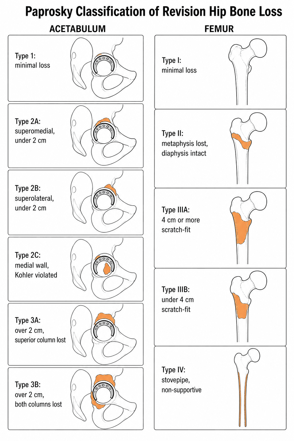
The acetabular system classifies bone loss into three types based on the integrity of the acetabular columns, the degree of superior and medial migration of the hip centre, and the status of the teardrop and Köhler line.
- Type 1
- Intact
- Type 2A
- Intact
- Type 2B
- Intact
- Type 2C
- Intact
- Type 3A
- Disrupted superior
- Type 3B
- Disrupted both columns
- Type 1
- Minimal
- Type 2A
- Less than 2 cm
- Type 2B
- Less than 2 cm
- Type 2C
- Minimal
- Type 3A
- Greater than 2 cm
- Type 3B
- Greater than 2 cm
- Type 1
- Intact
- Type 2A
- Intact
- Type 2B
- Intact
- Type 2C
- Absent
- Type 3A
- Absent
- Type 3B
- Absent
- Type 1
- Intact
- Type 2A
- Intact
- Type 2B
- Violated
- Type 2C
- Violated
- Type 3A
- Violated
- Type 3B
- Violated
- Type 1
- Greater than 50 percent
- Type 2A
- Greater than 50 percent
- Type 2B
- 30–50 percent
- Type 2C
- Less than 30 percent medial wall
- Type 3A
- Less than 30 percent
- Type 3B
- Less than 30 percent
- Type 1
- Hemispherical cup (cemented or uncemented)
- Type 2A
- Uncemented hemispherical ± augments
- Type 2B
- Bilobed/oblong cup or augments
- Type 2C
- Uncemented cup with medial augments or graft
- Type 3A
- Cage or custom triflange
- Type 3B
- Custom triflange or allograft-prosthesis composite
Columns • Teardrop • Köhler • Migration (CTKM)Acetabular Paprosky — Think Columns and Migration
Hook:CTKM: assess in order — Columns, Teardrop, Köhler, Migration. If both columns disrupted with migration over 2 cm, you are in Type 3.
Type 2A vs 2B: Both have intact columns and less than 2 cm superior migration, but in Type 2B the Köhler line is violated (medial wall deficiency) and host-bone contact drops to 30 to 50 percent. This distinction changes the reconstruction from a straightforward uncemented cup (2A) to one requiring augments or an oblong component (2B).
The table lists the features; examiners want the reading method. Paprosky graded the acetabulum off the AP pelvis by four radiographic criteria, each reflecting loss of a specific supporting structure — the more severe each is, the higher the type:
- Superior migration of the hip centre — and its DIRECTION. The amount of superior migration grades severity (mild in Type 2, marked — generally beyond about 3 cm above the superior obturator line — in Type 3). The direction localises the defect: superolateral migration implies the dome/anterosuperior rim is deficient but the columns are largely intact (the less severe pattern); superomedial migration implies posterior column and dome loss (the more concerning pattern).
- Ischial osteolysis — lysis of the ischium reflects bone loss of the inferior posterior column and posterior wall; increasing ischial lysis means a more deficient posterior column.
- Teardrop osteolysis — destruction of the teardrop reflects inferomedial / medial-wall bone loss.
- Köhler (ilioischial) line — is it violated? Migration of the component medial to Köhler's line signals medial-wall and anterior-column deficiency.
Read all four together: minimal change with the cup above an intact teardrop and Köhler line is Type 1–2; marked superior (especially superomedial) migration with teardrop and ischial lysis and a violated Köhler line is Type 3, and whether one column (3A) or both (3B) have failed sets the subtype.
Paprosky Femoral Classification
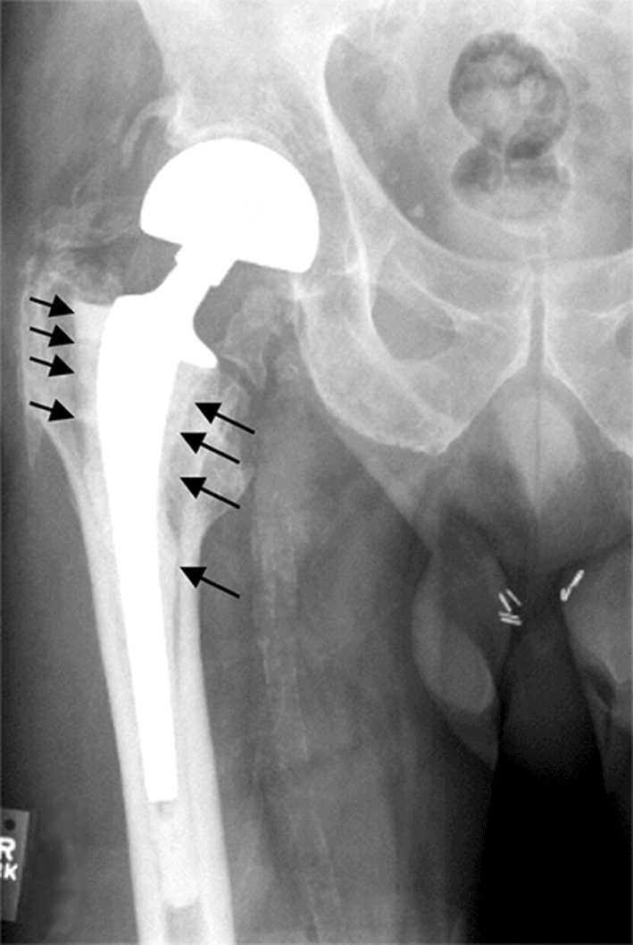
The femoral system grades diaphyseal bone stock and the ability to achieve distal scratch-fit fixation. The metaphysis is almost always deficient in revision surgery; what matters is the tube of intact diaphysis.
- Type I
- Minimal loss (intact)
- Type II
- Extensive cancellous loss
- Type IIIA
- Non-supportive
- Type IIIB
- Non-supportive
- Type IV
- Non-supportive
- Type I
- Intact
- Type II
- Intact
- Type IIIA
- ≥ 4 cm of intact cortical isthmus
- Type IIIB
- under 4 cm of usable isthmus
- Type IV
- Non-supportive (widened/stovepipe canal, thin cortices)
- Type I
- Not needed
- Type II
- Achievable
- Type IIIA
- Achievable (≥ 4 cm)
- Type IIIB
- Limited (under 4 cm)
- Type IV
- Not achievable
- Type I
- Primary-type (cemented or uncemented) stem
- Type II
- Extensively porous-coated or modular fluted tapered stem
- Type IIIA
- Modular fluted tapered stem (or extensively coated)
- Type IIIB
- Modular fluted tapered stem; APC if fixation marginal
- Type IV
- Allograft-prosthesis composite, impaction grafting, or megaprosthesis
Metaphysis → Diaphysis → IsthmusFemoral Paprosky — walk distal
Hook:Walk distal: metaphysis, then diaphysis, then the isthmus. The 4 cm scratch-fit threshold separates IIIA from IIIB; a non-supportive (stovepipe) isthmus is Type IV — not IIIB.
The critical threshold is 4 cm of diaphyseal scratch-fit. If you can get at least 4 cm of tight cylindrical fixation in the isthmus, a fluted tapered modular stem (e.g. Wagner, RECLAIM) will work — Type IIIA, and often IIIB. The distinction that matters most for reconstruction is whether the isthmus is supportive at all: a non-supportive, widened or stovepipe canal (diameter often over 20 mm, thin cortices) is Type IV, where distal scratch-fit is not achievable and you need an allograft-prosthesis composite, impaction grafting, or a tumour-type megaprosthesis. A marginal (under 4 cm) but still cylindrical isthmus is IIIB; an absent supportive isthmus is IV.
Reconstructive Algorithm
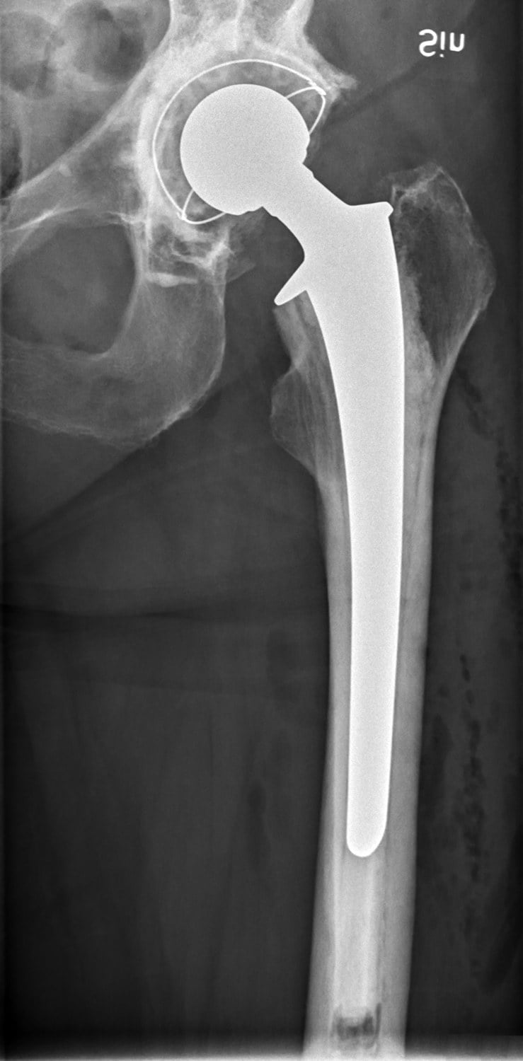
The Paprosky grade directly determines the reconstructive ladder. Choosing the wrong strategy for the defect grade is a common exam trap.
- First-Line Reconstruction
- Uncemented hemispherical cup
- Backup / Alternative
- Cemented cup with bone graft
- Key Principle
- Over 50 percent host-bone contact guarantees stable fixation
- First-Line Reconstruction
- Uncemented cup with porous metal augments
- Backup / Alternative
- Jumbo cup, oblong cup, or high hip centre
- Key Principle
- Restore the anatomic hip centre using augments to fill column defects
- First-Line Reconstruction
- Cup-cage construct or custom triflange
- Backup / Alternative
- Bulk structural allograft with cage
- Key Principle
- Custom triflange for young active patients; cage for lower-demand patients
- First-Line Reconstruction
- Standard-length uncemented or cemented stem
- Backup / Alternative
- Proximal modular replacement
- Key Principle
- Metaphyseal bone allows proximal load transfer
- First-Line Reconstruction
- Extensively porous-coated or modular fluted tapered stem
- Backup / Alternative
- Cylindrical extensively coated stem
- Key Principle
- Diaphyseal engagement bypasses the deficient metaphysis
- First-Line Reconstruction
- Modular tapered fluted stem (Wagner type)
- Backup / Alternative
- Extensively porous-coated stem
- Key Principle
- ≥4 cm isthmus gives rotational stability via the flutes
- First-Line Reconstruction
- Modular tapered fluted stem
- Backup / Alternative
- APC if distal fixation marginal
- Key Principle
- under 4 cm of usable isthmus — still cylindrical, so a tapered fluted stem usually engages
- First-Line Reconstruction
- Allograft-prosthesis composite or impaction grafting
- Backup / Alternative
- Megaprosthesis / tumour stem
- Key Principle
- Non-supportive isthmus — no scratch-fit; restore bone stock or replace the segment
Cup → Augments → Cage/Triflange · Stem → Fluted → APC/MegaReconstruction ladder memory aid
Hook:Each step up the ladder adds complexity and reduces survivorship — use the simplest reconstruction the defect allows. (Porous-metal augments have largely replaced structural allograft for Type 2 acetabula.)
Do not attempt an uncemented press-fit cup in a Paprosky Type 3 acetabulum without adjunctive fixation (cage, augment, or custom triflange). Less than 30 percent host-bone contact cannot achieve biological ingrowth, and early migration with catastrophic failure will follow.
Paprosky grades how much bone is missing; pelvic discontinuity is a different question — whether the superior (iliac) and inferior (ischiopubic) halves of the hemipelvis are dissociated through the acetabulum. It commonly co-exists with a Type 3B defect and changes the whole strategy, so you must look for it specifically.
- A lucent line or fracture crossing BOTH the anterior and posterior columns (the ilioischial and iliopubic lines).
- Medial translation of the inferior hemipelvis relative to the superior, with asymmetry/rotation of the obturator foramen.
- A break or offset in the ilioischial (Köhler) line, and visible separation on the CT acetabular rings.
- Acute (an intra-operative or recent fracture with healing potential and reasonable bone): treat like a fracture — ORIF of the posterior column with a plate plus a hemispherical cup, compressing across the discontinuity to let it heal.
- Chronic (an established non-union, usually with osteolysis and no healing potential): the construct must span/bridge the discontinuity — options are a cup-cage construct (porous cup with an ilioischial cage), a custom triflange (fixed to ilium, ischium and pubis), the acetabular distraction technique (a slightly oversized porous tantalum cup using the elastic recoil of the discontinuity for stability and ingrowth), or posterior-column plating plus a porous cup.
The exam point: a Type 3 acetabulum that also has a discontinuity is no longer a "fill the defect" problem — you either compress and heal it (acute) or bridge it (chronic).
Limitations and Modern Context
- Inter-observer reliability is moderate at best. The distinction between Type 2B and Type 3A is particularly subjective on plain radiographs. CT with 3D reconstruction significantly improves accuracy and is recommended for Type 2 and above.
- Paprosky does not account for pelvic discontinuity. A Type 3 acetabulum with pelvic discontinuity (radiolucent line crossing both columns, visible fracture through the acetabular rings on CT) requires additional stabilisation — a cup-cage construct, plating of the posterior column, or a custom triflange that bridges the defect.
- Custom triflange implants (CTAC / PSI) have largely replaced structural allograft for Type 3 acetabula in major revision centres, offering better early fixation and avoiding graft resorption. However, long-term registry data are still maturing.
- The femoral classification predated modern modular fluted tapered stems. Many Type 3A femurs previously requiring allograft-prosthesis composites can now be managed with modern fluted tapered stems (Wagner SL, RECLAIM, ZMR), potentially downgrading the surgical complexity.
- Paprosky does not incorporate infection status, patient age, or functional demand — all of which influence the final reconstructive choice. An infected Type 3B femur in an elderly low-demand patient may be best served by a cement spacer and staged megaprosthesis rather than an APC.
- AAOS and Paprosky systems are complementary, not interchangeable. The AAOS system describes the anatomical location of the defect (Type I–VI); Paprosky grades its severity. Examiners may ask for both.
Guidelines, Registries and Global Practice
- Classification Recommended
- AAOS + Paprosky used together
- Key Recommendation
- Classify with both systems; Paprosky to plan reconstruction
- Evidence Level
- Expert consensus
- Classification Recommended
- Paprosky preferred for surgical planning
- Key Recommendation
- Grade pre-operatively on CT; state Paprosky type in the operative plan
- Evidence Level
- Good practice point
- Classification Recommended
- Paprosky for acetabular and femoral grading
- Key Recommendation
- Paprosky grade determines the fixation strategy; document grade in the revision plan
- Evidence Level
- Expert consensus
- Classification Recommended
- Paprosky (with AAOS as a descriptive adjunct)
- Key Recommendation
- Custom implants/cup-cage for Type 3 acetabular defects; modular fluted tapered stems for femoral Type II–IIIB
- Evidence Level
- Level V
The National Joint Registry (UK), Australian Orthopaedic Association National Joint Replacement Registry (AOANJRR), and the American Joint Replacement Registry (AJRR) all capture revision burden data. Key findings relevant to Paprosky-guided reconstruction include:
- Porous metal augments for Type 2 acetabula demonstrate over 90 percent survivorship at 5 years across NJR and AOANJRR data, supporting their role as the workhorse for moderate defects.
- Custom triflange components for Type 3 acetabula show promising mid-term results (over 85 percent at 5 years in single-centre series), but registry capture is incomplete because many are classified as "other" or tumour implants.
- Modular fluted tapered stems have become the dominant femoral revision implant in all major registries, replacing extensively porous-coated cylindrical stems for Type 2 and 3A defects.
Custom triflange implants are readily available in the US, UK, and Australia but may be inaccessible in lower-resource settings, where structural allograft and cages remain the standard for Type 3 acetabula. In Scandinavia, the Lund classification is sometimes used alongside Paprosky for acetabular defects.
Viva practice
Exam Viva
Practise clinical reasoning and management decisions out loud
“A 72-year-old woman presents with progressive groin pain 12 years after a primary cemented total hip replacement. Radiographs show superior migration of the femoral head by approximately 3 cm, absent teardrop, and violation of the Köhler line. The femoral stem is loose with metaphyseal bone loss but intact diaphysis showing adequate calcar. Classify the bone loss and describe your reconstructive plan.”
“A 65-year-old man with a revised total hip arthroplasty presents with a loose femoral stem. The femoral canal is stovepipe in shape with a diameter over 20 mm, thin cortices, and a non-supportive isthmus. The metaphysis is completely absent. Classify the bone loss and discuss your reconstructive options, including the risks and benefits of each.”
Exam cheat sheet
Acetabular classification
- Type 1: intact columns, greater than 50 percent host bone — standard cup
- Type 2A: intact columns, intact Köhler, superior migration under 2 cm — uncemented cup ± augments
- Type 2B: intact columns, violated Köhler, 30–50 percent host bone — augments or oblong cup
- Type 2C: isolated medial wall defect — medial augments or graft
- Type 3A: disrupted superior column, migration over 2 cm — cage or custom triflange
- Type 3B: both columns disrupted — custom triflange or APC
Femoral classification (five types)
- Type I: minimal metaphyseal loss, intact diaphysis — primary-type stem
- Type II: extensive metaphyseal loss, intact diaphysis — extensively coated or modular fluted tapered stem
- Type IIIA: non-supportive metaphysis, ≥4 cm of intact isthmus — modular fluted tapered stem
- Type IIIB: under 4 cm of usable isthmus — fluted tapered stem (APC if marginal)
- Type IV: non-supportive/stovepipe isthmus — APC, impaction grafting, or megaprosthesis
- Threshold: 4 cm of diaphyseal scratch-fit separates IIIA from IIIB; a non-supportive isthmus is Type IV
Key clinical pearls
- Always classify both sides (acetabulum and femur) independently
- Use CT for pre-operative planning in Type 2 and above
- Check for pelvic discontinuity (separate entity from Paprosky grade)
- Porous metal augments have largely replaced structural allograft for Type 2 acetabula
- Modern fluted tapered stems reliably manage femoral Type II–IIIB; Type IV (non-supportive isthmus) still needs APC or megaprosthesis
Evidence
Acetabular defect classification and surgical reconstruction in revision arthroplasty: a 6-year follow-up evaluation
- 147 cemented acetabular components revised with cementless hemispherical press-fit cups (mean 5.7-year follow-up); defects typed 1–3 and reconstructed with particulate or structural allograft.
- Type 1 needed particulate graft; type 2A/B superior graft or cup superiorisation; type 2C medial-wall (wafer) graft; type 3A/B structural distal-femur or proximal-tibia allograft.
- Only 6 of 147 (4%) were unstable and warranted revision — and ALL six were type 3B.
Classifications in Brief: the Paprosky classification of femoral bone loss
- Concise review of the five-type Paprosky femoral classification (I, II, IIIA, IIIB, IV) based on metaphyseal and diaphyseal integrity.
- The 4 cm of intact diaphyseal scratch-fit distinguishes type IIIA from IIIB; a non-supportive isthmus defines type IV.
- Modern modular fluted tapered stems have expanded reconstructive options for severe (IIIA/IIIB) defects.
According to PubMed, the acetabular classification (147 revisions; all failures type 3B) is from Paprosky, Perona & Lawrence 1994 (DOI 10.1016/0883-5403(94)90135-x); the five-type femoral classification is described by Ibrahim & Fernando 2017 (DOI 10.1007/s11999-016-5012-z). Modular fluted tapered stems (97% 10-year survival) are validated by Wirtz et al. 2014 (DOI 10.3109/17453674.2014.958809); trabecular-metal augments for type IIIa by Sporer & Paprosky 2006 (PMID 16950067); and custom triflange components (12% failure at ~47 months in 1218 patients) by Broekhuis et al. 2023 (DOI 10.1530/EOR-22-0081).