Acute & Chronic Nail-Fold Infection
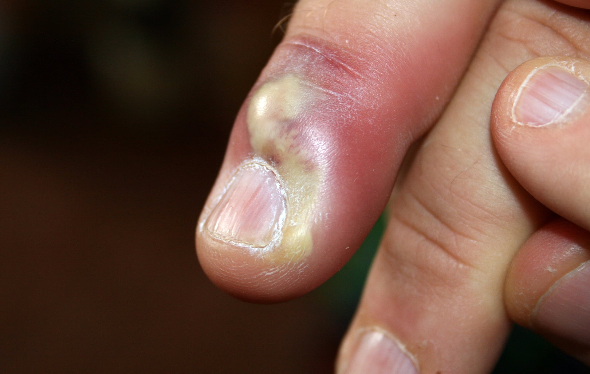
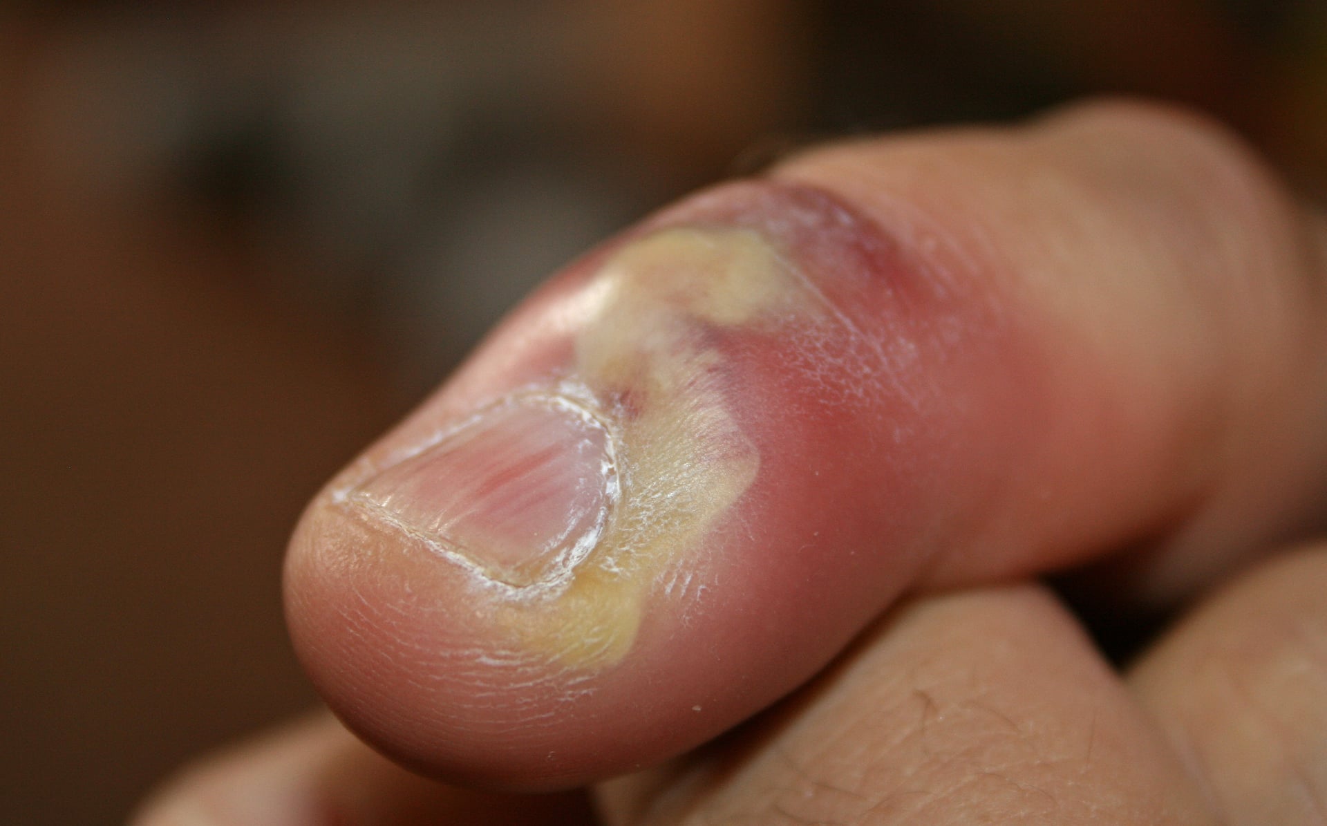
- Paronychia is an infection of the nail fold (perionychium) and is the most common infection of the hand.
- Acute paronychia is usually bacterial (Staphylococcus aureus most common, increasingly MRSA) and presents with rapid-onset pain, erythema and swelling of one nail fold, progressing to a visible abscess.
- Chronic paronychia (more than 6 weeks, often multiple digits) is NOT a simple bacterial abscess - it is an inflammatory/eczematous reaction to chronic moisture and irritants, with Candida albicans as a coloniser; it is treated as a dermatitis, not by repeated incision.
- Once pus is present, treatment is DRAINAGE - antibiotics alone will not cure an established abscess.
- A herpetic whitlow (HSV vesicles) can mimic acute paronychia but must NOT be incised - incision risks bacterial superinfection and viral spread; treat with aciclovir.
- Distinguish paronychia (nail fold) from a felon (closed pulp-space abscess of the fingertip) - different anatomy, different incision.
- “If pus has tracked under the nail base from one fold to the other it is a 'run-around' (horseshoe) abscess and needs eponychial elevation +/- partial nail removal.
- “Acute = one digit, pus, days. Chronic = multiple digits, no frank pus, weeks-months, in a 'wet hand' (cleaners, bartenders, diabetics).
- “Vesicles on an erythematous base + a history of recurrence = herpetic whitlow. Do not incise; Tzanck smear/PCR confirms; it is self-limiting (treat with aciclovir, especially if early).
Infection of the nail fold. Pus collects superficial to the nail plate, under the eponychium or lateral fold. Drain it. Antibiotics alone fail once pus is present.
Closed-space abscess of the pulp (volar fingertip), with its fibrous septa. Tense, throbbing, globally swollen pad. Different incision (volar/high-lateral) - drain to avoid skin necrosis and osteomyelitis.
Viral (HSV). Grouped vesicles on an erythematous base, intense pain, recurrent. Do NOT incise - self-limiting, treat with aciclovir; incision risks superinfection/spread.
Anatomy & Pathophysiology
The perionychium is the soft tissue surrounding the nail: the eponychium (proximal nail fold, the cuticle), the paronychium (lateral nail folds) and the hyponychium (the seal under the distal free edge). The eponychial fold continues as a thin layer over the germinal matrix. This continuity is why pus in one lateral fold can spread proximally under the eponychium and then across to the other fold - the "run-around" abscess. The seal between the cuticle and nail plate is the principal barrier; once it is breached (a hangnail, nail-biting, aggressive manicure, a splinter, an ingrowing nail) bacteria enter the potential space of the nail fold.
A breach of the cuticle seal allows skin flora into the nail fold. Staphylococcus aureus is the most common organism; MRSA is increasingly frequent and should be assumed in non-responders and in high-prevalence settings. Mixed flora (streptococci, and in nail-biters/oral contact anaerobes and Eikenella corrodens) also occur. Infection begins as a cellulitis of the fold (erythema, swelling, tenderness) and, if untreated, localises into an abscess that points at the fold margin or under the eponychium. Untreated pus can track under the nail (subungual abscess) or around the base (run-around / horseshoe abscess), and rarely into the pulp or to the distal phalanx.
Clinical Presentation
- Rapid onset (hours to days), usually one digit
- Throbbing pain, erythema, swelling of a single nail fold
- A visible collection of pus at the fold margin or under the cuticle
- Often a precipitant: hangnail, nail-biting, manicure, minor trauma, ingrowing nail
- Fluctuance and a "shiny", tense fold indicate a drainable abscess
- Insidious, more than 6 weeks, often several digits (dominant hand)
- Loss of the cuticle, boggy/retracted nail fold, intermittent discharge
- Nail dystrophy - transverse ridging, discolouration, surface irregularity
- Episodic acute flares superimposed on chronic change
- "Wet-hand" occupation, diabetes, immunosuppression, or systemic retinoid use
Assess how much pus and where (lateral fold, eponychium, subungual, run-around), and whether infection has spread to the pulp (felon) or there is lymphangitis/systemic upset. Always look for vesicles (herpetic whitlow) before reaching for a blade. In a chronic, non-healing, or atypical single-digit "paronychia" that resists treatment, think of the mimics: squamous cell carcinoma, amelanotic melanoma, pyogenic granuloma, chronic osteomyelitis, gout/tophi, sarcoid, and unusual organisms (mycobacteria, Candida in the immunosuppressed). Biopsy a chronic lesion that does not settle.
Investigations
Paronychia is a clinical diagnosis. Investigations are selective:
- Swab pus for Gram stain and culture/sensitivity at drainage - guides antibiotics and detects MRSA.
- Imaging is not routine; consider a radiograph if there is suspected foreign body, retained splinter, or chronicity/possible osteomyelitis of the distal phalanx.
- Bloods only if systemically unwell or in the immunocompromised/diabetic.
Management
No pus, no knife - but once there is pus, drain it. Early acute paronychia (cellulitis only) may settle with warm soaks and oral antibiotics. An established abscess requires drainage; antibiotics are an adjunct, not a substitute. Chronic paronychia is treated as a dermatitis - moisture avoidance and topical anti-inflammatories - not by repeated incisions.
Acute paronychia
Warm soaks, elevation, and a short course of an anti-staphylococcal oral antibiotic (e.g. flucloxacillin; cover MRSA - e.g. co-trimoxazole/doxycycline/clindamycin per local sensitivities - where prevalent or in non-responders). Many early cases resolve without surgery.
Drainage. Under digital (ring) block: lift the affected nail fold off the nail plate with a blunt instrument/scalpel held parallel to the nail to release pus; a small incision may be needed at the point of maximal fluctuance. Avoid cutting into or scarring the eponychium/germinal matrix. Send pus for culture; leave open or lightly pack; dressing and soaks afterwards.
If pus is under the nail base or has run around to the opposite fold, elevate the eponychial fold and perform partial (proximal) or complete nail-plate removal to deroof and drain the collection. Inadequate drainage is the usual reason for recurrence.
Reserve/continue oral antibiotics for surrounding cellulitis, lymphangitis, systemic features, or the immunocompromised/diabetic; tailor to culture. Antibiotics do not replace adequate drainage.
Chronic paronychia
The cornerstone is avoidance of moisture and irritants: keep hands dry, wear cotton-lined waterproof gloves for wet work, stop nail-biting/manicures, and use emollients. A topical corticosteroid for the inflammatory dermatitis is at least as effective as - and often better than - systemic antifungals; add a topical antifungal/anti-yeast if Candida is implicated. Treat any underlying diabetes.
For disease that fails conservative care, options include a course of systemic antifungal (if Candida-confirmed) and, in selected chronic cases, eponychial marsupialisation (excision of a crescent of skin over the proximal fold to allow drainage and re-establish a seal), with or without nail removal. Always biopsy the persistent atypical single-digit lesion before assuming benign disease.
Grouped vesicles on an erythematous base, severe pain out of proportion, and a recurrent history point to herpetic whitlow (HSV), not bacterial paronychia. Incision is contraindicated - it offers no benefit and risks bacterial superinfection and viral spread (including to the surgeon). It is self-limiting; manage with analgesia and a protective dressing, and consider aciclovir (most useful if started early or in recurrent/immunocompromised cases). Confirm with Tzanck smear/viral PCR if uncertain.
Complications
- Run-around (horseshoe) abscess from inadequate or late drainage
- Spread to the pulp (felon), flexor sheath, or distal phalanx (osteomyelitis)
- Nail dystrophy and permanent matrix damage in chronic disease
- Recurrence (often from persistent moisture exposure or missed subungual pus)
- Germinal matrix injury from an overzealous incision near the eponychium -> permanent nail deformity
- Inadequate drainage -> persistence/recurrence
- Missed mimic (SCC, melanoma) treated repeatedly as "infection" - the dangerous error
- Antibiotic overuse without drainage
Evidence & Key Studies
Fingertip Infections - anatomy and evidence-based management of paronychia and felon
- The fingertip is the most common site of hand infection; mismanagement carries serious consequences.
- Reviews the perionychial anatomy and the pathophysiology and treatment of acute and chronic paronychia, including the decision for surgical versus medical management, antibiotic choice, incision technique and aftercare.
- Emphasises recognising infectious, rheumatologic and oncologic conditions that mimic common fingertip infections.
Bacterial and viral infections of the nail unit: tips for diagnosis and management
- The nail unit is the most commonly affected area in hand infections, often triggered by mechanical or chemical trauma.
- Abscesses should always be drained, but herpetic whitlow may mimic an abscess and instead requires NON-operative treatment to prevent sequelae.
- A more conservative approach is advised for less severe bacterial infection and for subacute/chronic nail infection.
Evidence on this page is drawn from peer-reviewed literature indexed on PubMed. Where a percentage, organism or recommendation is stated, it reflects the cited reviews above. According to PubMed, acute paronychia is predominantly staphylococcal and chronic paronychia is best understood as an inflammatory barrier-failure dermatitis rather than a simple infection.
Clinical Decision Scenarios
Practise clinical reasoning and management decisions out loud
“A 34-year-old chef presents with a 3-day history of a painful, red, swollen left index finger nail fold. There is a visible collection of pus at the lateral fold. How do you manage this?”
“A 50-year-old bartender has a 4-month history of swollen, tender nail folds on three fingers of the dominant hand, with loss of the cuticles and ridged nails but no frank pus. How does your management differ?”
Mnemonics & Memory Aids
PUS
Hook:Acute paronychia in three letters: find the PUS, free the PUS, and Swab the PUS.
WET
Hook:Chronic paronychia lives on WET hands - dry them, calm the inflammation, and don't miss a tumour.
Definition
- Infection/inflammation of the nail fold (perionychium); the most common hand infection
- Acute = bacterial abscess (days, one digit); Chronic = inflammatory dermatitis (>6 weeks, multiple digits)
Acute - organisms & treatment
- S. aureus most common; cover MRSA in non-responders/high-prevalence settings
- Early cellulitis: soaks + oral anti-staph antibiotic; established abscess: DRAINAGE
- Run-around/subungual pus: elevate eponychium +/- partial or complete nail removal
Chronic - mechanism & treatment
- Eczematous reaction to moisture/irritants; Candida is a coloniser
- Barrier care (dry hands, gloves, emollients) + topical steroid +/- antifungal
- Topical steroid >= systemic antifungal; biopsy atypical/non-resolving lesions
Do not miss
- Herpetic whitlow (vesicles, recurrent) - do NOT incise; aciclovir
- Felon - pulp-space abscess, different anatomy/incision
- SCC / amelanotic melanoma masquerading as chronic paronychia