Lesser MTP Instability & Crossover Toe
- The plantar plate is a fibrocartilaginous structure and the PRIMARY static stabiliser of the lesser MTP joint.
- Degeneration/tear (most often the SECOND MTP, at the distal/phalangeal insertion laterally) causes progressive MTP instability and toe deformity.
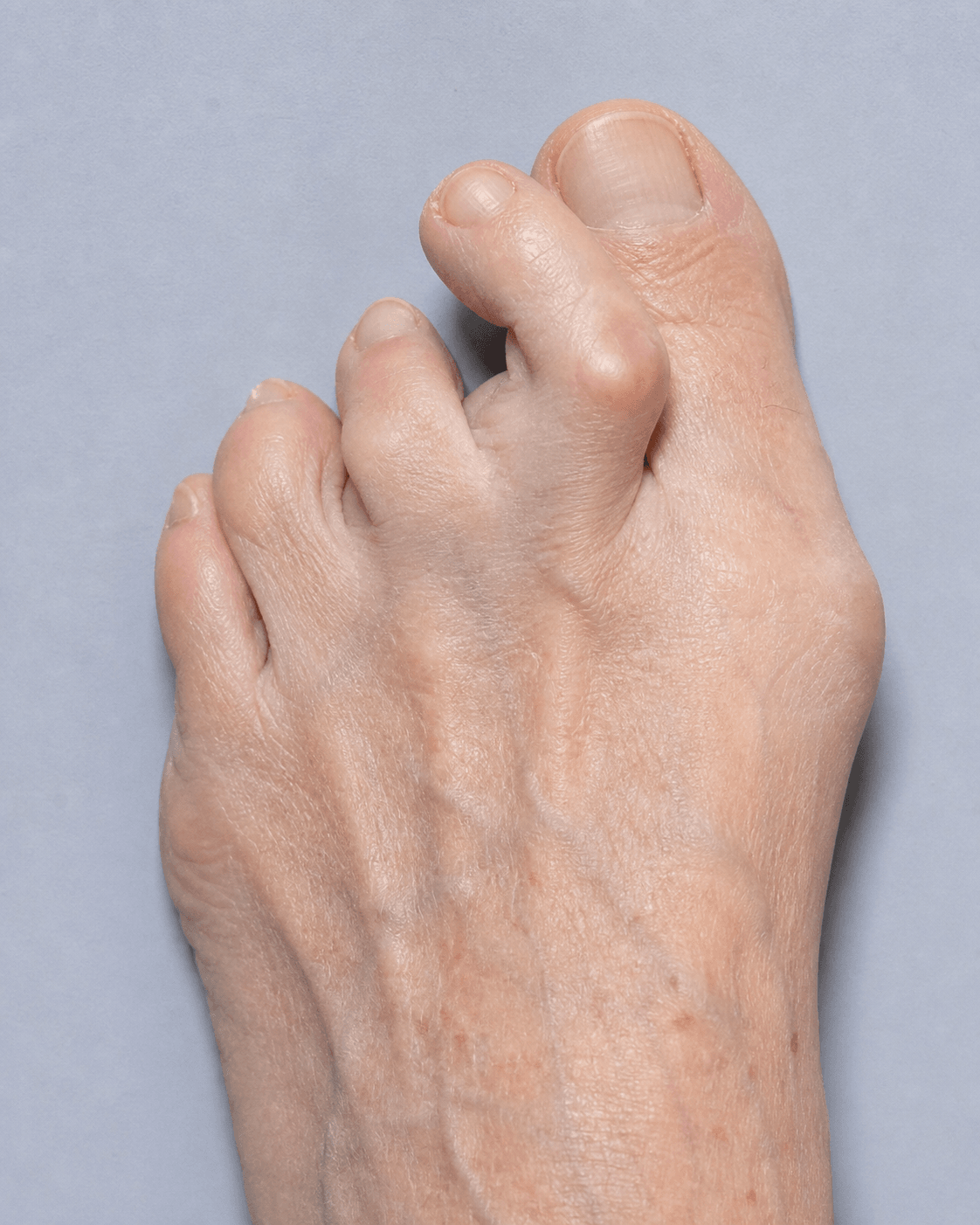
- It is the underlying cause of the CROSSOVER TOE deformity (medial/dorsomedial drift of the second toe).
- The plantar (dorsal) DRAWER test - dorsal translation of the proximal phalanx on the metatarsal head - is the key clinical sign of instability.
- It is frequently CONFUSED with a Morton's neuroma; MRI/ultrasound distinguishes them (and shows the tear).
- Treatment is staged: taping/orthoses early; direct plantar plate repair (usually via a dorsal approach with a Weil osteotomy) for instability that fails conservative care.
- “Plantar plate tear vs Morton's neuroma: plate disease causes MTP-joint pain, a positive drawer and toe deviation; a neuroma gives web-space pain, Mulder's click and toe-splaying - imaging settles it.
- “The tear classically begins at the LATERAL distal (phalangeal) insertion of the plantar plate.
- “A Weil osteotomy shortens/elevates the metatarsal to off-load and expose the joint for repair.
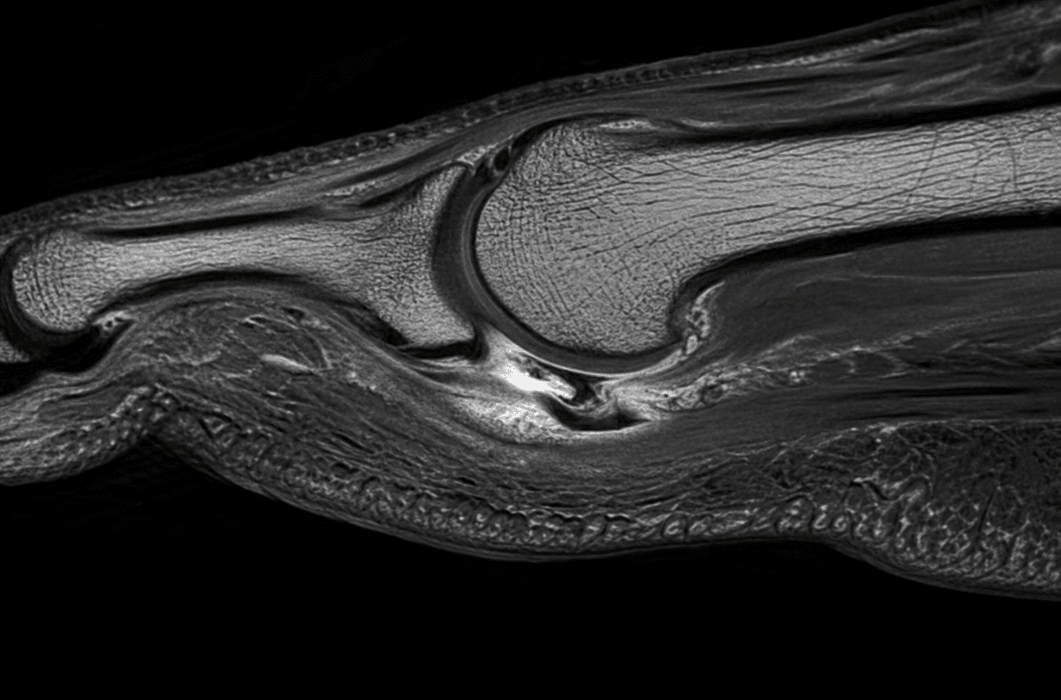
Pain localised to the MTP joint (plantar/under the metatarsal head), synovitis/swelling, toe deviation and a positive plantar drawer test. Progresses to crossover toe. The plantar plate is hypointense on MRI; a tear shows hyperintensity at the phalangeal insertion.
Pain in the web space (commonly 3rd), radiating to the toes, with a Mulder's click and toe-splaying, but a stable MTP joint and a negative drawer. Pericapsular oedema from a plate tear can mimic a neuroma on imaging - do not confuse them.
Anatomy & Pathophysiology
The plantar plate is a strong fibrocartilaginous structure on the plantar aspect of each lesser MTP joint. It originates loosely from the metatarsal neck and inserts firmly onto the base of the proximal phalanx, blending with the collateral ligaments, joint capsule and flexor sheath. Anatomical study shows it is the major stabiliser of the joint because of its central position and multiple attachments.
- Function: resists dorsal subluxation and hyperextension of the MTP joint, cushions load under the metatarsal head, and maintains toe alignment.
- Pathophysiology: chronic overload (e.g. long second metatarsal, hallux valgus transferring load, high heels) causes attritional degeneration and tearing, typically at the distal (phalangeal) insertion, laterally first. Loss of the plantar restraint allows dorsal and medial drift of the toe, ending in a crossover toe and ultimately MTP dislocation.
- The second MTP joint is most commonly affected.
Clinical Presentation & Assessment
History
- Plantar forefoot pain under the metatarsal head (often the second), with a feeling of "walking on a pebble" or sock bunching.
- Progressive swelling of the MTP joint and deviation of the toe (medial/dorsomedial), then crossover over the hallux.
- Often confused with, or coexists with, metatarsalgia and is mistaken for a Morton's neuroma.
Management
Non-operative
- First-line for early/lower-grade disease and the comorbid patient.
- Toe taping/strapping (plantarflexion taping to hold the toe down), stiff-soled shoes / rocker sole, metatarsal pads/orthoses to off-load the head, activity modification and NSAIDs.
- Corticosteroid injection is used cautiously (risk of further plate attenuation and deformity).
PLATEPlantar Plate Insufficiency
Hook:A failed PLATE drifts the toe up and over (crossover).
Evidence Base
Lesser MTP Instability: Plantar Plate Repair
- Prospective study; the plantar plate is the major stabiliser of the lesser MTP joint
- The second MTP joint was most commonly affected (63%); Grade III tears were most frequent
- Direct plantar plate repair through a dorsal approach with a Weil osteotomy and lateral soft-tissue reefing restored alignment
- AOFAS score improved from a mean of 52 to 92 points
Imaging of Plantar Plate Tear (vs Morton's Neuroma)
- Plantar plate degeneration/tear typically involves the second MTP joint at the lateral proximal phalangeal insertion
- It is frequently confused with a second web-space Morton's neuroma
- The normal plate is hypointense on MRI and hyperechoic on ultrasound; a tear shows hyperintensity (anechoic cleft on US), more conspicuous with dorsiflexion stress
- Pericapsular oedema/fibrosis from a tear should not be misread as a Morton's neuroma
Viva Scenarios
Practise clinical reasoning and management decisions out loud
“A patient has plantar pain under the second metatarsal head and the second toe is starting to ride up and toward the big toe. How do you assess and manage this, and how do you exclude a Morton's neuroma?”
Guidelines, Registries & Global Practice
Global Practice Picture
Plantar plate insufficiency is increasingly recognised internationally as the true pathology behind lesser MTP instability and crossover toe, supported by anatomical, imaging and surgical-outcome work (notably from Nery and Coughlin). The consistent principles: localise to the MTP joint and grade with the drawer test; confirm and differentiate from Morton's neuroma with MRI/ultrasound; manage conservatively first; and repair the plate (commonly with a Weil osteotomy) for persistent instability.
Side-by-Side Synthesis
- Plantar plate tear
- MTP joint (plantar to head)
- Morton's neuroma
- Web space
- Plantar plate tear
- Positive drawer; toe deviation
- Morton's neuroma
- Mulder's click; toe splaying
- Plantar plate tear
- Unstable
- Morton's neuroma
- Stable
- Plantar plate tear
- Plate hyperintensity/cleft at insertion
- Morton's neuroma
- Web-space mass
- Plantar plate tear
- Plantar plate repair + Weil osteotomy
- Morton's neuroma
- Neuroma excision/decompression
Key Facts
- Plantar plate = primary lesser-MTP stabiliser
- 2nd MTP most affected; tear at lateral phalangeal insertion
- Cause of crossover toe
- Coughlin clinical / Nery anatomical staging
Diagnosis
- MTP-joint pain (not web space)
- Positive plantar drawer (Lachman) test
- MRI/US: plate hyperintensity / cleft
- Exclude Morton's neuroma
Management
- Taping, stiff sole, metatarsal off-loading
- Cautious with steroid injection
- Direct plantar plate repair
- + Weil osteotomy (off-load/expose), correct hallux valgus