Forefoot, Midfoot, Hindfoot and Ankle Involvement | Surgical Reconstruction Principles
- Rheumatoid foot deformity is the result of synovitis-driven capsuloligamentous laxity, cartilage loss and tendon imbalance - not a primary muscle problem
- Forefoot reconstruction = first MTP arthrodesis (gold standard) + lesser MT head excision (Fowler)
- Hindfoot valgus in RA is driven by chronic posterior tibial tenosynovitis and spring ligament attenuation, not by PTTD as in the adult acquired flatfoot
- Triple arthrodesis is the reliable salvage for end-stage rheumatoid hindfoot; pantalar arthrodesis is reserved for combined ankle and hindfoot arthritis
- Total ankle replacement in RA is favourable because polyarticular disease loads the adjacent joints - arthrodesis transfers stress and accelerates STJ and midfoot arthritis
- Always examine the contralateral foot, footwear and the patient's DMARD/biologic status before planning surgery
- “Fowler procedure = excision of the lesser metatarsal heads through the metaphyseal neck; the older Hoffmann technique resected the metatarsal shafts and produced transfer lesions
- “First MTP arthrodesis (not excision) is the gold standard for the rheumatoid hallux - preserves push-off
- “A plantar incision gives the best access for MTP head excision and protects the dorsomedial cutaneous nerve
- “Triple arthrodesis in RA is best done in situ - aggressive realignment risks wound and fixation failure on a soft-tissue bed
Overview and Epidemiology
The foot and ankle are involved in over 90 percent of patients with rheumatoid arthritis at some point in the disease. The forefoot is the most common site of pain and deformity and the single most frequent reason an RA patient sees a foot and ankle surgeon. The hindfoot is the second most common region, and its involvement correlates with disease duration and seropositivity.
Who. Women are affected 2-3 times more often than men, and seropositive disease carries the highest foot burden. The foot is the initial presentation of RA in 15-20 percent of patients.
Where. Prevalence by region:
- Forefoot - symptoms in 70-80 percent of RA patients at 10-year follow-up
- Midfoot - 20-30 percent
- Hindfoot - valgus in 30-50 percent of established RA
- Ankle - 10-30 percent; less common than forefoot or hindfoot disease, but produces most disability
The forefoot dominates by prevalence, but the hindfoot dominates by functional impact.
What it costs the patient. Forefoot pain is the leading cause of walking disability in RA, and in long-standing disease plantar callosities and ulceration form over the prominent metatarsal heads. Footwear modification fails once fixed MTP dislocation is established. Fall risk rises with hindfoot valgus and ankle arthritis, and quality-of-life scores (HAQ, AOFAS) drop sharply with bilateral hindfoot disease.
Pathophysiology
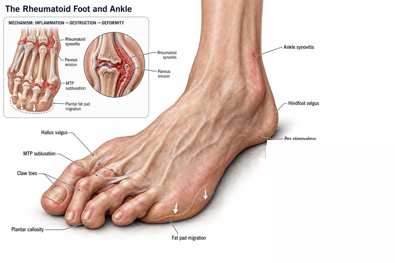
Synovitis drives every deformity. Chronic synovial inflammation is the primary driver of every deformity in the RA foot. Pannus, cytokines (TNF-alpha, IL-6, IL-17) and erosive enzymes destroy cartilage and ligament: the pannus erodes articular cartilage from the joint margins inward, weakens the collateral ligaments and plantar plate, and invades adjacent tendons, especially tibialis posterior, peroneus longus and the FDL and FHL to the toes. Once the static stabilisers fail, muscle imbalance, gravity and ground reaction force drive the foot into the classic RA deformities.
The forefoot. The MTP joint is the first casualty. Synovitis erodes its cartilage and capsule and attenuates the plantar plate and collateral ligaments, and plantar plate attenuation is the single most important step in forefoot collapse: the plate no longer restrains dorsiflexion, so the proximal phalanx drifts dorsally over the metatarsal head and the head is left plantar.
The fat pad and the claw. The plantar fat pad does not atrophy. It is dragged distally with the subluxing phalanx, so it still exists but no longer sits under the metatarsal heads, and painful callosities form over the prominent heads and often ulcerate. The long flexors (FDL, FHL) now pull obliquely: FDL slips dorsally, becoming a dorsiflexor of the PIP and a plantarflexor of the DIP, and the toe claws.
The first ray and the lesser toes. In the arthritic hallux valgus the first ray drifts medially and the first MTP subluxes laterally. The lesser toes crowd laterally and dislocate dorsolaterally.
The hindfoot. Chronic tenosynovitis and intrasubstance degeneration of tibialis posterior drive hindfoot valgus, and the tenosynovitis is present on ultrasound in 30-50 percent of RA feet. With the medial arch unsupported and the spring ligament elongated by chronic synovitis, the talar head uncovers medially, the navicular abducts, the calcaneus drifts into valgus and forefoot abduction follows. The foot flattens, yet this is a different disease entity from adult-acquired flatfoot deformity.
Around the hindfoot. Subtalar and talonavicular arthritis often coexist. Lateral impingement (calcaneofibular abutment) gives sinus tarsi pain, and gastrocnemius tightness contributes equinus.
The ankle. Marginal erosions and cartilage loss at the tibiotalar joint lead to varus or valgus collapse and an end-stage, painful, stiff ankle.
Classification and Types
The rheumatoid foot is described by region, by the severity of the forefoot deformity, and by disease activity at the time of surgery. Severity guides the choice between joint-preserving (early) and joint-sacrificing (late) surgery.
- Classic Deformity
- MTP subluxation, claw toes, hallux valgus
- Surgical Strategy
- 1st MTP arthrodesis + lesser MT head excision (Fowler)
- Key Point
- Plantar approach protects neurovascular bundle
- Classic Deformity
- Talonavicular arthritis and sag, naviculocuneiform OA and sag
- Surgical Strategy
- Isolated TN or NC fusion if focal
- Key Point
- Often progresses with hindfoot; address together
- Classic Deformity
- Valgus, subtalar and TN arthritis
- Surgical Strategy
- Triple arthrodesis (in situ)
- Key Point
- Treats deformity and pain reliably
- Classic Deformity
- Tibiotalar synovitis, joint space loss, arthritis
- Surgical Strategy
- TAA in low-demand patient, arthrodesis in young/heavy labourer
- Key Point
- TAA preserves adjacent joint motion
Clinical Assessment
History. Establish where the pain is (forefoot most commonly, then hindfoot or ankle) and how long the disease has run, since longer disease brings more deformity and disability. Record the current regimen (methotrexate, anti-TNF, rituximab, JAK inhibitors), previous forefoot or hindfoot surgery, and any hip or knee arthroplasty. A patient who can no longer tolerate off-the-shelf shoes has severe deformity; ask about current or recurrent ulceration of plantar callosities.
Examination. Always examine the contralateral foot and the footwear as well.
- Forefoot - MTP synovitis, dorsal subluxation, fixed or flexible claw, hallux valgus angle
- Callosities - plantar under the lesser metatarsal heads, and medial over the first metatarsal in hallux valgus
- Hindfoot - heel valgus, sinus tarsi tenderness, and tibialis posterior function (below)
- Ankle - the tibiotalar line, range of motion, and whether the pain is tibiotalar or subtalar
- Skin and nails - capillary refill, prior ulceration, onychomycosis
- Gait - antalgic, externally rotated, or vaulting over a stiff ankle
- Inspection - medial swelling and erythema along the tendon course (Henrys sign)
- Single-leg heel raise - the patient cannot invert the hindfoot and lock the midfoot; the heel does not invert
- Too many toes sign - standing behind the patient, more than 1-2 lateral toes are visible because of forefoot abduction
- First metatarsal rise test - when the hindfoot is corrected to neutral the first metatarsal head rises, indicating fixed forefoot supination
In RA, correlate these findings with disease activity and the patient's medical regimen before deciding between tendon reconstruction and triple arthrodesis.
Tibiotalar pain is felt anteriorly at the joint line, worsens with dorsiflexion, and the patient points to the front of the ankle. Subtalar pain is felt laterally in the sinus tarsi, worsens with inversion/eversion on a fixed tibia, and the patient points to the lateral side of the heel. Both commonly coexist in RA - a steroid injection into the symptomatic joint (with imaging control) is a useful diagnostic and therapeutic step before planning fusion or replacement.
- Pain location
- Dorsal MTP, plantar under MT head
- Discriminating finding
- Synovial thickening, marginal erosions
- Key test / imaging
- Ultrasound for active synovitis; MRI early
- Pain location
- Interdigital 2-3 or 3-4
- Discriminating finding
- Mulder sign, web-space tenderness
- Key test / imaging
- Diagnostic local anaesthetic injection
- Pain location
- Second MT head, adolescents
- Discriminating finding
- Subchondral collapse, sclerotic MT head
- Key test / imaging
- Standing X-ray, MRI if atypical
- Pain location
- Plantar 2nd MTP, swelling
- Discriminating finding
- Lachman test of MTP, drawer sign
- Key test / imaging
- Dynamic ultrasound, MRI
- Pain location
- Forefoot pain on activity
- Discriminating finding
- Pain on axial load, swelling
- Key test / imaging
- MRI gold standard; X-ray often late
- Pain location
- Acute hot, swollen MTP
- Discriminating finding
- Systemic features, very high CRP
- Key test / imaging
- Aspiration for cell count, culture
Rheumatoid arthritis is one of the commonest inflammatory causes of tarsal tunnel syndrome, and it is easy to miss because the symptoms are blamed on the joint disease. Two RA-specific mechanisms compress the posterior tibial nerve and its branches beneath the flexor retinaculum behind the medial malleolus:
- Flexor tenosynovitis (tibialis posterior, FDL and FHL), producing a space-occupying synovial mass in the tunnel
- The valgus hindfoot itself, which tents and stretches the nerve as the heel drifts laterally
Presentation. Burning or tingling pain in the plantar foot and toes, worse at night and after activity, with a positive Tinel sign behind the medial malleolus and symptoms reproduced by a sustained dorsiflexion-eversion manoeuvre.
Investigation. Nerve conduction studies and EMG confirm the lesion and localise it; ultrasound or MRI demonstrates the tenosynovial mass and excludes a ganglion or other space-occupying cause.
Treatment. Control the synovitis (DMARD optimisation, intra-articular or peritendinous steroid), correct the contributing valgus deformity, and perform a flexor tenosynovectomy with tarsal tunnel decompression (release of the flexor retinaculum) when compression is confirmed. Distinguish this focal entrapment from a diffuse peripheral neuropathy (DMARD-related, vasculitic or coexisting diabetic), which is non-focal, symmetrical and not relieved by decompression.
Investigations
Radiographs first. RA is largely a clinical and X-ray diagnosis, and standing films are the cornerstone. Take them weight-bearing:
- Foot - AP, lateral and oblique
- Ankle - AP, mortise and lateral
Templating for a first MTP arthrodesis or TAA is done on standing films, not supine.
What the films show. The hallmarks are marginal erosions, not the central erosions of OA, juxta-articular osteopenia rather than subchondral sclerosis, and symmetric joint space loss. Erosions appear first at the metatarsal heads and PIP joints, and the 5th metatarsal head is often the first site to show erosive change, which makes it a useful early sign. In established disease the subchondral bone is moth-eaten rather than sclerotic as in OA, and advanced disease forms cysts, occasionally leading to pathological fracture of a metatarsal head. Look also for hindfoot valgus and tibiotalar arthritis.
Ultrasound. It detects active synovitis when the radiographs are normal and confirms tibialis posterior tenosynovitis. Power Doppler grades synovial activity, guides injection and monitors the response to treatment, and because it is bedside, cheap and dynamic it can assess tibialis posterior dysfunction in real time.
MRI. It shows early RA change (bone marrow oedema, early erosions), tendon pathology including tibialis posterior tenosynovitis, spring ligament injury and sinus tarsi synovitis, and it is indicated for atypical pain. Before surgery it defines cartilage status before TAA, the extent of talar dome AVN and midfoot involvement.
CT. For planning an arthrodesis, for complex hindfoot deformity and after prior failed surgery.
Bloods and aspiration. CRP, ESR, full blood count, renal and liver function, and DMARD levels if available. Aspirate the joint if there is any suspicion of septic arthritis, especially in a patient on biologics.
Management Algorithm
Before any operation. Before the patient is listed, agree the medical regimen with rheumatology and assess the cervical spine.
Rheumatology liaison is essential. A simple phone call before listing the patient reduces cancellations and improves outcomes.
- Methotrexate - continue perioperatively in most patients; withdrawal increases RA flare and is not associated with higher infection
- Biologics (anti-TNF, IL-6, rituximab) - hold for 1-2 dosing cycles before and after surgery, and restart at 2 weeks once the wound is dry
- Prednisolone - taper to less than 10 mg/day if possible; high-dose steroids increase infection and impair wound healing
Before an RA patient is listed for any foot or ankle procedure under general anaesthesia, the cervical spine must be assessed for instability. This is a high-yield viva trap: the same disease destroying the foot also destroys the neck, and the danger is at intubation, not in the foot.
Why. Roughly half of patients with long-standing RA have radiographic cervical involvement. Atlantoaxial (C1-C2) subluxation is the commonest and most dangerous pattern: chronic synovitis erodes the transverse ligament and dens, and neck extension at intubation can drive the dens into the cord. Subaxial subluxation and basilar invagination (cranial settling) are the other patterns.
Screen. Neck pain, occipital headache, paraesthesiae or "electric shock" sensations, hand clumsiness, gait disturbance or sphincter change all suggest myelopathy.
Image. Request flexion-extension lateral cervical radiographs. An anterior atlanto-dental interval (ADI) over 3 mm indicates anterior atlantoaxial subluxation, but the posterior ADI (PADI, the space available for the cord) is the better surgical predictor: a PADI under 14 mm correlates with neurological risk and is a threshold for surgical stabilisation.
Act. Flag any instability to the anaesthetist (consider awake fibreoptic intubation, manual in-line stabilisation and avoidance of neck extension) and obtain a spinal or neurosurgical opinion before elective foot surgery if the patient is myelopathic or radiographically unstable.
Goal. Relieve plantar pain, restore plantigrade alignment and allow off-the-shelf footwear. The reconstruction combines a first MTP arthrodesis with Fowler excision of the lesser metatarsal heads.
The first ray. First MTP arthrodesis is the gold standard for the rheumatoid hallux: it preserves push-off and protects the lesser metatarsal heads from transfer overload. Keller excision arthroplasty (excision of the proximal phalanx base, no fixation) is reserved for low-demand, elderly or bedbound patients, because it sacrifices push-off, recovers more slowly and risks transfer metatarsalgia.
What fusion buys. A viva will press this. Against implant arthroplasty of the first MTP, the pooled comparative evidence shows no difference in pain (SMD 0.04, p=0.734). What fusion delivers is durable alignment, with better maintenance of the hallux valgus and 1st-2nd intermetatarsal angles, and that matters because a great toe that drifts back into valgus pushes the lesser rays with it. The cost is non-union, reported from 0% to 26% across series and around 10-15% in most. Argue for fusion on deformity control and push-off, not on analgesia.
Fusing the first MTP. Through a dorsal approach, prepare the joint with cup-and-cone reamers and fix it with a 6.5 mm compression screw plus a dorsal plate. Position the toe in 10-15 degrees of valgus and 15-20 degrees of dorsiflexion relative to the floor.
The lesser metatarsal heads. A plantar transverse approach (single, two or three incisions) is the gold standard for the lesser heads: it gives the best exposure, protects the dorsomedial cutaneous nerve and produces the most reliable pain relief. The dorsal approach is reserved for first MTP work and lesser PIP release. Resect through the metaphyseal neck, not the shaft, since shaft-level cuts cause the transfer lesions that plagued the older Hoffmann shaft-resection technique.
- Supine, tourniquet at the thigh or calf, foot on a bolster
- Longitudinal capsulotomy to open each MTP joint and release the capsule
- Excise metatarsal heads 2-5 through the plantar incisions, 1 cm distal to the distal metaphyseal flare, with an oscillating saw in a slight oblique plane (dorsal to plantar, lateral to medial) that leaves a short oblique cut
- Synovectomy of the remaining MTP joints and the intermetatarsal bursae
- PIP release or arthroplasty for a fixed claw; wire the lesser toes only if deformity is severe, otherwise soft-tissue release is sufficient
After surgery. Heel-weight-bearing in a stiff-soled shoe for 6 weeks, then extra-depth shoes with a rocker sole from 6-8 weeks.
Complications
- Incidence
- 10-20 percent in RA
- Risk Factors
- Active disease, biologics, smoking, steroids
- Management
- Local wound care, plastic surgery input, biologics hold
- Incidence
- 2-5 percent
- Risk Factors
- Active disease, poor nutrition, prior surgery
- Management
- DAIR, two-stage revision, suppressive antibiotics
- Incidence
- 10-15 percent
- Risk Factors
- Smoking, biologics, prior surgery, deformity
- Management
- Bone graft, revision fixation, bone stimulator
- Incidence
- 10-20 percent after Fowler
- Risk Factors
- Asymmetric MT resection, shaft-level cuts
- Management
- Orthosis, rocker sole, revision of second MT
- Incidence
- 5-10 percent
- Risk Factors
- Active synovitis post-op, poor fixation
- Management
- Bracing, revision fusion, biologics optimisation
- Incidence
- 5-10 percent at 5-10 years
- Risk Factors
- High BMI, malalignment, osteolysis
- Management
- Conversion to arthrodesis with allograft
- Incidence
- 30-50 percent at 10 years
- Risk Factors
- Ankle vs hindfoot fusion in adjacent joint
- Management
- Custom orthosis, fusion extension if symptomatic
Outcomes and Prognosis
- Pain Relief
- 85-90 percent good-excellent
- Satisfaction
- 80-85 percent patient satisfaction
- Complications
- Transfer metatarsalgia, wound
- Long-term
- Durable at 10 years in 80 percent
- Pain Relief
- 70-80 percent good
- Satisfaction
- Lower satisfaction, transfer lesions
- Complications
- Transfer metatarsalgia common
- Long-term
- Superseded by Fowler in most centres
- Pain Relief
- 85-90 percent fusion, 80 percent satisfied
- Satisfaction
- High satisfaction if no non-union
- Complications
- Non-union
- Long-term
- Stable at 10 years in 75-85 percent
- Pain Relief
- 85-90 percent good at 5 years
- Satisfaction
- High satisfaction in selected patients
- Complications
- Poly wear, loosening, cysts
- Long-term
- 90 percent survivorship at 5-10 years
- Pain Relief
- 90 percent fusion, reliable pain relief
- Satisfaction
- High satisfaction, loss of motion
- Complications
- Adjacent arthritis 30-50 percent
- Long-term
- Durable at 10 years in 80-90 percent
The best prognosis comes with disease in remission, a motivated patient off steroids with good nutrition, and a well-aligned first ray with a stable 1st MTP arthrodesis. Active disease, continued smoking, persistent hindfoot valgus after forefoot surgery alone, unrealistic footwear expectations and prior failed surgery all point to a poor result.
Forefoot reconstruction is a one-shot operation: revision of a failed Fowler is far harder than a primary procedure, so plan the alignment carefully the first time.
Guidelines, Registries & Global Practice
- RA prevalence is 0.5-1 percent worldwide, with seropositive disease carrying the highest foot burden
- Foot involvement occurs in 90 percent of patients at some point, with forefoot the most common site
- Racial variation is modest, but South Asian and African ancestry show more severe erosive disease
- Disease duration correlates strongly with foot deformity; deformity is rare in the first year but common after 5-10 years
- High-resource: combination biologic/DMARD therapy has reduced severe deformity; reconstruction is the dominant surgical workload
- Limited-resource: presentation with end-stage deformity is more common; off-the-shelf rocker-sole footwear and palliative Fowler-style surgery are the mainstay
- Universal principle: liaison with rheumatology to optimise DMARDs and biologics before surgery is the single biggest determinant of wound and infection outcomes
- Forefoot
- Early DMARD escalation, footwear and orthosis first
- Hindfoot
- Triple arthrodesis in situ for end-stage disease
- Ankle
- TAA in selected low-demand patients; arthrodesis remains an option
- Forefoot
- Combined 1st MTP arthrodesis + lesser MT head excision
- Hindfoot
- Triple arthrodesis, often with medial column fixation
- Ankle
- TAA for selected low-demand patients; arthrodesis in young high-demand
- Forefoot
- Plantar approach for MT head excision, 1st MTP arthrodesis
- Hindfoot
- In situ triple arthrodesis; avoid aggressive realignment in RA
- Ankle
- TAA or TTC nail depending on demand and deformity
- Forefoot
- Fowler procedure, 1st MTP arthrodesis; barefoot cultural challenges
- Hindfoot
- Triple arthrodesis; consider cultural and occupational demands
- Ankle
- TAA in selected centres; arthrodesis still common
There is no dedicated registry for rheumatoid foot and ankle surgery in the way there is for hip and knee arthroplasty. Most large registries (NJR, AJRR, AOANJRR) collect ankle replacement data, and the Swedish, Norwegian and New Zealand ankle registries provide the strongest survivorship data for TAA. The evidence base for forefoot and hindfoot surgery in RA is dominated by retrospective case series; systematic reviews are emerging but the level of evidence remains lower than for hip and knee arthroplasty.
Record in every RA foot and ankle consultation:
- Disease activity (CRP, ESR, DAS-28 where available)
- Current DMARD and biologic regimen
- Functional impact (HAQ, AOFAS, footwear tolerance)
- Examination of footwear, contralateral foot and adjacent joints
- Pre-operative DMARD and biologic plan documented with rheumatology
Operating on active RA without medical optimisation is a recurring source of poor outcomes, complaints and revision surgery. Always document the medical plan before listing the patient.
Controversies & Areas of Uncertainty
Modern third-generation TAA designs give 90 percent survivorship at 5-10 years in low-demand RA patients and preserve adjacent joint motion. Arthrodesis is more reliable in young, high-demand, heavy labourers and in severe deformity. There is no head-to-head randomised trial; choice is pragmatic and patient-led.
Continuation of methotrexate is supported by randomised evidence showing lower infection rates than withdrawal. Anti-TNF agents are typically held for one dosing cycle; the evidence for IL-6 inhibitors and JAK inhibitors is thinner. Always liaise with rheumatology rather than follow a single protocol.
Single-stage bilateral Fowler plus 1st MTP arthrodesis is described and accelerates recovery, but carries a higher wound and VTE risk. Most surgeons stage bilateral cases 3-6 months apart, particularly on biologics or in patients with poor soft tissues.
In RA, isolated talonavicular fusion is appealing because it preserves subtalar motion, but adjacent STJ arthritis is common and triple arthrodesis is more reliable long term. Decision is based on imaging, symptoms and the patient's age and demand.
MCQ Practice Points
Q: Which tendon is most commonly implicated in rheumatoid hindfoot valgus? A: Tibialis posterior. Chronic tenosynovitis and intrasubstance degeneration of the tibialis posterior tendon is the principal driver of hindfoot valgus in rheumatoid arthritis, often with concurrent spring ligament attenuation. The peroneus longus is a secondary contributor; the FHL and FDL are rarely the primary problem.
Q: What is the imaging hallmark of rheumatoid arthritis in the foot? A: Marginal erosions with juxta-articular osteopenia. RA erodes the joint margins first (where synovium reflects over the cartilage), producing an asymmetric, peripheral pattern of bone destruction. Osteoarthritis, in contrast, produces central erosions with subchondral sclerosis. The 5th MT head is often the first site to show erosive change in the foot.
Q: What is the typical cascade of rheumatoid forefoot deformity? A: MTP synovitis → plantar plate attenuation → dorsal subluxation of the proximal phalanx → plantar migration of the fat pad → clawing of the toes → prominent MT heads with plantar callosities. The first ray drifts medially and the hallux valgus intermetatarsal angle increases, pushing the lesser toes laterally and worsening their dislocation.
Q: What is the gold-standard surgical treatment for the end-stage rheumatoid forefoot? A: 1st MTP arthrodesis combined with Fowler excision of the lesser MT heads. The arthrodesis preserves push-off and protects the lesser MT heads from transfer overload. Keller arthroplasty is reserved for low-demand, elderly or bedbound patients.
Q: What is the operation of choice for end-stage rheumatoid hindfoot deformity? A: Triple arthrodesis (in situ). Triple arthrodesis reliably corrects valgus deformity and eliminates subtalar and talonavicular pain. It is performed in situ (not aggressively realigned) because RA soft tissues are poor and aggressive correction risks wound and fixation failure. Aggressive realignment and pantalar fusion are reserved for severe combined disease.
Q: When is total ankle replacement preferred over arthrodesis in rheumatoid arthritis? A: In the low-demand patient with preserved subtalar motion and no significant deformity. RA produces polyarticular disease, so arthrodesis transfers stress to a subtalar and midfoot that is already diseased. TAA preserves adjacent motion and reduces adjacent joint degeneration, but is contraindicated in severe deformity, talar AVN, Charcot and infection.
Exam Viva Scenarios
Practise clinical reasoning and management decisions out loud
“A 58-year-old woman with long-standing seropositive rheumatoid arthritis on methotrexate and etanercept presents with bilateral forefoot pain. She has painful plantar callosities under the second and third metatarsal heads, dorsal subluxation of the lesser MTP joints, fixed clawing of the second and third toes, and a hallux valgus deformity of 35 degrees. The metatarsal heads are prominent and the plantar fat pad has migrated distally. She has failed orthosis and footwear modification. How would you manage this?”
“A 62-year-old man with seropositive RA on methotrexate presents with severe hindfoot valgus, fixed pronation, sinus tarsi pain, and a stiff painful ankle. He has previously had a bilateral 1st MTP arthrodesis with Fowler excision and is now struggling to mobilise because of the hindfoot. Imaging shows combined subtalar, talonavicular and tibiotalar arthritis with significant valgus alignment. He is a low-demand retired office worker. How would you approach this?”
Epidemiology and Anatomy
- Foot/ankle involvement in over 90 percent of RA patients over the disease course
- Forefoot is the most common site; hindfoot valgus affects 30-50 percent of established RA
- Posterior tibial tenosynovitis drives hindfoot valgus, not adult-acquired PTTD
- Marginal erosions and juxta-articular osteopenia are the radiologic hallmarks
Forefoot Reconstruction
- Fowler = plantar approach, excise lesser MT heads at the metaphyseal neck
- 1st MTP arthrodesis is the gold standard for the rheumatoid hallux (10-15 degrees valgus, 15-20 degrees dorsiflexion)
- Keller reserved for low-demand, elderly or bedbound patients
- Plantar approach protects the dorsomedial cutaneous nerve and gives best pain relief
Hindfoot and Ankle
- Triple arthrodesis (in situ) is the workhorse for end-stage RA hindfoot
- TAA preferred over arthrodesis in low-demand RA with adjacent disease
- TTC nail for combined ankle and subtalar arthritis with deformity
- Pantalar fusion reserved for severe combined disease
Perioperative Management
- Continue methotrexate perioperatively; hold biologics 1-2 cycles
- Taper steroids to less than 10 mg/day if possible
- Rheumatology liaison is essential before listing the patient
- Optimise disease activity (CRP/ESR/DAS-28) before elective surgery
Complications to Avoid
- Wound healing problems in 10-20 percent; biologics and steroids increase risk
- Transfer metatarsalgia after asymmetric or shaft-level MT resection
- Recurrent deformity from persistent synovitis and inadequate fixation
- Adjacent joint arthritis after ankle or hindfoot arthrodesis (30-50 percent at 10 years)
Evidence Base and Key Trials
Surgical correction of rheumatoid forefoot deformities
- Retrospective series combining 1st MTP arthrodesis with lesser MT head excision for rheumatoid forefoot reconstruction
- High rates of pain relief and patient satisfaction at intermediate follow-up
- 1st MTP arthrodesis preserved push-off and protected the lesser MT heads from transfer overload
Effectiveness of the First Metatarsophalangeal Joint Arthrodesis Versus Arthroplasty for Rheumatoid Forefoot Deformity: A Systematic Review and Meta-Analysis of Comparative Studies
- 6 comparative studies, 337 patients (459 feet) - all compared lesser-ray resection arthroplasty PLUS either 1st MTP arthrodesis or 1st MTP arthroplasty, so the lesser rays are handled identically and only the great toe is in dispute
- PAIN WAS NO DIFFERENT between arthrodesis and arthroplasty (SMD 0.04, p=0.734) - the advantage of fusion in this meta-analysis is radiographic, not symptomatic
- Arthrodesis maintained the hallux valgus angle (SMD -0.439, p=0.002) and the 1st-2nd intermetatarsal angle (SMD -0.569, p<0.001) better, but with extreme heterogeneity (I-squared 96.6% and 98.2%)
- Non-union after arthrodesis ranged 0% to 26% across the included series - the price of the alignment advantage
- Reoperation 3-9.6% after arthrodesis and 4-11.6% after arthroplasty - overlapping descriptive ranges, not a tested difference
The foot function index is more sensitive to change than the Leeds Foot Impact Scale for evaluating rheumatoid arthritis patients after forefoot or hindfoot reconstruction
- Foot Function Index detected clinically meaningful change more sensitively than the Leeds Foot Impact Scale after RA forefoot and hindfoot reconstruction
Survivorship and long-term outcome of a consecutive series of 200 Scandinavian Total Ankle Replacement (STAR) implants
- 200 consecutive STAR implants in 184 patients by a single surgeon, 1993-2000, with genuinely long follow-up
- 32 implants failed and required revision (16%); mean time to revision 80 months, with a range of 2 to 257 months - failure is not confined to the early years
- Survival at 15.8 years, revision as endpoint, was 76.2% (95% CI 64.4 to 87.9) - a steady low-rate attrition rather than a plateau
- AOFAS hindfoot score improved from 28 to 61 - a large gain that still leaves patients well short of a normal ankle
- Among the 84 patients (87 ankles) alive at study end, 40 ankles were RA and 47 were OA, but NO separate survival analysis by diagnosis is reported - this series cannot tell you whether RA ankles did better or worse