A finding-first approach: never operate on a painful knee replacement without a diagnosis
- Periprosthetic joint infection is the diagnosis of exclusion in reverse — you exclude it first, actively, with serology and aspiration, not by impression.
- A pain-free interval followed by activity-related start-up pain suggests aseptic loosening; pain that never resolved after surgery suggests infection, instability or a diagnosis outside the knee.
- Radiographs must be weight-bearing AP, true lateral, skyline and long-leg alignment films; a supine AP alone will miss instability and coronal malalignment.
- Radiolucent lines matter only if they are progressive, greater than 2 mm, or circumferential — a stable non-progressive 1 mm line at the cement–bone interface is normal.
- Around a fifth of painful TKRs have an extrinsic cause: hip osteoarthritis, lumbar radiculopathy, vascular claudication, CRPS or neuroma — examine the hip and spine before ordering a CT.
- Revision surgery for unexplained pain is among the poorest of all revision indications, and there is a national number for it: across 24,540 first revisions in the UK registry, revision for unexplained pain gave a mean postoperative Oxford Knee Score of 25 with only 55 per cent responders. Be precise, though — STIFFNESS was worse still (score 24, 48 per cent responders). No diagnosis, no operation.
- Diagnose infection with the 2018 ICM WEIGHTED score, not a checklist: two positive cultures or a sinus tract are diagnostic on their own; otherwise synovial white cell count over 3,000, alpha-defensin and leukocyte esterase score 3 each, serum CRP over 1 mg/dL and D-dimer over 860 ng/mL score 2, ESR over 30 scores 1. Six or more is infected; 2 to 5 is INCONCLUSIVE and defers the answer to intraoperative findings.
- Alpha-defensin is two different tests. The laboratory assay pools at 95 per cent sensitivity and 97 per cent specificity; the lateral flow cassette used at the bedside is significantly worse at 85 and 90 per cent, and performs no better than a leukocyte esterase strip. A negative cassette is weak reassurance, not an exclusion.
- “Say it out loud: 'My priority is to exclude infection, then define whether the pain is intrinsic or extrinsic to the knee.'
- “Mid-flexion instability is invisible on static radiographs — it is a clinical diagnosis of a raised joint line and femoral component downsizing.
- “Anterior knee pain with a well-fixed prosthesis and internal rotation of the tibial tray on CT is component malrotation until proven otherwise.
- “A hot bone scan in the first 12 months after TKR is uninterpretable — periprosthetic remodelling is expected for up to a year.
Normal CRP and ESR reduce but do not abolish the probability of infection — low-virulence organisms such as Cutibacterium acnes and coagulase-negative staphylococci frequently give normal serology. If the story is atypical, aspirate.
Thin, non-progressive lucent lines under the tibial tray are seen in many asymptomatic, well-functioning knees. Loosening requires a progressive or greater than 2 mm circumferential lucency, component migration, or subsidence on serial films.
Referred pain from hip osteoarthritis presents as anteromedial knee and thigh pain with pain-free knee examination and restricted, painful hip rotation. Lumbar stenosis gives buttock-to-calf pain worsened by walking with a normal knee exam.
Revision without a diagnosis reliably fails. If every investigation is negative, the correct answer is structured non-operative management, review of the pain phenotype and re-evaluation — not an exchange of polyethylene 'to see'.
Recognising the Pattern
The presentation. Pain in a knee that has undergone total knee arthroplasty, persisting or recurring beyond the expected recovery window of approximately three months. It is a pattern, not a diagnosis: the task is to convert "painful knee replacement" into a mechanism.
Confirming the pattern is genuine and localised.
- Establish the temporal profile: was there ever a pain-free interval? Pain that never went away versus pain after a good result is the single most powerful branch point in the entire differential.
- Establish the character: start-up pain settling after a few steps (loosening); giving way and swelling on stairs or slopes (instability); pain only on stairs and rising from a chair with anterior localisation (patellofemoral or malrotation); rest and night pain with warmth (infection).
- Establish localisation: point tenderness at the tibial tubercle, medial joint line, or a Tinel sign over the infrapatellar branch of the saphenous nerve each redirects the differential.
- Confirm the pain is in the knee: full hip examination with logroll and internal rotation, straight-leg raise, distal pulses. Pain reproduced by hip rotation with a benign knee is a hip problem.
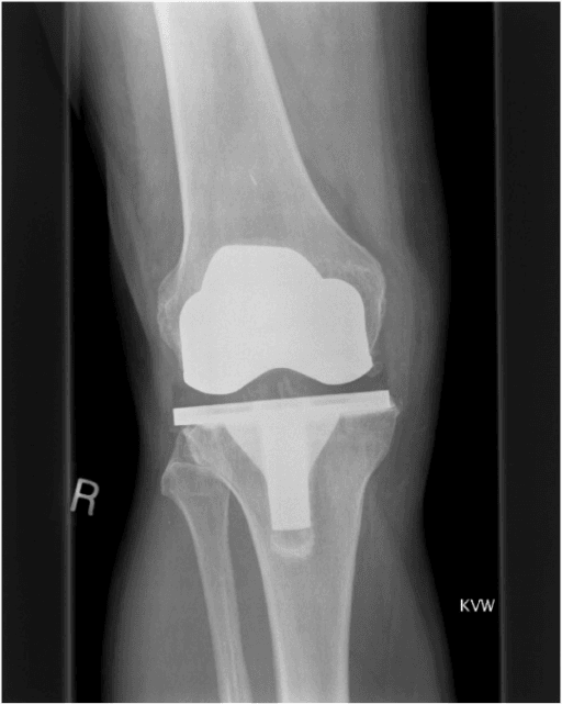
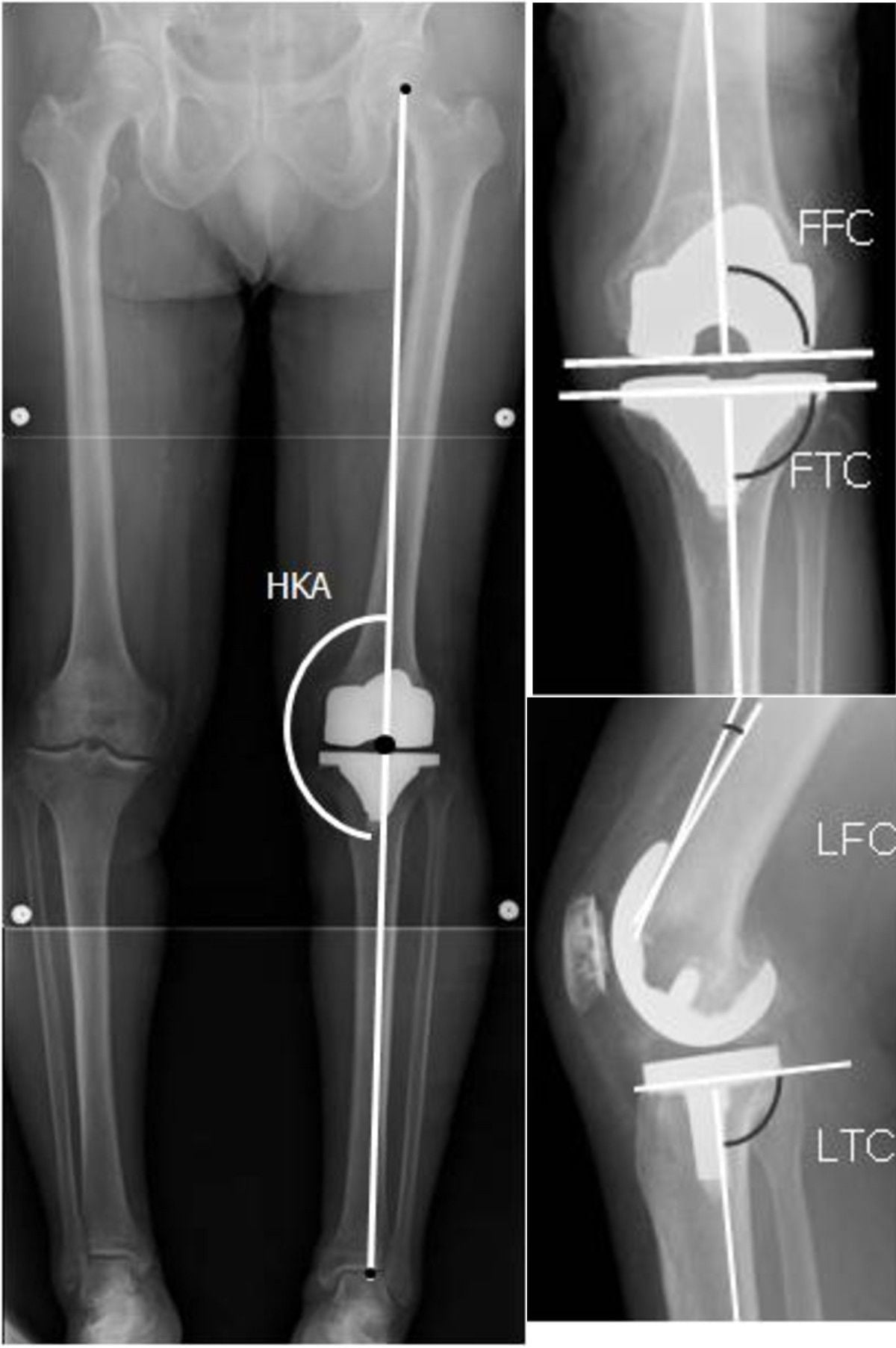
- Confirm on adequate imaging: weight-bearing AP, true lateral with the femoral condyles superimposed, skyline at 30 degrees, and a full-length hip-to-ankle alignment film. Serial films compared with the immediate post-operative radiograph are worth more than any single film.
How to describe it in a viva.
"These are weight-bearing AP and lateral radiographs of a cemented total knee replacement. I would first want the immediate post-operative films for comparison and a long-leg alignment view. I am assessing component position and alignment, the cement–bone interfaces on both sides for progressive or circumferential lucency, evidence of subsidence or migration, the patella and joint line height, and the periprosthetic bone for osteolysis or periosteal reaction. My clinical priority in any painful knee replacement is to exclude periprosthetic joint infection with CRP, ESR and aspiration before attributing the pain to a mechanical cause."
What mimics the pattern — false positives.
- Normal early post-operative pain within the first three months, with progressive improvement and no systemic features. This is not a painful TKR.
- Residual stiffness and soft-tissue soreness from an under-rehabilitated but well-fixed, well-aligned knee.
- Referred pain from the ipsilateral hip or the L3–L4 nerve roots — the knee itself is entirely comfortable on examination.
- Vascular claudication — reproducible walking distance, absent pulses, relief with standing still rather than sitting.
- Radiographic pseudo-lucency from beam obliquity: a lucent line that disappears on a properly tangential repeat view was never there.
- Metal artefact on cross-sectional imaging simulating osteolysis when artefact reduction sequences have not been used.
Next Investigation
The correct instinct in the viva is sequential and cheap-to-expensive: history and examination, then plain films with comparison, then CRP and ESR, then aspiration, and only then cross-sectional or nuclear imaging directed at a specific hypothesis. Candidates who leap to MRI or bone scan first lose marks.
Do not pass a needle through overlying cellulitis — it risks seeding a previously uninfected joint. Treat the soft-tissue infection, and if a septic joint is still suspected, discuss urgent theatre-based sampling rather than a blind bedside tap. Equally, do not give empirical antibiotics before sampling in a stable patient: it destroys the diagnostic yield of culture.
The Differential
- Typical age / setting
- Any age; diabetes, obesity, immunosuppression, prolonged wound ooze, prior revision
- Discriminating feature
- No pain-free interval, or new pain after a bacteraemic event; rest and night pain, warmth, effusion; stiffness out of proportion
- What confirms it
- Aspiration: synovial white cell count and neutrophil differential, culture; raised CRP and ESR; alpha-defensin; multiple intra-operative tissue samples
- Typical age / setting
- Elderly, osteoporotic, low-energy fall; notched anterior femoral cortex
- Discriminating feature
- Sudden pain and deformity after a discrete injury in a previously good knee
- What confirms it
- Radiographs in two planes; CT if occult or intra-articular extension suspected
- Typical age / setting
- Any; after manipulation, lateral release, or in inflammatory arthropathy
- Discriminating feature
- Inability to perform a straight-leg raise with an extensor lag; palpable gap
- What confirms it
- Lateral radiograph showing patella alta or baja; ultrasound or MRI with metal suppression
- Typical age / setting
- Older patient with coexisting degenerative hip or spine disease
- Discriminating feature
- Knee examination entirely benign; pain reproduced by hip internal rotation or by straight-leg raise, not by knee stress
- What confirms it
- Hip radiographs and diagnostic intra-articular hip local anaesthetic injection; MRI lumbar spine
- Typical age / setting
- Typically beyond 5 years; heavier, more active patients; malaligned constructs
- Discriminating feature
- Genuine pain-free interval then progressive start-up and weight-bearing pain that settles with rest
- What confirms it
- Serial radiographs showing progressive or greater than 2 mm circumferential lucency, subsidence or change in component position; CT if equivocal
- Typical age / setting
- Any time from early; raised joint line, downsized femur, imbalanced gaps
- Discriminating feature
- Giving way descending stairs, recurrent effusion, sense of the knee 'letting go'; often normal static radiographs
- What confirms it
- Examination in extension, mid-flexion and 90 degrees; stress radiographs; lateral film showing joint line elevation and patella baja
- Typical age / setting
- Persistent from the outset; anterior knee pain and patellar maltracking
- Discriminating feature
- Anterior pain on stairs and rising from a chair with a well-fixed, well-aligned prosthesis on plain films
- What confirms it
- Rotational CT protocol: femoral component to the transepicondylar axis, tibial tray to the tibial tubercle (Berger method)
- Typical age / setting
- Beyond 7–10 years; young, active, higher-demand patients
- Discriminating feature
- Effusion, crepitus and asymmetric joint space on weight-bearing views with often modest pain
- What confirms it
- Weight-bearing radiographs showing asymmetric polyethylene thickness; CT to map lytic lesions
- Typical age / setting
- First 6–12 months; poor early rehabilitation, prolonged wound problems
- Discriminating feature
- Pain at the extremes of a restricted arc with a well-fixed prosthesis; loss of flexion rather than instability
- What confirms it
- Documented range of motion, exclusion of infection, examination under anaesthesia
- Typical age / setting
- 6–24 months after posterior-stabilised designs
- Discriminating feature
- Palpable and audible clunk as the knee extends through approximately 30–45 degrees
- What confirms it
- Clinical demonstration; skyline view for tilt and subluxation; arthroscopic confirmation of a fibrous nodule
- Typical age / setting
- Any; more common in smaller, female knees
- Discriminating feature
- Localised, reproducible point tenderness over a palpable prosthetic edge, usually medial
- What confirms it
- Skyline and oblique radiographs or CT demonstrating component overhang greater than 3 mm
- Typical age / setting
- Weeks to months post-operatively; anxious or previously sensitised patients
- Discriminating feature
- Allodynia, vasomotor and sudomotor changes, pain wholly disproportionate to findings
- What confirms it
- Clinical diagnosis by the Budapest criteria after exclusion of infection and mechanical causes
- Typical age / setting
- Any; after a midline or medial parapatellar approach
- Discriminating feature
- Focal Tinel sign at the scar with a discrete area of dysaesthesia
- What confirms it
- Diagnostic local anaesthetic block abolishing the pain
- Typical age / setting
- Rare; documented nickel or cobalt sensitivity, persistent effusion
- Discriminating feature
- Recurrent sterile effusion with a bland aspirate and normal serology after all else excluded
- What confirms it
- Diagnosis of exclusion; patch testing and lymphocyte transformation testing are supportive only
- Typical age / setting
- Elderly arteriopath, smoker; or prior deep vein thrombosis
- Discriminating feature
- Fixed claudication distance, absent pedal pulses, calf rather than joint-line pain
- What confirms it
- Ankle–brachial pressure index, arterial duplex; venous duplex for post-thrombotic pain
Narrowing It Down
- 1Step 1 — Was there ever a pain-free interval?
Ask it first, before examining anything, and pin down the date the pain started relative to the operation.
Pain that never settled points to infection, instability, malrotation, an unrecognised extrinsic cause, or the wrong operation for the original pain. A genuine period of good function followed by new pain points to aseptic loosening, wear and osteolysis, late haematogenous infection, periprosthetic fracture, or patellar clunk. This single answer halves the differential before you touch the patient.
- 2Step 2 — Exclude infection actively, and SCORE it
Send CRP and ESR in every case. If either is raised, the story is atypical, there was no pain-free interval, or there was prolonged wound drainage, aspirate for synovial white cell count with differential, alpha-defensin and culture held for extended incubation. Stop antibiotics for at least two weeks first.
Apply the 2018 ICM weighted score rather than a checklist. Two positive cultures or a sinus tract are diagnostic alone. Otherwise synovial white cells over 3,000, alpha-defensin and leukocyte esterase score 3 each; serum CRP over 1 mg/dL and D-dimer over 860 ng/mL score 2; ESR over 30 scores 1. Six or more is infected. A score of 2 to 5 is INCONCLUSIVE — it does not mean aseptic, it means the answer is deferred to intraoperative histology, purulence and culture, and the operation must be planned accordingly.
- 3Step 3 — Is the pain intrinsic or extrinsic to the knee?
Examine the hip through a full range with logroll, perform a straight-leg raise and a neurological examination of the limb, and palpate distal pulses. Consider a diagnostic intra-articular hip local anaesthetic injection.
Reproduction of pain by hip rotation with an unremarkable knee examination, or a dermatomal distribution, redirects investigation entirely. Approximately one in five painful replacements is explained here — which is why the hip and spine are examined before a CT is ordered.
- 4Step 4 — Interrogate the radiographs against the immediate post-operative films
Retrieve the immediate postoperative series and compare component position, cement mantle, joint line height and coronal alignment side by side.
Look for progressive or greater than 2 mm circumferential lucency, subsidence, change in component position, osteolysis, asymmetric polyethylene thickness on weight-bearing views, patella baja and periprosthetic fracture. A single film in isolation answers very little; a series answers most of the fixation question.
- 5Step 5 — Define the mechanical phenotype clinically
Assess stability in full extension, at 30 degrees of mid-flexion and at 90 degrees of flexion, then assess extensor mechanism integrity, patellar tracking and the presence of a clunk.
Coronal laxity in extension indicates collateral or extension gap imbalance; laxity at 90 degrees with recurrent effusion and stair difficulty indicates flexion instability; symptomatic laxity at 30 degrees with a raised joint line and downsized femur indicates mid-flexion instability. These findings are almost entirely clinical — the static radiograph will not make them for you.
- 6Step 6 — Use cross-sectional imaging to answer a specific question
Decide the question before ordering the scan, and state it on the request.
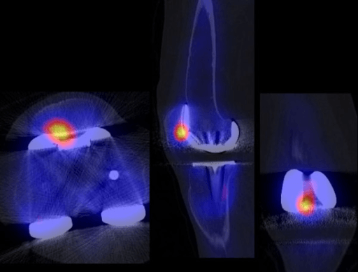
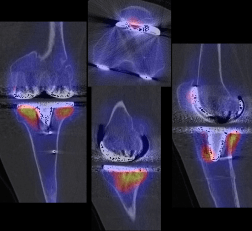
Rotational CT when anterior knee pain or maltracking coexists with a well-fixed, well-aligned prosthesis — measure femoral rotation against the transepicondylar axis and tibial tray rotation against the tibial tubercle. CT with metal artefact reduction to map osteolysis or confirm equivocal loosening. Nuclear imaging is a late resort and is uninterpretable within twelve months of implantation.
- 7Step 7 — If no diagnosis, do not operate
Say so explicitly to the patient, and give the number rather than an opinion. Reassess the pain phenotype, consider central sensitisation and CRPS, involve pain services and physiotherapy, and re-image after an interval.
Across 24,540 first revision knee arthroplasties in the UK National Joint Registry, revision for unexplained pain left a mean Oxford Knee Score of 25 with only 55 PER CENT of patients improving enough to count as responders — among the poorest results of any indication, with only stiffness worse (24 points, 48 per cent). Set that against a re-revision risk that compounds and a serious 90-day complication rate of several per cent. Serial radiographs at six-month intervals may yet reveal progressive loosening that was not apparent — a diagnosis found late is still better than an operation done blind.
MCQ Practice Points
Q: A patient with a painful but structurally sound knee replacement asks to be revised. What do you tell them?
A: Give the registry figure rather than an opinion. Across 24,540 first revision knee arthroplasties in the UK National Joint Registry, revision for unexplained pain left a mean postoperative Oxford Knee Score of 25 — a knee that still hurts — with only 55 per cent of patients improving enough to count as responders. Only revision for stiffness did worse (24 points, 48 per cent). Against a compounding re-revision risk and a serious 90-day complication rate of several per cent, the arithmetic does not favour operating. Caveat worth stating: this is observational registry data in which "unexplained pain" is a surgeon-completed field, so the group includes missed low-grade infection and undiagnosed malrotation — which is an argument for looking harder, not for operating.
Q: How do the 2018 ICM criteria diagnose periprosthetic joint infection?
A: By a weighted score, not a checklist. Major criteria — two positive cultures or a sinus tract — are diagnostic alone. Otherwise: synovial white cell count over 3,000/µL, alpha-defensin (signal-to-cutoff over 1) and leukocyte esterase (++) score 3 each; serum CRP over 1 mg/dL and D-dimer over 860 ng/mL score 2 each; ESR over 30 mm/h scores 1. Six or more is infected. A score of 2 to 5 is INCONCLUSIVE — not aseptic — and defers to intraoperative findings (histology 3, purulence 3, single positive culture 2). Sensitivity 97.7 per cent against MSIS 79.3 per cent at similar specificity. Note the circularity in its derivation: the PJI cohort was defined using MSIS major criteria, so some of that sensitivity gain is built in.
Q: Alpha-defensin is quoted at 95 per cent sensitivity. Is that the test in your theatre?
A: Probably not. The laboratory immunoassay pools at sensitivity 0.95 and specificity 0.97. The lateral flow cassette used at the point of care pools at 0.85 and 0.90 — significantly lower (p = 0.019) — and the authors note its performance is comparable to a leukocyte esterase strip costing a fraction as much. A negative cassette is weak reassurance, not an exclusion. Read the meta-analysis's own warning too: significant conflicts of interest were identified in five of the eleven included studies.
Q: How many patients are dissatisfied after total knee replacement?
A: Fewer than the familiar "one in five" implies. Across 208 studies and 95,560 patients, 83 per cent of studies reported satisfaction above 80 per cent, with the range across studies 80 to 100 per cent; instructional reviews quote "up to 19 per cent" unsatisfied, which is the pessimistic bound. Two caveats that matter more than the number: only 13 per cent of studies used a validated satisfaction instrument, so the whole literature rests largely on unvalidated ordinal scales; and the commonest preoperative predictor of dissatisfaction is anxiety or depression — modifiable before surgery in a way that implant choice is not.
Q: What single historical feature most reliably separates aseptic loosening from infection and instability?
A: The presence of a genuine pain-free interval. Loosening characteristically follows years of good function; infection, instability, malrotation and extrinsic causes typically produce pain that was never resolved by the index operation.
Q: Which radiographic feature makes a periprosthetic lucent line pathological?
A: Progression on serial films, a width greater than 2 mm, circumferential extent around the component, or associated subsidence and change in component position. A thin, stable, non-progressive line is a common incidental finding in asymptomatic knees — which is why the immediate postoperative films must be retrieved for comparison.
Q: Which axis is used to assess femoral component rotation on CT, and which landmark for the tibia?
A: The femoral component is referenced to the surgical transepicondylar axis; the tibial tray is referenced to the medial third of the tibial tubercle using the Berger method. Combined internal rotation correlates with patellofemoral maltracking and anterior knee pain.
Q: Why is a triple-phase bone scan unhelpful in the first year after knee replacement?
A: Periprosthetic bone remodelling produces increased uptake for up to twelve months, and often longer around the tibia, so a positive scan cannot distinguish physiological remodelling from loosening or infection. Its principal value is a NEGATIVE scan in the later unexplained painful knee.
Q: A patient has recurrent effusions and difficulty descending stairs with radiographs showing a raised joint line and patella baja. What is the diagnosis?
A: Flexion or mid-flexion instability arising from an over-resected distal femur, a downsized femoral component and an elevated joint line. Management is revision aimed at restoring the joint line and balancing the flexion and extension gaps, not simply a thicker polyethylene insert.
Exam Viva Scenarios
Practise clinical reasoning and management decisions out loud
“You are shown weight-bearing AP and lateral radiographs of a cemented total knee replacement in a 74-year-old woman. She had eight excellent years and now describes pain on standing from a chair that eases after a dozen steps. There is a 3 mm lucency around the tibial tray, wider than on films taken two years ago.”
“You are shown radiographs of a total knee replacement in a 62-year-old man, two years post-operatively. The components look well positioned and well fixed with no lucency. He has never been comfortable since surgery, with anterior pain on stairs and when rising from a chair, and his patella tilts laterally on the skyline view.”
“You are shown a radiograph of a well-fixed total knee replacement in a 68-year-old diabetic man. Five good years, then two weeks of escalating pain, a warm effusion and difficulty weight-bearing, three weeks after treatment for a dental abscess.”
Opening line in any viva
- Exclude periprosthetic joint infection first, actively, in every case
- Then decide: intrinsic or extrinsic to the knee
- Then define the mechanical phenotype: fixation, alignment, stability, extensor mechanism
- No diagnosis means no revision
History discriminators
- Pain-free interval present then lost — loosening, wear, late infection, fracture
- Never pain-free — infection, instability, malrotation, wrong diagnosis, extrinsic cause
- Start-up pain easing with walking — aseptic loosening
- Giving way on stairs with effusions — flexion instability
- Anterior pain rising from a chair — patellofemoral or malrotation
- Rest and night pain with warmth — infection
Imaging essentials
- Weight-bearing AP, true lateral, skyline at 30 degrees, hip-to-ankle alignment
- Always compare with the immediate post-operative films
- Pathological lucency: progressive, greater than 2 mm, or circumferential
- Rotational CT for anterior pain with well-fixed components
- Bone scan uninterpretable within 12 months of implantation
Infection work-up sequence
- CRP and ESR in every patient
- Aspiration off antibiotics for at least two weeks
- Synovial white cell count with neutrophil differential plus culture with extended incubation
- Alpha-defensin and synovial CRP for equivocal cases
- Multiple separate intra-operative tissue samples at surgery
Extrinsic causes not to miss
- Hip osteoarthritis — pain on hip rotation, benign knee exam, confirm with diagnostic injection
- Lumbar radiculopathy or stenosis — dermatomal pain, positive straight-leg raise
- Vascular claudication — fixed walking distance, absent pulses, abnormal ankle–brachial index
- Infrapatellar saphenous neuroma — focal Tinel sign, abolished by local block
- Complex regional pain syndrome — allodynia and vasomotor change, Budapest criteria
Management triggers
- Acute infection, well-fixed implants, short symptom duration — debridement with modular polyethylene exchange
- Chronic infection, loose implants or sinus — staged revision
- Progressive loosening with infection excluded — revision with stems, augments, cones as AORI defect dictates
- Confirmed malrotation — component revision, not lateral release
- Instability — restore joint line and balance gaps; increase constraint only when collaterals are deficient
- No diagnosis — non-operative management, pain pathway review, re-image after an interval
Evidence Base
Patient-Relevant Outcomes Following First Revision Total Knee Arthroplasty, by Diagnosis: A National Joint Registry Analysis
- 24,540 first revision knee arthroplasties from the UK National Joint Registry, January 2009 to June 2019, linked to Hospital Episode Statistics, national PROMs and death registrations; 54 per cent female, 62 per cent White, mean age 69, with implant survivorship followed to 11 years
- REVISION FOR UNEXPLAINED PAIN GAVE AMONG THE POOREST RESULTS OF ANY INDICATION: mean postoperative Oxford Knee Score 25 with only 55 PER CENT of patients classed as responders. Revision for STIFFNESS was worse still - Oxford Knee Score 24 and 48 per cent responders
- Two-year re-revision ranged from 2.7 per cent (95 per cent CI 1.9 to 3.4) after revision for progressive arthritis to 16.3 PER CENT (15.2 to 17.4) after revision for infection
- Ninety-day mortality was highest after revision for fracture (3.6 per cent) and infection (1.8 per cent), and under 0.5 per cent for every other indication
- Serious medical complications within 90 days: 21.8 per cent after revision for fracture, 12.5 per cent for infection, 4.3 per cent for progressive arthritis
The 2018 Definition of Periprosthetic Hip and Knee Infection: An Evidence-Based and Validated Criteria
- Multi-institutional derivation across 3 academic centres using stringently defined cohorts - 684 PJI cases meeting MSIS major criteria and 820 aseptic one-stage revisions that did not fail within 2 years - then EXTERNALLY VALIDATED on 222 PJI and 200 aseptic patients
- MAJOR criteria, diagnostic on their own: two positive cultures, or a sinus tract
- MINOR criteria are WEIGHTED, not counted. Serum: CRP over 1 mg/dL and D-dimer over 860 ng/mL score 2 each, ESR over 30 mm/h scores 1. Synovial: white cell count over 3,000/microlitre, alpha-defensin (signal-to-cutoff over 1) and leukocyte esterase (++) score 3 each, polymorphonuclear percentage over 80 scores 2, synovial CRP over 6.9 mg/L scores 1
- A preoperative aggregate of 6 or more is INFECTED; 2 to 5 is inconclusive and requires intraoperative findings - positive histology 3, purulence 3, a single positive culture 2 - with a combined total of 6 or more infected, 4 to 5 inconclusive, and 3 or less not infected
- Sensitivity 97.7 per cent against MSIS 79.3 per cent and the earlier International Consensus Meeting definition 86.9 per cent, at a similar specificity of 99.5 per cent
Alpha-Defensin and the Synovasure Lateral Flow Device for the Diagnosis of Prosthetic Joint Infection: A Systematic Review and Meta-Analysis
- 11 eligible studies, quality-assessed with QUADAS (median score 13 of 14) and pooled with a bivariate model
- THE LABORATORY ASSAY AND THE POINT-OF-CARE TEST ARE NOT THE SAME TEST. Laboratory alpha-defensin at a 5.2 mg/L threshold: pooled sensitivity 0.95 (95 per cent CI 0.91 to 0.98), specificity 0.97 (0.95 to 0.98)
- LATERAL FLOW CASSETTE: pooled sensitivity 0.85 (0.74 to 0.92), significantly lower than the laboratory assay (p equals 0.019), with specificity 0.90. THE SPECIFICITY DIFFERENCE WAS NOT SIGNIFICANT (p equals 0.47) - the cassette's weakness is that it MISSES infection, not that it over-calls it
- The authors state the lateral flow device's pooled results are COMPARABLE TO THE LEUKOCYTE ESTERASE STRIP, a test costing a fraction as much
- SIGNIFICANT CONFLICTS OF INTEREST WERE IDENTIFIED IN FIVE OF THE ELEVEN STUDIES; the authors conclude further work is needed before widespread adoption of the cassette
Patient Satisfaction After Total Knee Replacement: A Systematic Review
- 208 studies including 95,560 patients who had undergone total knee replacement, screened from 1,219 records
- EIGHTY-THREE PER CENT OF STUDIES REPORTED SATISFACTION ABOVE 80 PER CENT, with the overall range across studies 80 to 100 per cent
- ONLY 27 STUDIES (13 PER CENT) USED A VALIDATED SATISFACTION INSTRUMENT; satisfaction was most often measured on an unvalidated ordinal scale
- The literature is young as well as weak: 112 of the 208 studies (53.8 per cent) had been published in the preceding three years, so the evidence base was expanding faster than its methods were maturing
- The commonest reported predictor of satisfaction was postoperative patient-reported FUNCTION; relief of pain and function together were the paramount determinants
- The commonest PREOPERATIVE predictor of dissatisfaction was anxiety or depression
Evaluation of the Painful Total Knee Arthroplasty
- Instructional review setting out a structured algorithmic approach to the painful knee replacement
- States that up to 19 PER CENT of patients are unsatisfied with the outcome of total knee arthroplasty
- Emphasises that history and thorough clinical examination identify the aetiology, with investigations SUPPLEMENTING rather than replacing them
- Ancillary testing covers conventional laboratory analysis, imaging and diagnostic injections
- Notes emerging novel laboratory markers, RNA and DNA-based tests and newer imaging as developing tools