Nerve Root Compression | Dermatomal Patterns | Disc Herniation
- L5 root = EHL weakness (big toe dorsiflexion), L4-L5 disc most common
- S1 root = Ankle reflex ABSENT, calf weakness (heel walk), L5-S1 disc
- L4 root = Knee jerk reduced, quad weakness, anterior thigh numbness
- Conservative first: 6-12 weeks for most cases. Most recover by a year, but quote it as Peul's 95% in BOTH arms rather than '90% improve without surgery' - 39% of his conservative arm was operated
- Cauda Equina: Saddle anesthesia + urinary retention = EMERGENCY surgery
- “L5 root has NO reliable reflex - motor testing (EHL) is key
- “Leg pain WORSE than back pain distinguishes radiculopathy from axial LBP
- “SLR positive at 30-70° raises intrathecal pressure, reproduces leg pain
- “Crossed SLR (positive in contralateral leg) = high specificity for disc herniation
Overview and Epidemiology
Lumbar radiculopathy is a clinical syndrome caused by compression or irritation of a lumbar nerve root, producing pain, sensory change or motor weakness in the distribution of that root. "Sciatica" is narrower: radicular pain along the sciatic nerve distribution (L4-S3), typically the buttock and posterior or lateral leg. True radiculopathy means a neurological deficit (weakness, numbness or reflex change) in addition to the pain.
Who. Radiculopathy accounts for 5-10% of patients with low back pain and peaks at 30-50 years, when disc degeneration begins, with a slight male predominance (1.5:1). The L5 root, from the L4-L5 disc, is the most commonly affected and S1, from the L5-S1 disc, the second. Bilateral involvement is rare and should make you consider cauda equina or a central disc.
Natural history. The majority of disc herniations causing radiculopathy improve without surgery, and that is the basis for a conservative trial in most cases. But the question the patient is actually asking is not whether they will recover but when, and that is what the trials answer.
- Weber (PMID 6857385), a controlled prospective study of 126 randomised patients with ten years of observation: surgery gave a statistically significantly better result at one year; by four years the operated patients were still better but the difference was no longer statistically significant, and only minor changes occurred over the remaining six years
- SPORT reached a similar place, with between-group differences favouring surgery that were small and not statistically significant on intent-to-treat, in a trial with very high crossover in both directions; both groups improved substantially over two years
- Peul: 95% perceived recovery at one year in both arms, with 39% of the conservative arm operated at a mean of 18.7 weeks
The honest framing. Discectomy mostly buys earlier relief rather than a better destination, which turns the consent conversation into how much a year of pain is worth to this particular patient, not whether surgery "works". For the claim that the two paths reach a similar destination, cite Peul or Weber, not SPORT, whose authors state that bidirectional crossover leaves conclusions about superiority or equivalence unwarranted. The widely quoted "90% improve" is a round number with no single study behind it: a reasonable summary of the direction of travel, not a figure to defend under questioning. It says nothing about how long recovery takes, and "90% recover without surgery" ignores the 39% of Peul's conservative arm who were operated; the honest statement is that most patients recover within a year whichever path they take.
Anatomy and Biomechanics
Root numbering. In the lumbar spine each nerve root exits below the correspondingly numbered pedicle: the L4 root leaves between L4 and L5. At any disc there are therefore two roots to think about, the one exiting at that level and the one traversing the disc on its way to exit one level lower.
- Exiting Root
- L3
- Traversing Root
- L4
- Exiting Root
- L4
- Traversing Root
- L5
- Exiting Root
- L5
- Traversing Root
- S1
Which root the herniation catches. Most herniations are posterolateral and compress the traversing root, the one that continues down to exit below the next level. A far-lateral or foraminal herniation catches the exiting root instead, and a large central herniation threatens the cauda equina. The same disc therefore produces a different radiculopathy depending on where the fragment sits, and the surgical corridor changes with it.
- Location
- Lateral recess
- Root Affected
- Traversing root
- Example
- L4-L5 herniation → L5 root
- Clinical Features
- Classic radiculopathy, SLR positive
- Location
- Neural foramen
- Root Affected
- Exiting root
- Example
- L4-L5 herniation → L4 root
- Clinical Features
- One level up, may be missed on MRI
- Location
- Far lateral
- Root Affected
- Exiting root
- Example
- L4-L5 herniation → L4 root
- Clinical Features
- Femoral stretch test may be positive
- Location
- Central canal
- Root Affected
- Multiple roots (cauda equina)
- Example
- Large central L4-L5 → multiple roots
- Clinical Features
- Bilateral symptoms, cauda equina risk
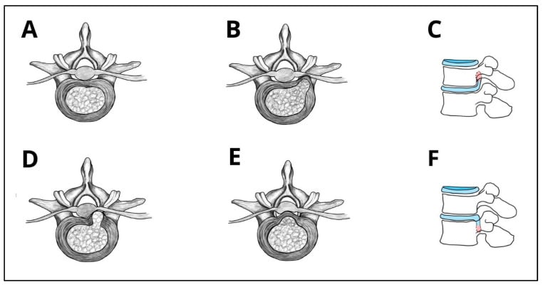
Pathophysiology
Mechanical compression. The most common cause is disc herniation: the nucleus pulposus protrudes through a tear in the annulus fibrosus and the disc material compresses the root directly against the bony canal. The terminology describes the shape of what has escaped, and morphology is a separate descriptor from location:
- Description
- Symmetric, circumferential extension
- Description
- Focal, base wider than apex
- Description
- Apex wider than base, through annulus
- Description
- Free fragment, separated from disc
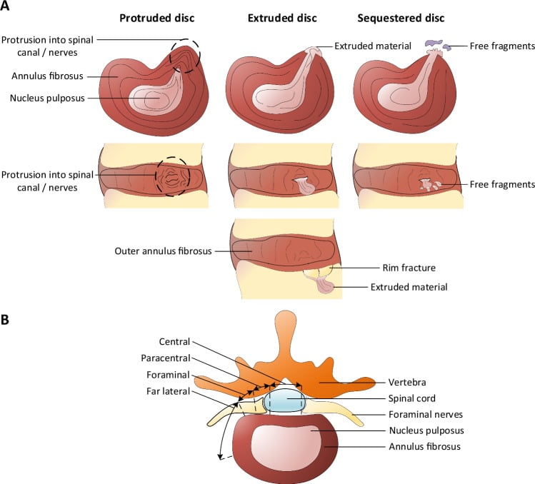
Chemical inflammation. The nucleus pulposus contains inflammatory mediators (phospholipase A2, TNF-alpha) that inflame and sensitise the root, which is why some small herniations cause severe symptoms while large herniations may be asymptomatic. Extrusion into the epidural space also exposes nucleus pulposus to vascular and immune mechanisms, so large extruded fragments can resorb; fragment size alone therefore does not mandate surgery without red flags or progressive deficit.
Other causes. Disc herniation is the usual culprit, but each of the other causes has a pattern of its own.
The most common cause (90%). The nucleus pulposus herniates through an annular tear and compresses the root mechanically and chemically. Risk factors are repetitive flexion-rotation loading, heavy lifting, vibration exposure (truck drivers), smoking (disc nutrition) and genetic factors. It peaks at 30-50 years, when the disc is still hydrated enough to herniate; older patients are more likely to have stenosis.
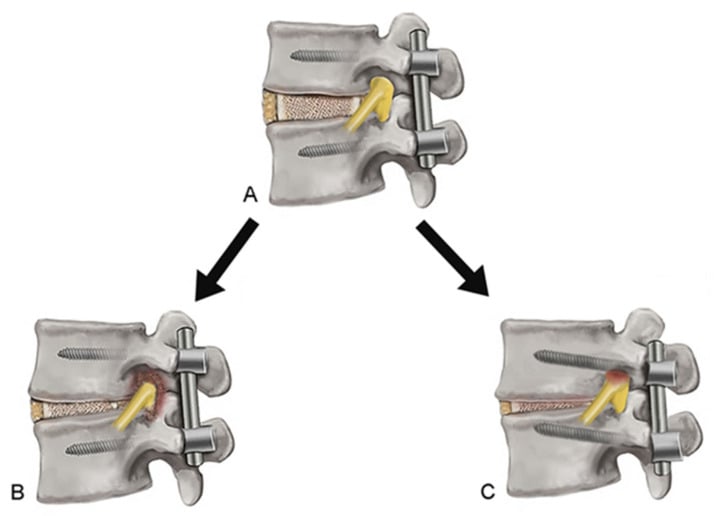
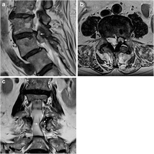
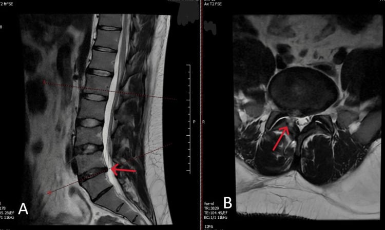
Not every radiculopathy is discogenic. Any mass at the root, a facet synovial cyst, a tumour or a haematoma, can mimic a disc, and the management differs: a synovial cyst is excised, sometimes with fusion of the unstable segment, not treated by discectomy. Synovial cysts arise from arthritic facet joints and are common in the elderly, and like a disc fragment the cyst compresses the root in the lateral recess. The clinical diagnosis rests on symptoms and examination; imaging reveals the cause of the compression.
Radicular versus referred pain. Radicular pain follows a dermatomal distribution and is sharp, shooting or electric. Referred pain is duller and more diffuse and does not follow a root pattern.
Sensitisation. Chronic compression lowers the root's threshold for pain signalling, which is why minor movements can trigger severe radicular symptoms.
Classification
By root level. Each root has a motor, sensory and reflex signature, and the disc level follows from the root. The table is the summary; the paragraphs beneath it are how to elicit each sign.
- Disc Level
- L2-L3
- Motor Weakness
- Hip flexion, knee extension
- Sensory Distribution
- Anterior thigh
- Reflex
- Knee jerk (L3-L4 arc)
- Disc Level
- L3-L4
- Motor Weakness
- Tibialis anterior, Quadriceps
- Sensory Distribution
- Medial leg, medial foot
- Reflex
- Knee jerk (reduced)
- Disc Level
- L4-L5
- Motor Weakness
- EHL, Hip abduction, Tibialis posterior
- Sensory Distribution
- Lateral leg, dorsum foot, 1st web space
- Reflex
- None reliable
- Disc Level
- L5-S1
- Motor Weakness
- Gastrocnemius, Peroneals, Gluteus maximus
- Sensory Distribution
- Lateral foot, sole, posterior calf
- Reflex
- Ankle jerk (ABSENT)
L4 (L3-L4 disc). Weakness of knee extension (quadriceps) shows as difficulty rising from a chair or climbing stairs, with tibialis anterior affected too. Sensory loss covers the anterior thigh and the medial leg to the medial malleolus and medial foot, and the knee jerk (the L3-L4 reflex arc) is reduced or absent. It is less common than L5 or S1; consider a femoral nerve palsy in the differential, and the femoral stretch test (reverse SLR) may be positive.
L5 (L4-L5 disc, the most common). EHL weakness, the inability to dorsiflex the big toe, is the most specific finding: have the patient lift the big toe against resistance while you stabilise the foot. Tibialis posterior (inversion) and the hip abductors are also affected. Sensory loss covers the lateral leg, the dorsum of the foot, the great toe and the first web space, which is pathognomonic. There is no reliable reflex for L5, which is why motor testing is essential.
S1 (L5-S1 disc). Gastrocnemius weakness means the patient cannot heel raise and struggles with push-off in walking; the peroneals and gluteus maximus share the root. A single heel raise is more sensitive than testing calf power directly, and the patient should manage 10 single-leg heel raises. Sensory loss covers the posterior calf, lateral foot, sole and small toe, and the ankle jerk is absent, which is highly specific for S1.
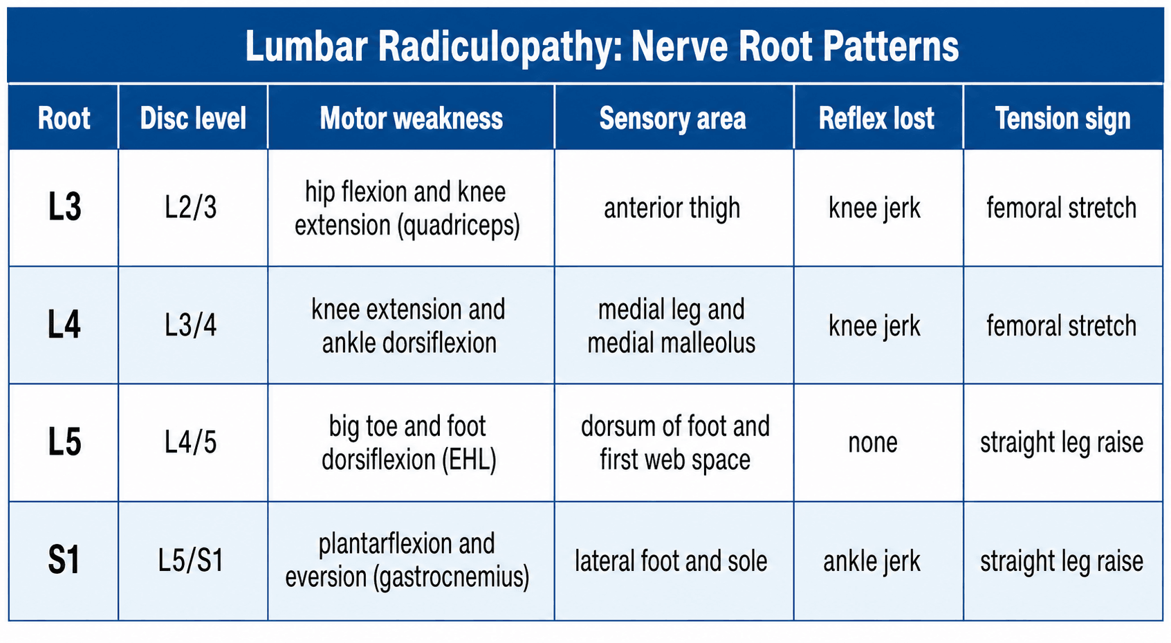
L5L5 vs S1 Radiculopathy
Hook:L5 Lifts toe up, S1 Stands on tiptoe down
Severity. Motor weakness is graded on the MRC scale, and the grade carries a management implication:
- Description
- Normal power
- Clinical Implication
- No motor deficit
- Description
- Movement against resistance
- Clinical Implication
- Mild weakness - conservative
- Description
- Movement against gravity only
- Clinical Implication
- Moderate weakness - consider surgery
- Description
- Movement with gravity eliminated
- Clinical Implication
- Severe weakness - surgery recommended
- Description
- Flicker of contraction
- Clinical Implication
- Near complete deficit - urgent surgery
- Description
- No contraction
- Clinical Implication
- Complete deficit - emergency surgery
Clinical Presentation
The pain. Leg pain worse than back pain is the key feature. It is sharp, shooting or electric, follows the dermatome, and is aggravated by sitting, coughing, straining and forward flexion and relieved by standing, lying and walking, unlike stenosis.
Duration and progression. The time course points to the cause:
- Acute onset suggests disc herniation
- Gradual onset may indicate stenosis
- Progressive weakness is concerning
- Bilateral symptoms suggest central pathology
Red flags. Cauda equina syndrome is the diagnosis not to miss.
Any combination of: saddle anaesthesia, urinary retention or incontinence, faecal incontinence, bilateral leg weakness, sexual dysfunction. Requires URGENT MRI and surgical decompression within 24-48 hours.
Cauda equina syndrome is staged by bladder status, which drives both urgency and prognosis. CESS (suspected): bilateral radicular symptoms with some saddle or sphincter symptoms but no objective sphincter compromise. CES-I (incomplete): altered urinary sensation, poor stream or straining to void, but voluntary micturition control is retained. CES-R (retention): painless urinary retention with overflow incontinence and an insensate bladder. CES-I carries a substantially better functional prognosis than CES-R, so the aim is to recognise and decompress at the incomplete stage — established painless retention is a poor-prognosis marker, never a threshold to wait for.
A compressive lesion at the thoracolumbar junction (around L1-L2, where the spinal cord terminates) produces a conus medullaris syndrome, distinct from a true cauda equina (below L2) lesion. Conus: tends to be sudden and symmetrical, with early and prominent bladder, bowel and saddle dysfunction, relatively mild symmetric leg weakness, and mixed upper and lower motor neuron signs (preserved or brisk ankle jerks, up-going plantars). Cauda equina: often more gradual and asymmetrical, with prominent radicular leg pain, asymmetric lower-limb weakness, pure lower motor neuron signs (areflexia), and bladder dysfunction appearing comparatively later. Both are decompression emergencies, but recognising the level and pattern guides imaging and counselling.
Examination. Watch the patient walk in: an antalgic gait, a list away from the painful side (sciatic scoliosis) and limited lumbar flexion. Palpate for paravertebral muscle spasm and sciatic notch tenderness. Then test each root's motor, reflex and sensory signature as set out in the classification section, and finish with the tension tests.
- Technique
- Raise straight leg, hip flexed, knee extended
- Positive Finding
- Pain 30-70° in radicular distribution
- Sensitivity/Specificity
- 90% sensitive, 26% specific
- Technique
- SLR of the unaffected leg reproduces pain in the affected leg
- Positive Finding
- Pain in contralateral leg; indicates a large disc tenting the dura
- Sensitivity/Specificity
- Low sensitivity, HIGH specificity (90%)
- Technique
- Prone, extend hip with knee flexed
- Positive Finding
- Pain in anterior thigh
- Sensitivity/Specificity
- Positive for L4, L3 radiculopathy
- Technique
- Seated, chin to chest, extend knee
- Positive Finding
- Reproduces radicular pain
- Sensitivity/Specificity
- Tension test for dura
Investigations
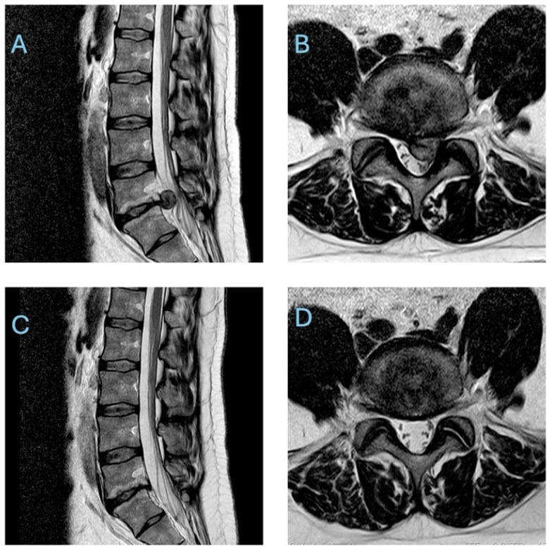
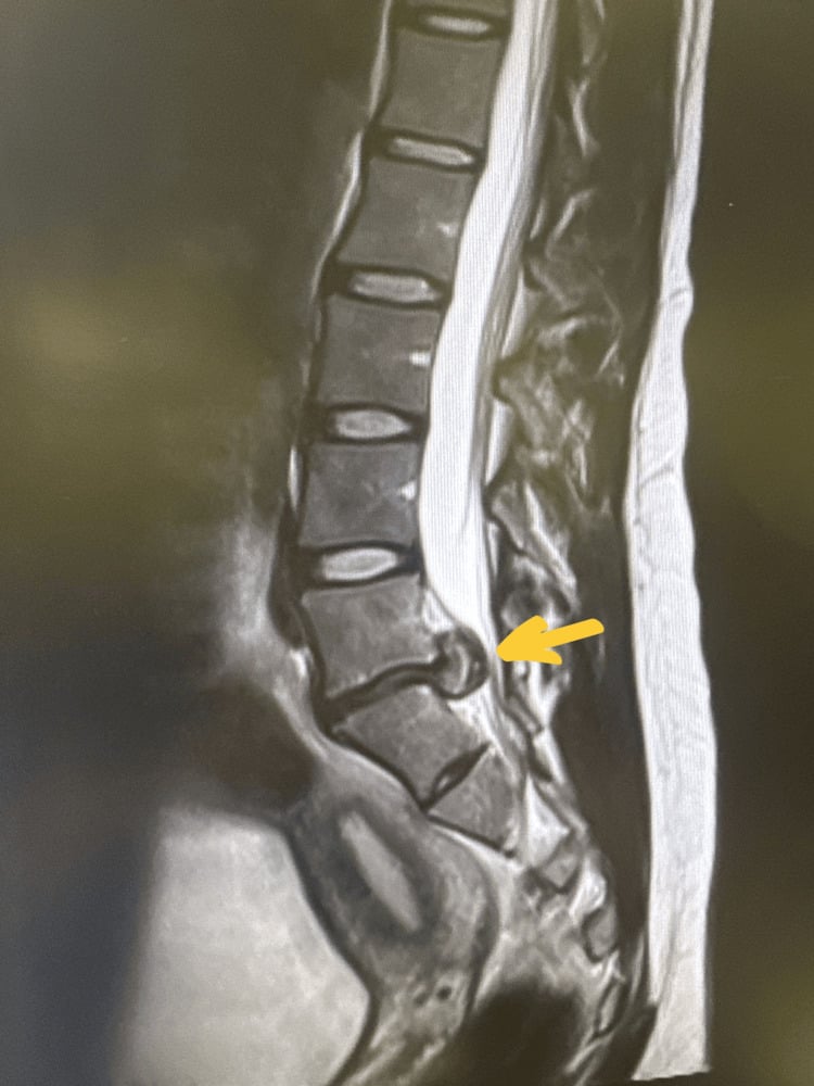
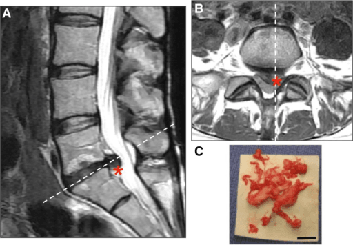
MRI is the gold standard. Each sequence answers a different question, and the sagittal and axial images are read together: the sagittal scan gives the level, the axial scan gives the laterality, and the two predict the symptomatic root before any operation.
- What It Shows
- Disc degeneration (dark disc), herniations, spinal alignment
- What It Shows
- Nerve root compression, lateral recess stenosis
- What It Shows
- Anatomy, fat in foramen (should be bright), bone marrow
- What It Shows
- Bone marrow oedema, infection, tumour
When to image. The standard recommendation is to wait 6 weeks before MRI if there are no red flags, because many herniations resolve spontaneously. Image early for:
- Cauda equina syndrome (urgent)
- Progressive neurological deficit
- Suspected tumour or infection
- Severe, unremitting pain
- Prior malignancy
CT myelography is the alternative when MRI is contraindicated (pacemaker, severe claustrophobia). It shows contrast around the nerve roots but gives less soft-tissue detail.
Electrodiagnostics. EMG and nerve conduction studies earn their place when the clinical picture is unclear, to differentiate radiculopathy from peripheral neuropathy, for medicolegal documentation, or for pre-operative confirmation. Denervation changes (positive waves, fibrillations) appear at 3 or more weeks, reduced recruitment signals chronic nerve injury, and a normal study does not rule radiculopathy out because it may simply be too early.
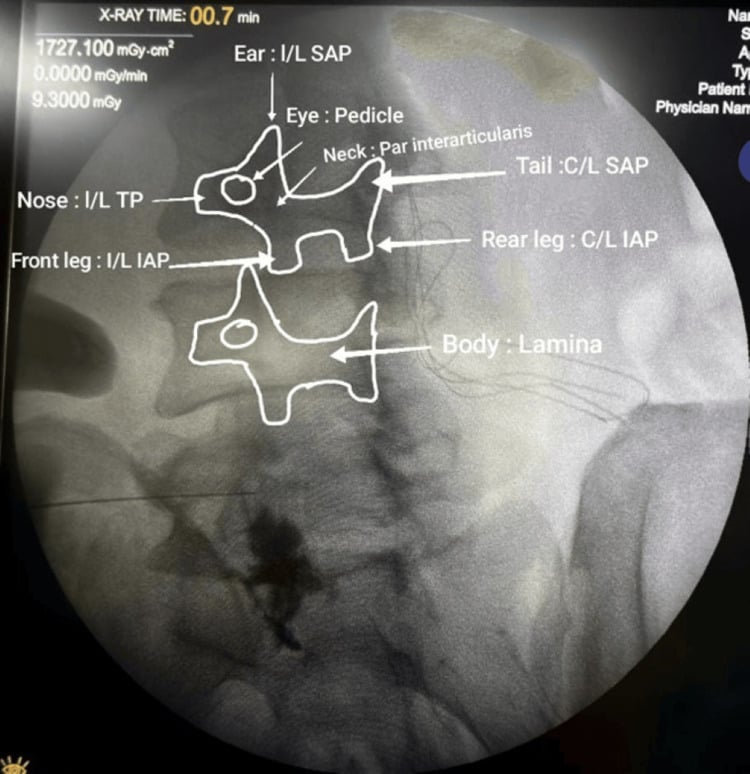
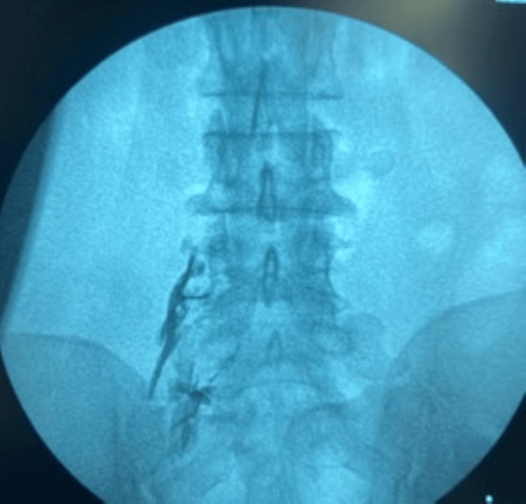
Diagnostic injections. A selective nerve root block can be diagnostic when it is unclear which root is symptomatic: relief with the block confirms that root as the pain generator, and it is used diagnostically only when short-lived relief reproduces the clinico-radiological level. Epidural steroid injection is more therapeutic than diagnostic; it may provide short-term relief and help avoid surgery.
Differential Diagnosis
Leg pain is not always radicular. The examiner wants to see you actively exclude mimics before committing to a disc-based diagnosis. The single most useful discriminator is whether the pain is genuinely dermatomal and reproduced by neural tension, versus regional, mechanical or vascular.
- Pain Pattern
- Dermatomal, leg worse than back
- Key Discriminator
- Positive SLR / femoral stretch, motor-sensory in one root
- Confirmatory Test
- MRI concordant with level
- Pain Pattern
- Bilateral buttock/leg, worse standing/walking
- Key Discriminator
- Relieved by flexion (shopping-cart sign), pulses normal
- Confirmatory Test
- MRI central canal stenosis
- Pain Pattern
- Calf cramp with exertion, fixed claudication distance
- Key Discriminator
- Relieved by standing still, absent pulses, no postural change
- Confirmatory Test
- ABPI reduced, arterial duplex
- Pain Pattern
- Groin/anterior thigh, rarely below knee
- Key Discriminator
- Pain on hip rotation (FABER/FADIR), no neuro deficit
- Confirmatory Test
- Hip radiograph, intra-articular block
- Pain Pattern
- Lateral hip/thigh
- Key Discriminator
- Point tenderness over trochanter, normal neurology
- Confirmatory Test
- Resisted abduction, ultrasound
- Pain Pattern
- Glove-and-stocking or single peripheral nerve
- Key Discriminator
- Does not follow a single root; NCS abnormal distally
- Confirmatory Test
- EMG/NCS
- Pain Pattern
- Buttock to posterior thigh
- Key Discriminator
- Tenderness deep to gluteus, pain on resisted external rotation
- Confirmatory Test
- Diagnosis of exclusion, MRI normal canal
- Pain Pattern
- Night pain, constant, non-mechanical
- Key Discriminator
- Red flags: weight loss, fever, malignancy history, age extremes
- Confirmatory Test
- MRI with contrast, inflammatory markers
A truly positive straight leg raise (reproducing radicular leg pain at 30-70 degrees, worsened by ankle dorsiflexion) shifts you strongly toward a nerve-root cause. Pain only in the back, or only at extremes of range, is not a positive SLR and should make you reconsider a mechanical or hip source.
Management
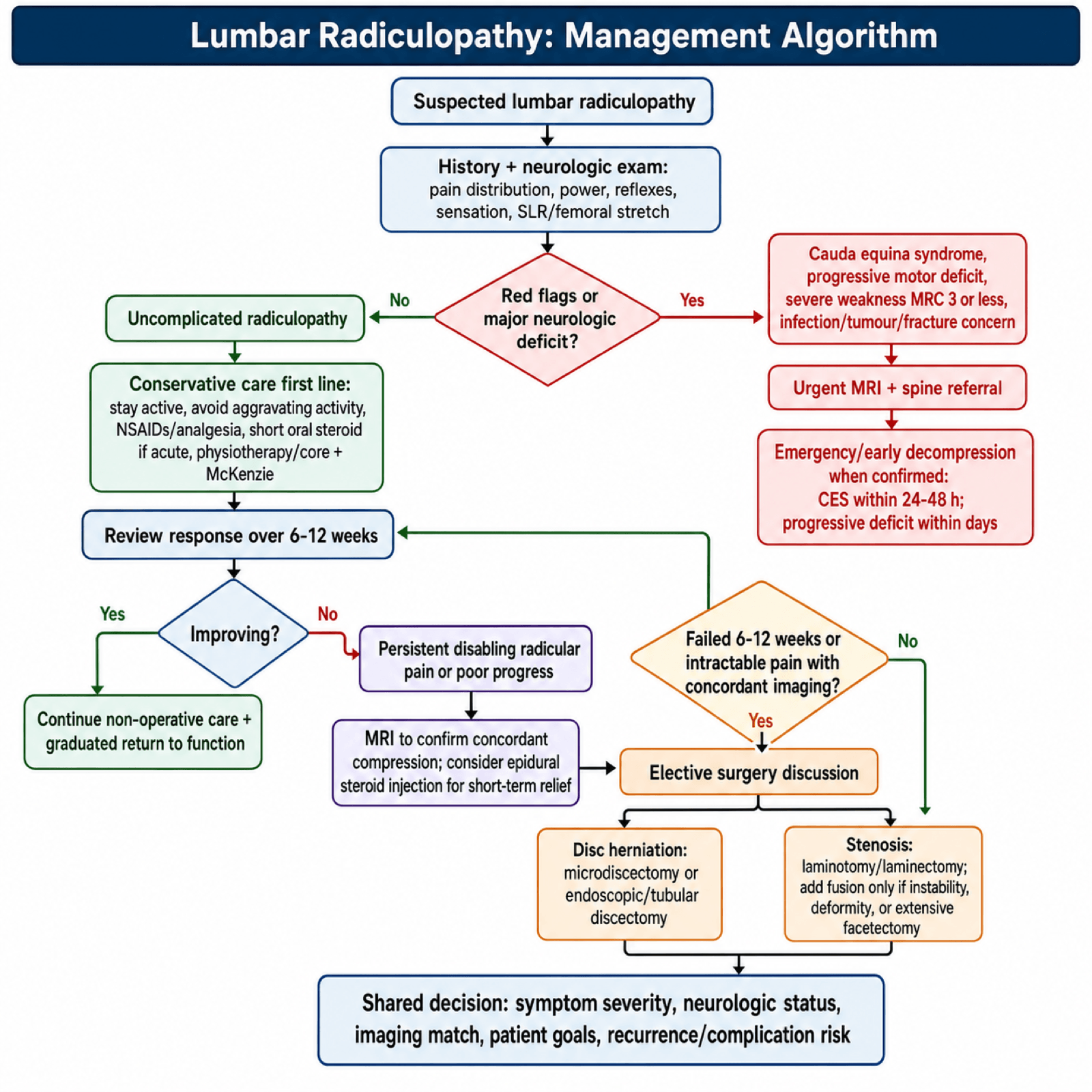
Conservative care first. Surgery is not mandatory for uncomplicated disc herniation, and conservative care remains a reasonable first-line choice: most patients without red flags start with a 6-12 week conservative trial before surgery is considered. The evidence behind each component varies:
- Evidence
- Strong
- Details
- Avoid aggravating activities, NOT bed rest
- Evidence
- Strong
- Details
- First-line pharmacotherapy
- Evidence
- Moderate
- Details
- Short course (6 days) may help acute phase
- Evidence
- Strong
- Details
- Core strengthening, McKenzie extension
- Evidence
- Moderate
- Details
- Roughly half of operative candidates avoided surgery, durable to 5 years (Riew) - note this was a nerve-root block, not an epidural, and the steroid component made no difference at 5 years
When to operate. Cauda equina syndrome, a progressive neurological deficit and intractable pain requiring intravenous analgesia are the exceptions that override the conservative trial; otherwise surgery is elective once the trial has failed.
- Timing
- URGENT (24-48 hours)
- Timing
- Early (within days)
- Timing
- Early consideration
- Timing
- Elective
- Timing
- Elective; earlier if it requires IV analgesia
Microdiscectomy is the gold standard for disc herniation: a small incision, limited laminotomy and flavectomy, removal of the herniated fragment, and preservation of as much normal disc as possible. It relieves leg pain in 85-95% and gives faster recovery than conservative care; the long-term convergence with conservative care is set out under natural history, and its complications are tabulated in the complications section.
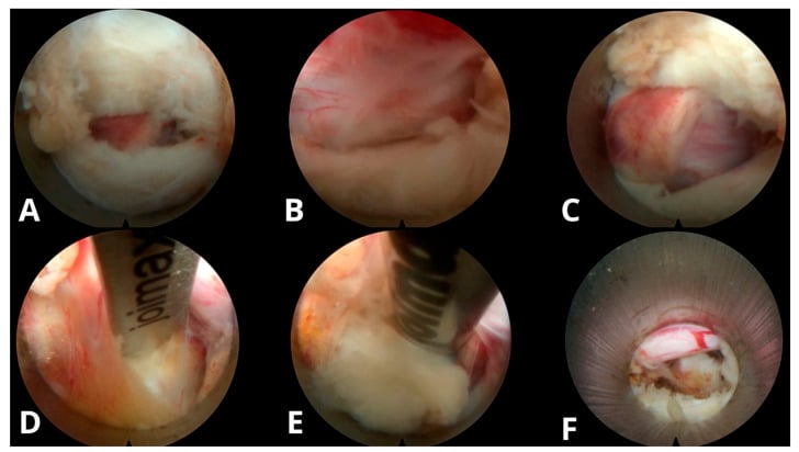
Minimally invasive discectomy. A tubular retractor or endoscope through a smaller incision, with less muscle damage. The advantages are less tissue trauma, faster recovery, less blood loss and outpatient surgery in some cases; the costs are a steeper learning curve, limited visualisation and, in some series, possibly a higher recurrence rate. Outcomes are comparable to open microdiscectomy for appropriately selected patients.
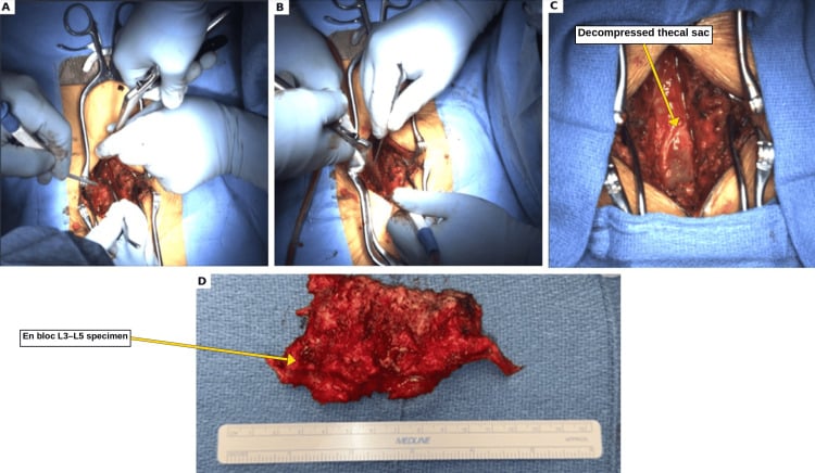
Decompression for stenosis. Laminectomy or laminotomy is the operation for radiculopathy from spinal stenosis rather than disc herniation: the lamina, ligamentum flavum and medial facet are removed to free the roots, preserving enough facet to avoid iatrogenic instability unless fusion is planned. Add fusion where there is instability: spondylolisthesis, extensive facetectomy or deformity. Claudication symptoms improve in 70-80%.
After surgery. Routine early MRI is unnecessary after an uncomplicated recovery. Obtain it for persistent or recurrent deficit, severe pain, or suspected haematoma, infection, wrong-level or incomplete decompression.
Management Algorithm
Complications
Of conservative management. Recovery that is delayed rather than managed brings prolonged disability, a chronic pain syndrome, muscle atrophy from disuse and psychological consequences (depression, anxiety). The graver failure is the missed cauda equina: a progressive neurological deficit and permanent bladder and bowel dysfunction.
Intraoperative complications, and how each is prevented or managed:
- Incidence
- 1-7%
- Prevention/Management
- Careful technique, primary repair, fibrin glue
- Incidence
- 0.1-0.3%
- Prevention/Management
- Intraoperative imaging, confirm with X-ray
- Incidence
- 0.1-0.5%
- Prevention/Management
- Adequate visualisation, gentle retraction
- Incidence
- Very rare
- Prevention/Management
- Avoid plunging instruments anteriorly
Postoperative.
- Incidence
- 5-10%
- Management
- May require revision surgery
- Incidence
- 1-2%
- Management
- Antibiotics, possible washout
- Incidence
- Rare
- Management
- Urgent decompression if symptomatic
- Incidence
- 10-40%
- Management
- Multidisciplinary management
Dural tear. An incidental durotomy may present later as a postoperative pseudomeningocele or a persistent CSF leak. Recognise the postural headache, wound drainage or recurrent neural symptoms, and manage according to tear size, watertight closure, symptoms and persistence rather than imaging alone.
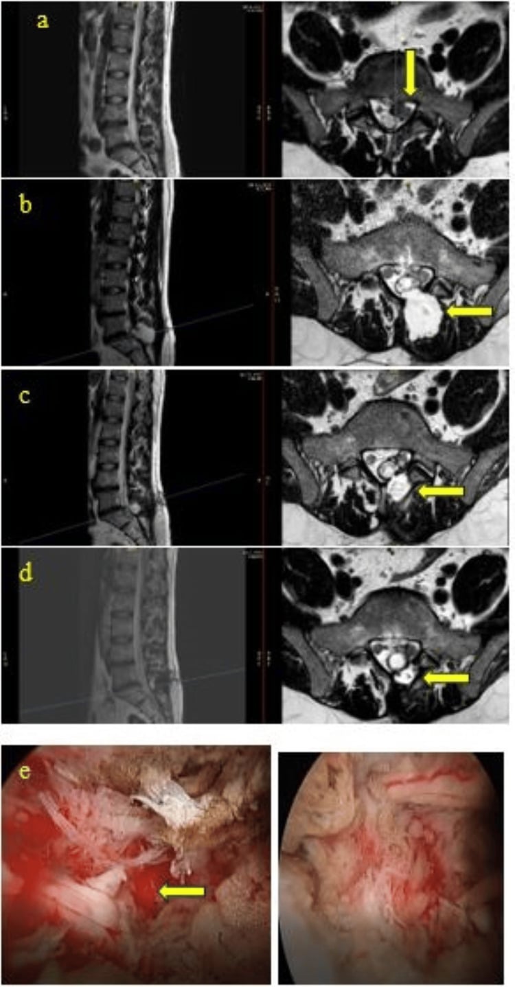
Recurrent herniation at the same level is likelier with a large annular defect, younger age, male sex, smoking and heavy lifting. Treatment is revision discectomy, or fusion if recurrent.
Failed back surgery syndrome is persistent pain after lumbar spine surgery, from a wrong diagnosis, incomplete decompression, new pathology or scar tissue. Management is spinal cord stimulation and a multidisciplinary pain programme.
Adjacent segment disease is rare after discectomy alone and more common after fusion.
Guidelines, Registries & Global Practice
Global Epidemiology
- Lifetime prevalence of sciatica/radicular leg pain is commonly cited at roughly 10-40%, with annual incidence around 1-5% in adult populations.
- Radiculopathy accounts for roughly 5-10% of patients presenting with low back pain.
- Peak incidence is in the fourth and fifth decades, when the disc is still hydrated enough to herniate; in older adults degenerative foraminal and central stenosis becomes a relatively more common cause.
- L4-L5 and L5-S1 together account for the large majority of herniations, making L5 and S1 the most frequently affected roots worldwide.
Side-by-Side Guideline Comparison
- Imaging Stance
- Only if it will change management; not routine in primary care
- Injections
- Consider epidural LA + steroid for acute severe sciatica
- Surgery Threshold
- Decompression when conservative care fails AND imaging is concordant
- Imaging Stance
- MRI is the imaging of choice for suspected herniation
- Injections
- Transforaminal ESI an option for short-term relief
- Surgery Threshold
- Discectomy for persistent concordant radiculopathy failing 6+ weeks conservative care
- Imaging Stance
- MRI to confirm level and laterality before surgery
- Injections
- Reasonable adjunct/diagnostic tool
- Surgery Threshold
- Urgent surgery for CES or progressive deficit; otherwise elective
- Imaging Stance
- Avoid early imaging without red flags
- Injections
- Modest, mainly short-term benefit
- Surgery Threshold
- Red flags (CES, progressive motor loss) override the conservative trial
The areas of genuine agreement are larger than the disagreements: avoid early imaging without red flags, offer a conservative trial of around 6-12 weeks for uncomplicated cases, and decompress urgently for cauda equina or progressive deficit. Differences are mostly of emphasis - the relative weight placed on injections and the exact conservative-trial duration.
Registry and Outcome Notes
- Spine surgery registries (for example the Norwegian NORspine registry and several national spine outcome databases) consistently report substantial improvement in leg pain and disability after microdiscectomy, with reoperation/recurrence rates in the order of 5-15% over medium-term follow-up.
- Registry data reinforce the RCT message: minimally invasive and open microdiscectomy yield comparable patient-reported outcomes, so technique choice is driven by expertise rather than a proven outcome advantage.
High- vs Limited-Resource Practice Variation
- Typical Pathway
- Early MRI when red flags present, ready access to ESI and elective microdiscectomy, emergency MRI for suspected CES
- Rationale / Constraint
- Imaging and theatre access readily available
- Typical Pathway
- Greater reliance on clinical diagnosis and conservative care; selective use of MRI; CT myelography where MRI is scarce
- Rationale / Constraint
- MRI access and cost limit early imaging; clinical examination carries more diagnostic weight
A clinically confident diagnosis of single-level radiculopathy can be made and managed conservatively on history and examination alone; advanced imaging is mandatory before surgery and for any suspicion of cauda equina, infection or tumour.
Controversies and Areas of Uncertainty
Early surgery clearly speeds recovery (Peul, Leiden trial), but 1-year outcomes match watchful waiting. Whether earlier surgery prevents long-term residual deficit in those with motor weakness remains debated.
Benefit is real but mainly short-term and modest. Their role as a surgery-sparing tool (Riew) versus a temporising measure of limited durability is still contested across guidelines.
The 48-hour signal from meta-analysis (Ahn) should not be read as permission to wait. Most surgeons decompress as soon as feasible; incomplete CES (CES-I) carries a better prognosis than retention (CES-R), and the data are retrospective.
Long-term RCT data show no clinically meaningful difference. Endoscopic and tubular techniques offer perioperative advantages in selected hands but no proven superiority in patient-reported outcomes.
Additional unsettled questions include: the value of routine EMG/NCS (rarely changes management in clinically clear cases), how aggressively to operate on isolated painless motor weakness, and whether sequestrated fragments resorb often enough to justify prolonged conservative care even with large herniations.
Viva Scenarios
Practise clinical reasoning and management decisions out loud
“A 38-year-old male presents with 6 weeks of right leg pain radiating to the dorsum of the foot. He has weakness lifting his big toe. MRI shows L4-L5 right posterolateral disc herniation.”
“The same patient now presents to emergency with bilateral leg weakness, difficulty voiding, and numbness around the perineum.”
“A 45-year-old female presents with 8 weeks of left posterior calf pain and numbness on the sole of her foot. Ankle jerk is absent on the left. SLR is positive at 45 degrees.”
“A 52-year-old male has 4 weeks of anterior thigh pain and weakness climbing stairs. SLR is negative, but femoral stretch test is positive. MRI shows L3-L4 far lateral disc herniation.”
ROOT PATTERNS
- L4: Knee extension weak, reduced knee jerk, medial leg
- L5: EHL weakness, NO reflex, dorsum foot/1st web space
- S1: Calf weakness, ABSENT ankle jerk, lateral foot/sole
KEY CLINICAL FEATURES
- Leg pain WORSE than back pain
- Dermatomal distribution of symptoms
- SLR positive at 30-70 degrees
- Crossed SLR = high specificity for disc
DISC MECHANICS
- Posterolateral herniation affects TRAVERSING root
- Far lateral herniation affects EXITING root (one level up)
- L4-L5 PL = L5 root; L4-L5 FL = L4 root
- Central herniation = risk of cauda equina
CAUDA EQUINA EMERGENCY
- Saddle anesthesia + urinary retention
- Bilateral leg symptoms
- MRI + surgery within 24-48 hours
- Delayed surgery = permanent deficits
MANAGEMENT PEARLS
- Conservative first: 6-12 weeks. Most recover by a year - Peul's 95% perceived recovery, but in BOTH arms, with 39% of his conservative arm operated. Do not quote '90% improve without surgery'
- Microdiscectomy: 85-95% leg pain relief
- Surgery faster; similar long-term (cite Peul or Weber - NOT SPORT, which disclaims both superiority and equivalence)
- Nerve-root block: about half of operative candidates avoided surgery (29 of 55, Riew), and the deferral held at 5 years in 17 of 21 followed - but bupivacaine ALONE did as well as bupivacaine plus steroid, so the corticosteroid is not the active ingredient
Evidence Base
SPORT - Surgery vs Nonoperative Care for Lumbar Disc Herniation (RCT)
- Multicentre RCT, 501 surgical candidates with imaging-confirmed disc herniation and radiculopathy of at least 6 weeks (mean age 42, 42% women)
- Intent-to-treat differences favoured surgery for all outcomes but were small and not statistically significant for the primary measures
- Crossover was high: 50% assigned to surgery had it by 3 months, while 30% assigned to nonoperative care also had surgery
- Both groups improved substantially over 2 years; large crossover precluded firm conclusions of superiority or equivalence by ITT
Surgery vs Prolonged Conservative Treatment for Sciatica (Leiden Trial)
- RCT of 283 patients with severe sciatica for 6-12 weeks: early surgery vs prolonged conservative care with surgery if needed
- 39% of the conservative arm eventually required surgery (mean 18.7 weeks)
- Early surgery gave faster leg-pain relief and faster perceived recovery (HR 1.97, 95% CI 1.72-2.22)
- At 1 year the probability of perceived recovery was 95% in BOTH groups
Lumbar Disc Herniation - Controlled Prospective Study, 10-Year Observation
- Landmark trial - but note the design: of 280 patients with radiculographically verified herniation, only the 126 with an UNCERTAIN surgical indication were randomised. A further 67 with an indisputable indication were operated and 87 with no indication were treated conservatively, both without randomisation
- Statistically significant advantage for surgery at 1 year
- By 4 years the operated group was still better but the difference was no longer significant; minimal change thereafter to 10 years
- The randomised question is therefore specifically 'what if you are unsure?' - which is the useful clinical question, but it means the result does not transfer to patients with a clear indication either way