Gold Standard for Lumbar Disc Herniation | 90-95% Success | Minimal Morbidity
- Concordant radiculopathy required - clinical picture must match imaging level
- L4-5 herniations compress L5 nerve (traversing root), L5-S1 compress S1
- Failed 6-12 weeks conservative treatment is standard indication
- Cauda equina syndrome is emergency requiring surgery within 24-48 hours
- Recurrence 5-10% at same level, 5% at different level
- “Posterolateral herniation = traversing root (L4-5 = L5 nerve)
- “Far lateral/foraminal = exiting root (L4-5 = L4 nerve)
- “CES: urinary retention, saddle anesthesia, bilateral leg symptoms
- “SPORT trial: Surgery faster recovery but similar 4-year outcomes
Overview and Epidemiology
Lumbar microdiscectomy is the most commonly performed spinal operation worldwide: the herniated disc material compressing a nerve root is removed through a minimally invasive approach. It remains the gold standard surgical treatment for a symptomatic lumbar disc herniation that has not responded to conservative management.
History. Mixter and Barr described open discectomy in 1934. Caspar and Yasargil brought the operating microscope to spine surgery in the 1970s, and the modern operation achieves decompression through a 2-3 cm incision with minimal tissue disruption.
Who. Lumbar disc herniation affects 1-3% of the population, with a peak incidence between 30 and 50 years. Approximately 10% of symptomatic patients ultimately require surgery.
Where. L4-5 and L5-S1 together account for 95% of herniations:
- L4-5 - 40-50%, the most common level overall
- L5-S1 - 40-50%, the second most common
- L3-4 - 5%
The natural history of lumbar disc herniation is generally favourable: 90% of patients improve with conservative treatment alone. Surgery accelerates recovery but does not change the long-term outcome for most patients (SPORT trial).
Anatomy and Pathophysiology
The canal at disc level. The key structures are the dural sac containing the cauda equina, the epidural fat and veins, and the ligamentum flavum posteriorly. Two roots are at risk at every level: the traversing root, descending to exit at the foramen one level below, and the exiting root, leaving through the foramen at this level.
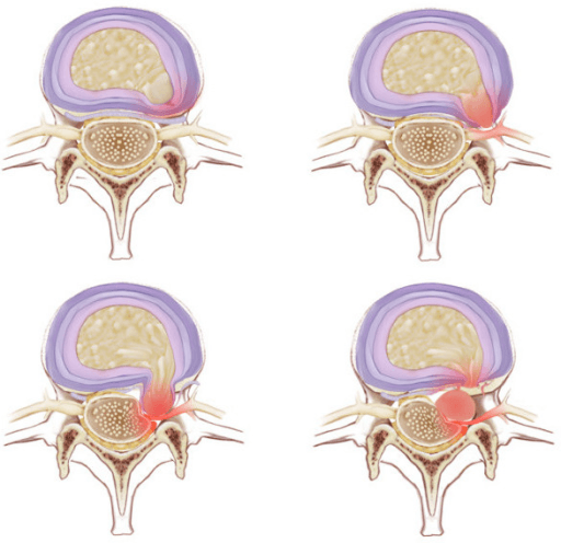
Which root a herniation compresses. A posterolateral herniation, the most common, lies in the path of the traversing root; a foraminal or far lateral (extraforaminal) herniation lies in the path of the exiting root; a central herniation may cause bilateral symptoms or cauda equina syndrome. So at L4-5 a posterolateral fragment compresses L5 and a far lateral fragment compresses L4.
- Posterolateral Herniation
- L4 traversing root
- Far Lateral Herniation
- L3 exiting root
- Clinical Distinction
- Posterolateral is more common
- Posterolateral Herniation
- L5 traversing root
- Far Lateral Herniation
- L4 exiting root
- Clinical Distinction
- Most common level overall
- Posterolateral Herniation
- S1 traversing root
- Far Lateral Herniation
- L5 exiting root
- Clinical Distinction
- Second most common
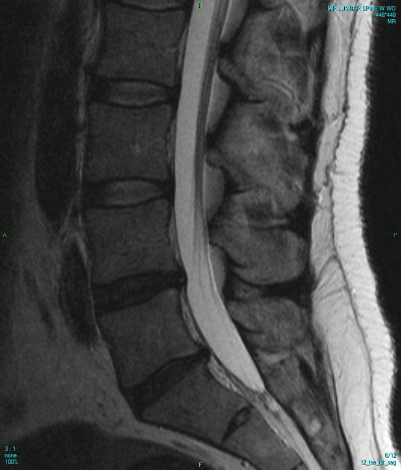
Why the root hurts. Compression is mechanical and chemical. Direct pressure on the root, venous congestion and ischaemia account for the mechanical part; the nucleus pulposus is itself inflammatory, releasing phospholipase A2 and TNF-alpha, and may cause symptoms without any mechanical compression.
Classification Systems
The herniation is described three ways: by morphology, by location in the canal, and by the Modic endplate changes.
The Michigan State University classification describes morphology, and morphology guides the surgical approach.
- Description
- Focal bulge, base wider than dome
- PLL Integrity
- Intact
- Fragment Containment
- Contained by annulus
- Description
- Dome wider than base, continuous with disc
- PLL Integrity
- Torn
- Fragment Containment
- Through annulus but attached
- Description
- Free fragment, no disc continuity
- PLL Integrity
- Torn
- Fragment Containment
- Completely separated
Clinical Assessment
Who does well. The good surgical candidate has leg pain that dominates the back pain, in a dermatomal distribution that matches the disc level, with positive tension signs (straight leg raise, femoral stretch), an MRI that correlates with the examination, a failed 6-12 week conservative trial, and is motivated with realistic expectations.
Who does badly. Any of the following should make you hesitate:
- Dominant axial back pain without radiculopathy
- A non-dermatomal pain pattern
- Imaging that does not correlate with the symptoms
- Significant psychosocial factors, pending litigation or workers' compensation, or other secondary gain
- A minimal conservative treatment trial
Neurological examination. The root is localised by motor, sensory and reflex findings taken together.
- Motor
- Hip flexion, knee extension
- Sensory
- Anterior thigh
- Reflex
- None reliable
- Motor
- Knee extension, ankle dorsiflexion
- Sensory
- Medial leg/foot
- Reflex
- Patellar (knee jerk)
- Motor
- Great toe extension (EHL), hip abduction
- Sensory
- Lateral leg, dorsum foot
- Reflex
- None reliable
- Motor
- Ankle plantar flexion, hip extension
- Sensory
- Lateral foot, posterior calf
- Reflex
- Achilles (ankle jerk)
Tension signs.
- Straight leg raise - positive between 30 and 70°, worse with ankle dorsiflexion
- Crossed straight leg raise - raising the unaffected leg reproduces pain on the affected side; highly specific
- Femoral stretch test - for L2-L4 radiculopathy
- Bowstring sign - popliteal pressure during the straight leg raise reproduces the pain
Red flags. Cauda equina syndrome is the one that cannot wait. The other red flags, each of which demands urgent evaluation, are a progressive motor deficit, fever or other signs of infection, a history of malignancy and unexplained weight loss.
Large central disc herniation can cause cauda equina syndrome - a surgical emergency. Features: urinary retention or incontinence (retention is the most sensitive), saddle anaesthesia (S2-S5), faecal incontinence with reduced anal tone, bilateral progressive leg weakness. Incomplete CES has a better prognosis than complete. Decompress as soon as it can be done safely and competently. Read the widely quoted "24 to 48 hours" as an outer limit and not as a target: Ahn's meta-analysis (PMID 10851100) found significantly better recovery of sensory, motor, bladder and bowel function in patients decompressed within 48 hours versus after 48 hours, and found no additional benefit from operating inside 24 hours compared with 24 to 48 hours - but it pools 42 retrospective series in which the patients operated later were also those with less complete syndromes, so it cannot be used to justify planned delay. The clinically decisive distinction is not the clock but whether the syndrome is incomplete (altered perineal sensation, difficulty voiding, preserved sensation of bladder fullness) or has progressed to established painless retention with overflow incontinence - the incomplete syndrome is the one where prompt surgery still changes the outcome, so it is the one that must not be left overnight for a list.
The most sensitive feature of cauda equina syndrome is urinary retention - specifically inability to void with a distended bladder. Always measure a post-void residual if the syndrome is suspected; more than 100-200 ml is concerning.
The differential. A herniated disc should only be implicated when the clinical syndrome, the dermatomal distribution and the imaging level are concordant; otherwise these mimics must be actively excluded before any operation is offered.
- Distinguishing Features
- Older patient, neurogenic claudication, relief with flexion
- Key Investigation
- MRI showing bony/facet stenosis rather than soft disc
- Distinguishing Features
- Stocking distribution, bilateral, no dermatomal pattern
- Key Investigation
- Nerve conduction studies, HbA1c
- Distinguishing Features
- Groin or lateral hip pain, pain on hip rotation, no neurology
- Key Investigation
- Hip examination, hip radiograph, diagnostic injection
- Distinguishing Features
- Buttock pain, no clear dermatome, tender sciatic notch
- Key Investigation
- Clinical, MRI to exclude disc; diagnosis of exclusion
- Distinguishing Features
- Night pain, fever, weight loss, history of malignancy
- Key Investigation
- MRI with contrast, inflammatory markers
- Distinguishing Features
- Calf pain with walking, absent pulses, relief with standing
- Key Investigation
- ABPI, arterial duplex
Investigations
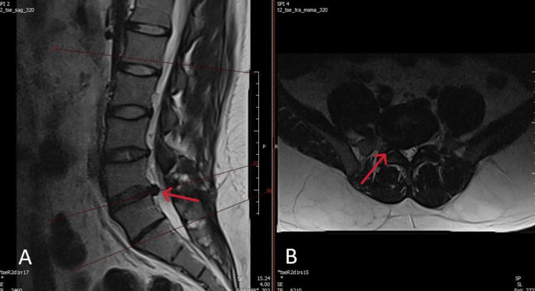
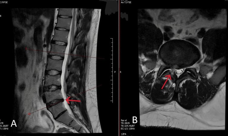
MRI is the gold standard. The standard protocol is sagittal T1 and T2 with axial T2 at each level; consider gadolinium when a recurrent disc must be told from scar. It shows the herniation's location and size, root compression and displacement, foraminal stenosis, disc degeneration (Pfirrmann grading) and Modic endplate change, and it helps distinguish between the herniation types.
Correlation is the point. Incidental disc abnormalities are common, present in 30-40% of asymptomatic individuals, so the imaging level must match the clinical level and clinical correlation is mandatory before surgery.
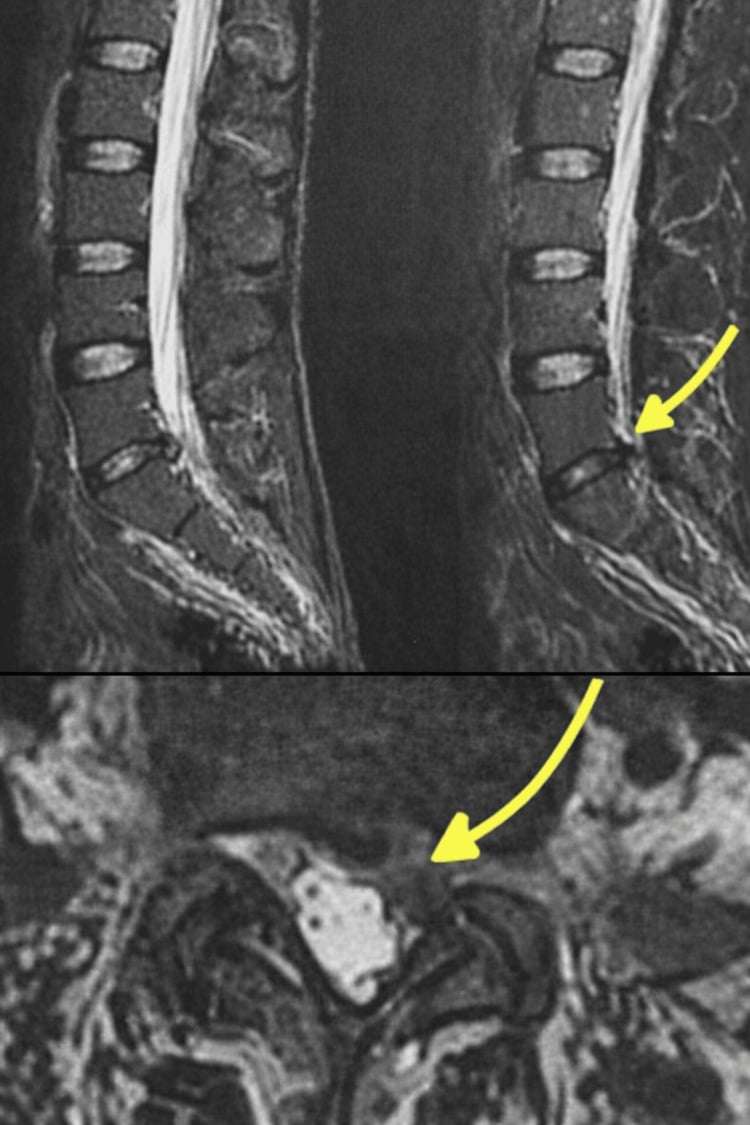
CT shows bony anatomy well, is less sensitive for a soft disc herniation, and may be used if MRI is contraindicated, as in patients with pacemakers. CT myelography adds intrathecal contrast: it is better for bony stenosis, shows dynamic compression, and is used when MRI is contraindicated or equivocal.
Electrodiagnostics. EMG and nerve conduction studies confirm radiculopathy, distinguish it from peripheral neuropathy, localise the level when imaging is equivocal, and assess chronicity and severity. They are most useful when the clinical and imaging findings are discordant. Timing matters: the changes take 3-4 weeks to develop, so an acute herniation may have a normal EMG and false negatives are possible early. Indications:
- Atypical presentation
- Multi-level disease on imaging
- Suspected peripheral neuropathy
- Medicolegal documentation
Plain radiographs have a limited role in disc herniation: alignment, instability and degenerative change, with flexion-extension views for instability.
Selective nerve root block is both diagnostic and therapeutic. It is helpful when imaging shows multi-level disease, because it confirms the symptomatic level before surgery.
Management
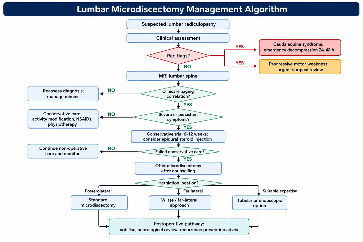
First line. Activity modification avoiding the aggravating positions, NSAIDs and muscle relaxants, physiotherapy, and time, because the natural history is favourable. Beyond that, an epidural steroid injection, an oral corticosteroid taper or a nerve root block can be added; the injection is considered when symptoms are severe.
How long. The standard trial is 6-12 weeks. It may be shortened by a progressive deficit, and cauda equina syndrome gets no conservative trial at all.
Surgical Technique
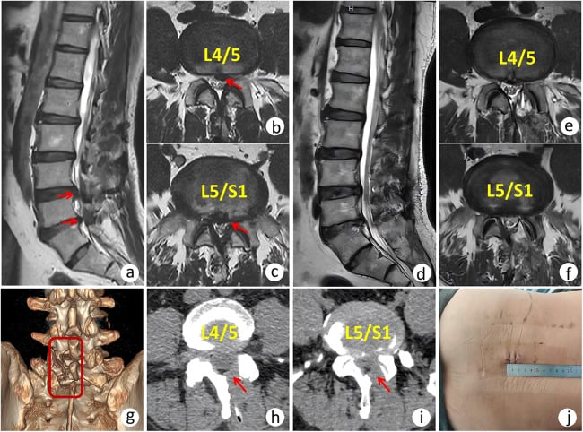
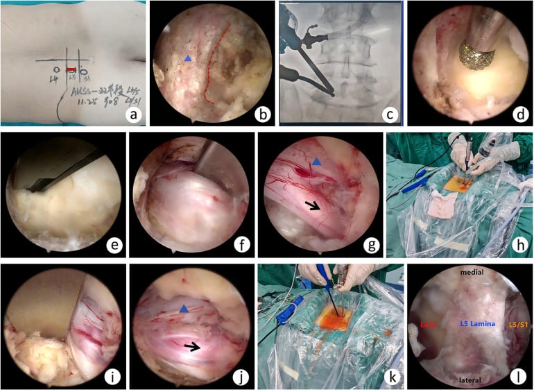
Positioning. Prone on a Wilson frame or Jackson table with the hips flexed to flatten the lumbar lordosis and the abdomen free to reduce venous pressure; eyes protected, arms positioned.
Level. Fluoroscopy is mandatory. Mark the incision preoperatively and verify the level with intraoperative imaging.
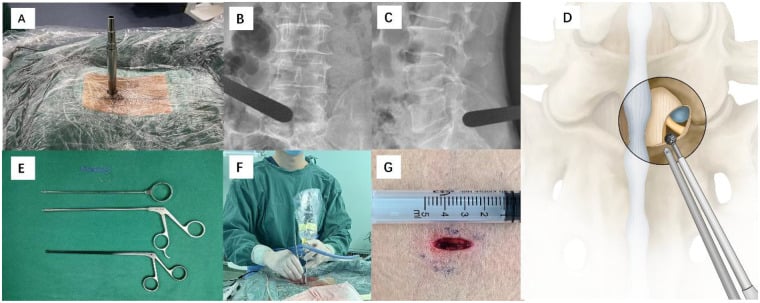
Incision and exposure.
- Midline incision (2-3 cm) centred over the disc level
- Dissect through the subcutaneous tissue
- Incise the fascia paramedian on the symptomatic side
- Elevate the muscle subperiosteally off the spinous process and lamina
- Identify the interlaminar window
- Confirm the level with fluoroscopy
Landmarks. The spinous process of the upper vertebra, the interlaminar space and the medial facet joint orientate the exposure. The approach should preserve the majority of the facet joint to prevent instability.
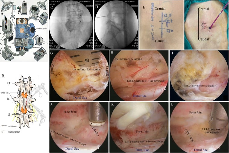
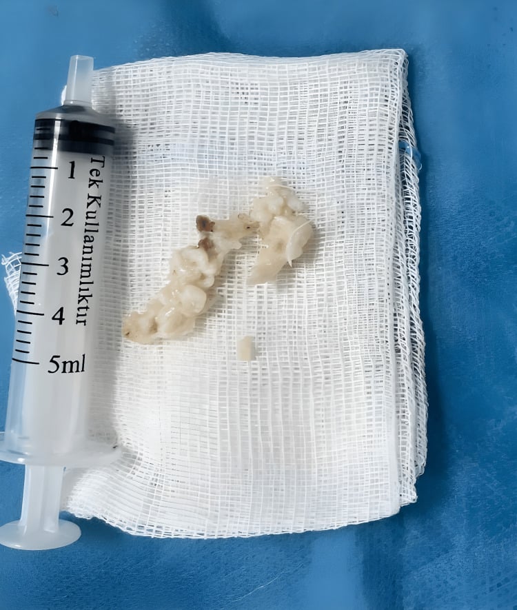
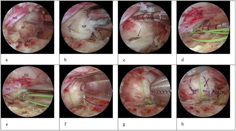
Dural tear. Repair primarily with 4-0 or 5-0 suture if possible; the alternatives are a dural sealant (fibrin glue, DuraSeal) and a fat graft or muscle patch. Bed rest for 24-48 hours postoperatively; a persistent leak may need a lumbar drain.
Haemostasis. Bipolar cautery for the epidural veins, avoiding monopolar near neural structures, haemostatic agents (Gelfoam, Surgicel) as needed, and a dry field before closure.
A conjoined (anomalous) nerve root - two adjacent roots sharing a common dural sheath/origin or running an anomalous course - is present in a small but important minority of patients and is a classic microdiscectomy pitfall:
- It can be mistaken for a herniated fragment or a swollen root, tempting the surgeon to "decompress" or retract it - risking a traction nerve injury or a dural tear at the axilla where the two roots share a sheath.
- The conjoined root is less mobile and cannot be retracted as far, so the working corridor to the disc is reduced; forcing retraction is the danger.
- Suspect and look for it preoperatively on MRI/CT (an asymmetric, enlarged or doubled root shadow, a flat-shouldered lateral recess), so it is recognised, not discovered under retraction.
If a conjoined root is found, widen the bony decompression (more medial facet undercut/laminotomy) to create room and work around it gently rather than mobilising it, and remove the disc through the available window. Exam point: an immobile, "too-big" or doubled root at microdiscectomy may be a conjoined root - recognise it (ideally pre-op), do NOT retract aggressively, and enlarge the bony exposure instead.
Complications
- Incidence
- 1-2% primary, 5-10% revision
- Prevention
- Careful technique, identify dura early
- Management
- Primary repair, sealant, bed rest
- Incidence
- Less than 1%
- Prevention
- Gentle retraction, visualisation
- Management
- Observation, steroids if needed
- Incidence
- Rare but serious
- Prevention
- Fluoroscopic confirmation
- Management
- Intraoperative correction, documentation
- Incidence
- Very rare (0.01-0.05%)
- Prevention
- Anterior awareness, depth control
- Management
- Immediate vascular surgery consult
- Incidence
- Rare
- Prevention
- Meticulous haemostasis
- Management
- Urgent decompression if symptomatic
Early. Wound infection in 1-2%, a CSF leak after a dural tear, recurrent herniation and persistent radiculopathy. Late. Recurrent herniation, adjacent segment disease, chronic pain, and instability, which is rare with a limited laminotomy.
Recurrent herniation. The recurrence rate is 5-10% at the same level and 5% at a different level. Risk factors:
- Obesity (BMI above 25)
- Smoking
- A larger annular defect
- Disc degeneration
- Occupational factors
- Diabetes
When it recurs. Trial conservative treatment again and repeat the imaging with MRI and gadolinium before any decision between revision microdiscectomy and fusion.
MRI with gadolinium distinguishes a recurrent disc (no enhancement) from postoperative scar tissue (enhances). Scar is not an indication to reoperate.
Revision microdiscectomy. The indications are a recurrent disc herniation (not scar), new symptoms at the same level, and failure to improve or worsening after the first operation. Epidural fibrosis increases the dural tear risk (5-10%).
When to fuse instead. Fusion is considered for:
- Significant instability
- Multiple recurrent herniations
- Concomitant spondylolisthesis
- A large annular defect with disc space collapse
- A significant back pain component
Postoperative Care
Most patients mobilise the same day - a microdiscectomy is commonly a day case. Record a fresh neurological examination, because it is the comparator against which any later deficit is judged, and new or worsening weakness after operation is the finding that mandates urgent re-imaging rather than reassurance. Multimodal analgesia, and encourage ambulation actively rather than permitting it.
Discharge when stable - day surgery or a single overnight stay. Beyond wound care and activity advice, the patient must leave knowing which symptoms mean return immediately: new or worsening leg weakness, any difficulty passing urine, numbness in the saddle area, or a positional headache with clear fluid from the wound. A recurrent herniation and a post-operative haematoma both present as leg pain that is getting worse rather than better, which is the pattern that distinguishes them from ordinary operative discomfort.
Activity. Walking and sitting start at once; everything else is staged.
- Timeline
- Immediate
- Restrictions
- Encouraged, gradually increase
- Timeline
- Immediate
- Restrictions
- Limit prolonged sitting initially
- Timeline
- 2-4 weeks
- Restrictions
- Less than 5 kg initially, gradually increase
- Timeline
- 1-2 weeks
- Restrictions
- Off narcotics, comfortable sitting
- Timeline
- 2-4 weeks
- Restrictions
- Gradual return
- Timeline
- 6-12 weeks
- Restrictions
- Depends on demands
- Timeline
- 3-6 months
- Restrictions
- Surgeon clearance required
Rehabilitation. Physiotherapy covers core strengthening, delayed until 2-4 weeks, with flexibility work, posture education and ergonomic training. The lifestyle conversation is weight optimisation, smoking cessation, proper lifting technique and activity modification.
Follow-up.
- 2 weeks - wound check, early progress
- 6 weeks - clinical assessment, activity progression
- 3 months - outcome assessment
- 12 months - final follow-up, if needed
Recovery after microdiscectomy is typically rapid - most patients notice immediate leg pain relief on waking from surgery. Back pain and residual numbness may take longer to improve.
Outcomes and Prognosis
Success rates.
- Leg pain relief - 90-95%
- Back pain improvement - 70-80%
- Return to work - 80-90%
- Patient satisfaction - 80-85%
SPORT. Surgery gave faster improvement in the first 3 months; by 4 years the surgical and conservative groups had similar outcomes, both having improved substantially, and cross-over rates were high enough to complicate interpretation. Patient preference therefore carries real weight in the decision, and conservative treatment remains a reasonable option.
Prognostic factors.
- Favourable - leg pain more than back pain, short symptom duration, clear imaging correlation, no previous surgery, an extruded or sequestered disc, good psychosocial status
- Unfavourable - predominant back pain, long symptom duration, previous failed surgery, workers' compensation, depression or anxiety, obesity and smoking
Workers' compensation. Outcomes are generally inferior and higher recurrence rates are reported. A multidisciplinary approach is recommended, documentation must be clear, and expectations managed carefully.
Guidelines, Registries & Global Practice
Global Epidemiology and Burden
Lumbar disc herniation affects an estimated 1-3% of the population, with a lifetime prevalence of sciatica of 13-40%. Peak incidence is in the fourth and fifth decades, and L4-5 and L5-S1 together account for around 95% of symptomatic herniations. Discectomy is consistently the most commonly performed spinal operation in high-income health systems. The natural history is favourable: roughly 90% of symptomatic patients improve without surgery, and the central message of the SPORT randomised trial (Weinstein, JAMA 2006) and the Leiden sciatica trial (Peul, NEJM 2007) is that surgery accelerates recovery rather than changing the eventual outcome for most patients.
Side-by-Side Guideline Comparison
- Conservative trial
- Recommend a structured nonoperative period for radiculopathy without red flags
- Surgical recommendation
- Discectomy offered for persistent radiculopathy with concordant imaging after conservative failure; faster relief than continued nonoperative care
- Evidence basis
- Graded recommendations from RCT evidence (SPORT, Peul)
- Conservative trial
- Encourage activity, avoid prolonged bed rest; consider epidural for acute severe sciatica
- Surgical recommendation
- Consider decompression for sciatica when non-surgical treatment has not resolved symptoms and imaging correlates
- Evidence basis
- Systematic review with health-economic modelling
- Conservative trial
- Conservative care for 6-12 weeks unless progressive deficit or CES
- Surgical recommendation
- Microdiscectomy is the reference standard; minimally invasive techniques are equivalent in outcome
- Evidence basis
- RCT and registry synthesis
- Conservative trial
- Conservative trial with clear escalation pathway
- Surgical recommendation
- Urgent pathway and emergency decompression for suspected cauda equina syndrome
- Evidence basis
- Consensus standards plus medicolegal guidance
All four bodies converge on the same principles: a time-limited conservative trial for uncomplicated radiculopathy, imaging-clinical concordance before surgery, and emergency decompression for cauda equina syndrome. Differences are largely in emphasis and the structure of referral pathways rather than in the indication itself.
Registry and Practice-Variation Evidence
Large national spine registries (for example the Swedish Swespine register, the British Spine Registry and the Norwegian NORspine register) collect patient-reported outcomes after discectomy and consistently report leg-pain relief and satisfaction in the region of 80-90% at 1 year, mirroring the trial literature. Registry data also document substantial geographic variation in discectomy rates that is not explained by disease prevalence, underscoring the influence of local practice and patient preference highlighted by SPORT. A Dutch surveillance survey (Arts, J Neurosurg Spine 2008) found unilateral transflaval discectomy to be the dominant technique, with surgeons expecting minimally invasive approaches to carry higher recurrence and percutaneous laser decompression to be least effective.
Service Delivery and Workforce
Microdiscectomy is performed by both neurosurgeons and orthopaedic spine surgeons across public and private settings in many health systems, reflecting the shared spinal workload. Elective practice follows the international guidance summarised above, with emphasis on appropriate patient selection and an adequate conservative trial before surgery. Day-case or overnight pathways are increasingly used for uncomplicated cases, supported by the procedure's low morbidity and rapid recovery profile. Smoking cessation is encouraged in view of the association between smoking and reherniation.
Medicolegal Considerations
Key documentation points for medicolegal protection include recording of red-flag assessment, documentation of concordance between symptoms and imaging, the consent discussion (including recurrence risk and the possibility of dural tear), and the level-confirmation process (pre-operative marking and intra-operative fluoroscopy). Cauda equina syndrome cases require meticulous documentation of timing: when symptoms began, when the patient presented, when imaging was obtained and when surgery was performed.
MCQ Practice Points
High-Yield MCQ Topics
Q: L4-5 posterolateral disc herniation typically affects which nerve root?
A: The L5 nerve root (traversing root). Posterolateral herniations affect the traversing root, which exits one level below. Far lateral herniations at L4-5 would affect L4 (the exiting root).
Q: What is the most sensitive clinical feature of cauda equina syndrome?
A: Urinary retention (specifically inability to void with a distended bladder). Post-void residual more than 100-200ml is concerning. Other features include saddle anesthesia (S2-S5) and bilateral leg symptoms.
Q: What MRI finding distinguishes recurrent disc herniation from epidural fibrosis (scar tissue)?
A: Gadolinium enhancement pattern. Scar tissue ENHANCES (it is vascular). Recurrent disc does NOT enhance (it is avascular). This distinction is critical as scar tissue does not benefit from surgery.
Q: What did the SPORT trial show about lumbar microdiscectomy for disc herniation?
A: Surgery provides faster initial recovery (advantage at 3 months) but 4-year outcomes are similar between surgical and conservative treatment. Both groups improved substantially. The natural history of disc herniation is generally favorable.
Q: A far lateral L4-5 disc herniation affects which nerve root?
A: The L4 nerve root (exiting root). Unlike posterolateral herniations that affect the traversing root one level below, far lateral/foraminal herniations affect the exiting root at the same level.
Clinical Decision Scenarios
Practise clinical reasoning and management decisions out loud
“A 35-year-old man presents with 3 months of right leg pain radiating to the dorsum of the foot and great toe. He has weakness of great toe extension. MRI shows L4-5 right posterolateral disc herniation. He has failed 8 weeks of physiotherapy and two epidural injections. How would you manage this patient?”
“A 50-year-old woman presents with sudden onset bilateral leg weakness, urinary retention requiring catheterization, and saddle anesthesia. MRI shows large central L4-5 disc herniation. How do you manage her?”
“You perform an L5-S1 microdiscectomy and encounter a dural tear during removal of the ligamentum flavum. Describe your management.”
“A 45-year-old man is 18 months post L4-5 microdiscectomy with excellent initial result. He now has recurrent right leg pain identical to his original presentation. MRI shows enhancement around the previous surgical site. How do you proceed?”
Nerve Root Anatomy
- Posterolateral herniation = traversing root (L4-5 = L5)
- Far lateral/foraminal = exiting root (L4-5 = L4)
- L4: knee extension, patellar reflex
- L5: EHL (great toe extension), no reflex
- S1: plantar flexion, Achilles reflex
Cauda Equina Syndrome
- Emergency - surgery within 24-48 hours
- Most sensitive: urinary retention
- Saddle anesthesia (S2-S5)
- Bilateral leg weakness
- Incomplete better prognosis than complete
SPORT Trial
- Surgery faster initial improvement
- 4-year outcomes similar
- Both groups improved substantially
- Natural history favorable for most
Surgical Technique
- Prone on Wilson frame, fluoroscopy for level
- Laminotomy, preserve facet
- Protect dura and traversing root
- Remove loose fragments only (limited discectomy)
- Dural tear 1-2% primary, 5-10% revision
Recurrence
- 5-10% at same level
- Risk factors: obesity, smoking, large defect
- MRI + gadolinium: scar enhances, disc does not
- Revision vs fusion decision based on instability
Outcomes
- 90-95% leg pain relief
- 70-80% back pain improvement
- Day surgery or overnight stay
- Return to work: sedentary 2-4 weeks, physical 6-12 weeks
Evidence-Based Practice
SPORT Randomised Trial (Weinstein et al., JAMA 2006)
- Randomised trial of open discectomy vs nonoperative care; 501 surgical candidates (mean age 42 years, 42% women) with imaging-confirmed herniation and at least 6 weeks of radiculopathy
- Both groups improved substantially over 2 years on SF-36 bodily pain, physical function and modified Oswestry Disability Index
- Intent-to-treat between-group differences consistently favoured surgery but were small and not statistically significant for the primary outcomes
- Crossover was extensive: only 50% assigned to surgery had surgery by 3 months, while 30% assigned to nonoperative care crossed to surgery
SPORT 8-Year Results (Lurie et al., Spine 2014)
- Combined randomised (501) and observational (743) SPORT cohorts across 13 spine clinics in 11 US states, followed to 8 years; cumulative non-adherence was extensive - 49% assigned to nonoperative care received surgery, against 60% of those assigned to surgery
- As-treated analysis showed durable surgical treatment effects: bodily pain 10.9, physical function 10.6 and Oswestry Disability Index -11.3 in favour of surgery
- Secondary outcomes (sciatica bothersomeness, satisfaction, self-rated improvement) were significantly better with surgery in intent-to-treat analysis
- Little to no degradation of outcomes in either group from 4 to 8 years
Sequestrectomy vs Microdiscectomy RCT (Barth et al., Spine 2008)
- Single-centre RCT of 84 patients randomised to standard microdiscectomy or microscopic sequestrectomy (free-fragment removal only)
- Reherniation rates did not differ at 2 years: 10.5% (discectomy) vs 12.5% (sequestrectomy), P=1.0
- The deterioration was directional and clinically meaningful: self-rated outcomes WORSENED over the 2 years after microdiscectomy while they improved after sequestrectomy, so the difference emerged over time rather than being present initially
- By 2 years the difference reached significance for use of analgesics, performance and overall outcome, favouring sequestrectomy
Limited vs Aggressive Discectomy Review (McGirt et al., Neurosurgery 2009)
- Systematic review of 60 cohorts (13,359 patients) comparing limited fragment removal with aggressive discectomy and curettage
- Reported reherniation was HIGHER after limited discectomy (mean 7%, range 2-18%) than aggressive discectomy (mean 3.5%, range 0-9.5%)
- Long-term recurrent back or leg pain was 2.5-fold LOWER after limited discectomy (11.6%) vs aggressive discectomy (27.8%)
- Illustrates the core trade-off: limited removal protects against axial pain but accepts a higher reherniation risk
Early Surgery vs Conservative Care RCT (Peul et al., NEJM 2007)
- 283 patients with 6-12 weeks of severe sciatica randomised to early microdiscectomy or prolonged conservative care with surgery if needed
- Leg-pain relief and perceived recovery were significantly faster with early surgery (recovery hazard ratio 1.97, 95% CI 1.72-2.22)
- No significant difference in disability scores over the first year (P=0.13)
- By 1 year the probability of perceived recovery was 95% in BOTH groups; 39% of the conservative arm ultimately had surgery