Treatment of Spinal Stenosis | Neurogenic Claudication | With/Without Fusion Decision
- Neurogenic claudication = bilateral leg symptoms with walking, relief with flexion/sitting
- Shopping cart sign = flexion reduces symptoms by increasing canal diameter
- Laminectomy vs fusion - add fusion if instability, deformity, or extensive facet resection
- Preserve more than 50% of facets to maintain stability without fusion
- SPORT showed surgery superior to conservative at 2-4 years for stenosis
- “Neurogenic vs vascular claudication: flexion relief vs position-independent
- “Preserve facets (more than 50%) to avoid post-laminectomy instability
- “Fusion indications: spondylolisthesis, scoliosis, more than 50% facet removal
- “SPORT: Surgery better than conservative for stenosis
Overview
Lumbar laminectomy is the removal of the lamina (the posterior arch) and the ligamentum flavum to decompress the spinal canal. It is the definitive treatment for symptomatic lumbar spinal stenosis that has not responded to conservative management, and it is performed alone or with a fusion depending on stability considerations.
Where the operation came from. Laminectomy has been performed since the early 1900s. Modern technique emphasises a limited decompression that preserves the stabilising structures, and the transition from wide laminectomy to targeted decompression has improved outcomes and reduced post-laminectomy instability.
Who has it. Lumbar spinal stenosis is the most common indication for spine surgery in patients over 65 years, and its prevalence rises with age as degenerative change accumulates. L4-5 is the most commonly affected level.
The SPORT trial showed that surgical treatment of lumbar spinal stenosis produces better outcomes than conservative treatment at 2-4 years. That contrasts with disc herniation, where the long-term outcomes of surgical and conservative treatment are similar.
Pathophysiology and Mechanisms
What narrows the canal. Central stenosis is assembled from several degenerative contributions, and the ligamentum flavum is the most significant of them:
- Ligamentum flavum hypertrophy
- Facet joint hypertrophy
- Disc bulging
- Spondylolisthesis, if present
Where the compression sits decides which neural element is caught, and that is the basis of the anatomical classification in the next section.
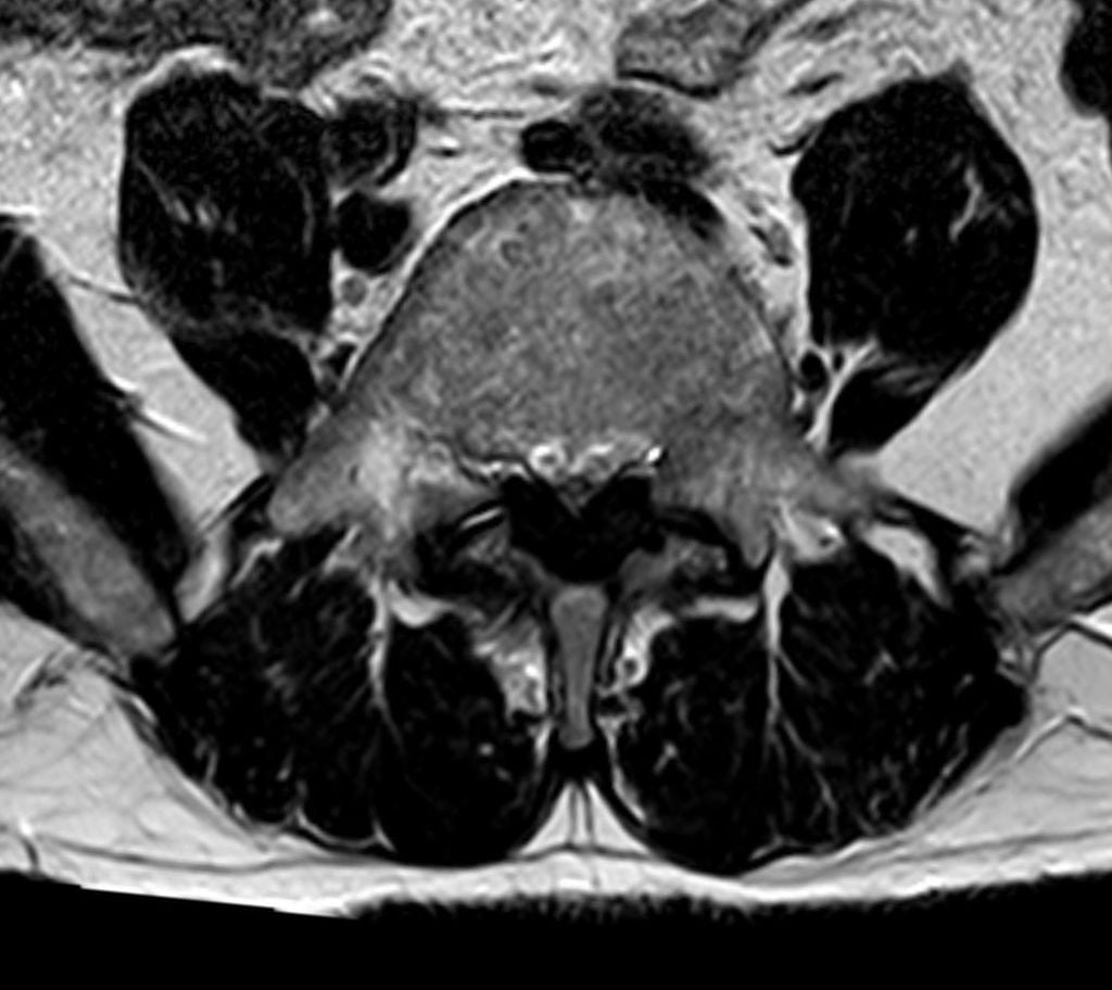
Why walking hurts and flexion helps. Standing and walking extend the lumbar spine, which reduces the canal diameter; in the confined space the veins become congested and the neural elements become ischaemic with ambulation. Flexion opens the posterior canal, reduces the bulge of the facet joints and ligamentum flavum and increases the central canal area by 10-20%, which is why the symptoms ease when the patient sits or leans forward. The classic posture is the patient leaning on a shopping cart.
The facets and stability. The facet joints are the primary posterior stabilisers and resist extension and rotation. Removing more than 50% of the facets bilaterally is the accepted working rule for instability risk, and it should be quoted for what it is: a surgical convention, not a measured threshold. Abumi's graded-facetectomy study (PMID 2267608), the source usually invoked for it, reports no percentage of resected joint at all. Its categories are anatomical: a medial facetectomy leaves range of motion essentially unchanged, whereas a total facetectomy destabilises the segment even when unilateral, the effect appearing in axial rotation with extension and lateral bending unaffected even after bilateral total resection.
How to use the rule. The 50 per cent figure is a sound operational guide; just do not present it as a derived number. The evidence-based version of the statement is anatomical: a medial facetectomy is biomechanically safe, whereas taking a facet in its entirety destabilises the segment even unilaterally and should prompt consideration of fusion at the index operation. Extensive facet removal without fusion leads to progressive instability, recurrent stenosis and poor outcomes.
Classification Systems
Anatomical Classification of Stenosis
- Location
- Spinal canal
- Root Affected
- Cauda equina (multiple)
- Surgical Target
- Laminectomy, ligamentum flavum
- Location
- Subarticular zone
- Root Affected
- Traversing root
- Surgical Target
- Medial facetectomy, undercut
- Location
- Neural foramen
- Root Affected
- Exiting root
- Surgical Target
- Foraminotomy, may need fusion
The anatomical classification guides the surgical approach according to where the compression occurs.
Clinical Assessment
The history. Neurogenic claudication is bilateral leg symptoms brought on by walking and relieved by flexion or sitting; vascular claudication is often unilateral. Separating the two is the central task of the consultation, and the discriminators are worth knowing as a set.
- Neurogenic
- Bilateral, buttock to legs
- Vascular
- Calves, unilateral or bilateral
- Neurogenic
- Aching, paraesthesias, weakness
- Vascular
- Cramping, tightness
- Neurogenic
- Variable walking distance
- Vascular
- Fixed walking distance (claudication distance)
- Neurogenic
- Flexion/sitting (shopping cart)
- Vascular
- Standing rest (no need to sit)
- Neurogenic
- Better (flexed posture)
- Vascular
- Worse (more work)
- Neurogenic
- Worse (extended posture)
- Vascular
- Better
- Neurogenic
- Good (flexed)
- Vascular
- Limited (cardiovascular)
- Neurogenic
- Normal
- Vascular
- Diminished or absent
Examination. The neurological examination is often normal at rest, so examine motor power, sensation and reflexes and then watch the patient walk: a wide-based gait may be present, and a positive Romberg test points to proprioceptive loss. Provoke the symptoms by walking, noting the distance, and by extension, which may reproduce them; relief with flexion supports a neurogenic cause. Examine the circulation as well: the dorsalis pedis and posterior tibial pulses, the ankle-brachial index (ABI) if there is concern, and the skin for hair loss and trophic change.
Patient selection. The table sets out who is likely to benefit from the operation and who is not.
- Poor surgical candidate
- Vascular claudication (needs vascular surgery)
- Poor surgical candidate
- Imaging does not correlate
- Poor surgical candidate
- Minimal functional limitation
- Poor surgical candidate
- Predominant back pain without leg symptoms
- Poor surgical candidate
- Severe medical comorbidities
- Poor surgical candidate
- Unrealistic expectations
The differential. Leg pain on walking has several causes, and the discriminating feature and the test that settles it are different for each.
- Distinguishing Features
- Bilateral, flexion relief (shopping cart), normal pulses, variable distance
- Key Investigation
- MRI lumbar spine
- Distinguishing Features
- Calf cramp, fixed distance, rest relief without flexion, absent pulses, trophic skin
- Key Investigation
- Ankle-brachial index, duplex
- Distinguishing Features
- Dermatomal, often unilateral, positive straight-leg raise, present at rest
- Key Investigation
- MRI lumbar spine
- Distinguishing Features
- Groin/buttock pain, restricted internal rotation, worse weight-bearing
- Key Investigation
- Pelvis/hip radiograph
- Distinguishing Features
- Stocking sensory loss, burning, distance-independent, often diabetic
- Key Investigation
- Nerve conduction studies, HbA1c
- Distinguishing Features
- Lateral hip tenderness, pain on direct pressure, not position-dependent canal
- Key Investigation
- Clinical, ultrasound
The shopping cart sign is pathognomonic for neurogenic claudication. Vascular and neurogenic claudication can coexist in the elderly, so check the pulses and the ankle-brachial index even when stenosis is confirmed on MRI.
Investigations
MRI is the gold standard for evaluating stenosis. The standard protocol is sagittal T1 and T2, axial T2 at each level and STIR for oedema. Read it for the central canal diameter (less than 10mm is concerning), the thickness of the ligamentum flavum, facet hypertrophy, the contribution of disc bulging, and lateral recess and foraminal stenosis; it provides the anatomical detail for surgical planning and identifies the primary pathology. Imaging findings are common in asymptomatic elderly people, so clinical correlation is mandatory, and in multi-level disease the worst level may need to be identified.
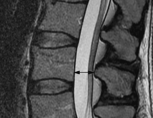
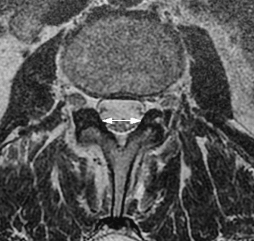
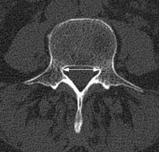
CT gives excellent bony detail, shows facet arthropathy and osteophytes, is useful for surgical planning and may be used when MRI is contraindicated, as in the patient with a pacemaker. CT myelography adds intrathecal contrast: it is better for dynamic assessment, shows functional stenosis, is useful in multi-level disease or when the MRI is equivocal, and is required when MRI is contraindicated.
Plain radiographs. Standing AP and lateral films assess alignment and spondylolisthesis, and full-spine films are taken if scoliosis is suspected. Flexion-extension views assess dynamic instability, which a supine MRI cannot show, and the criteria are translation of more than 4mm or a change in angulation of more than 10-15°. An unstable segment means fusion should be considered with the decompression, so the finding affects surgical planning significantly.
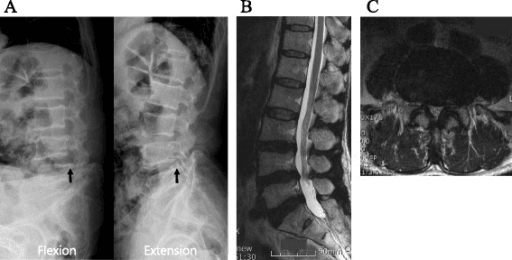
Electrodiagnostics (EMG/NCS) distinguish stenosis from peripheral neuropathy, document radiculopathy and are useful in multi-level disease. Vascular studies, the ankle-brachial index, duplex ultrasound and CT angiography if needed, exclude peripheral vascular disease as the cause of the leg symptoms.
Management
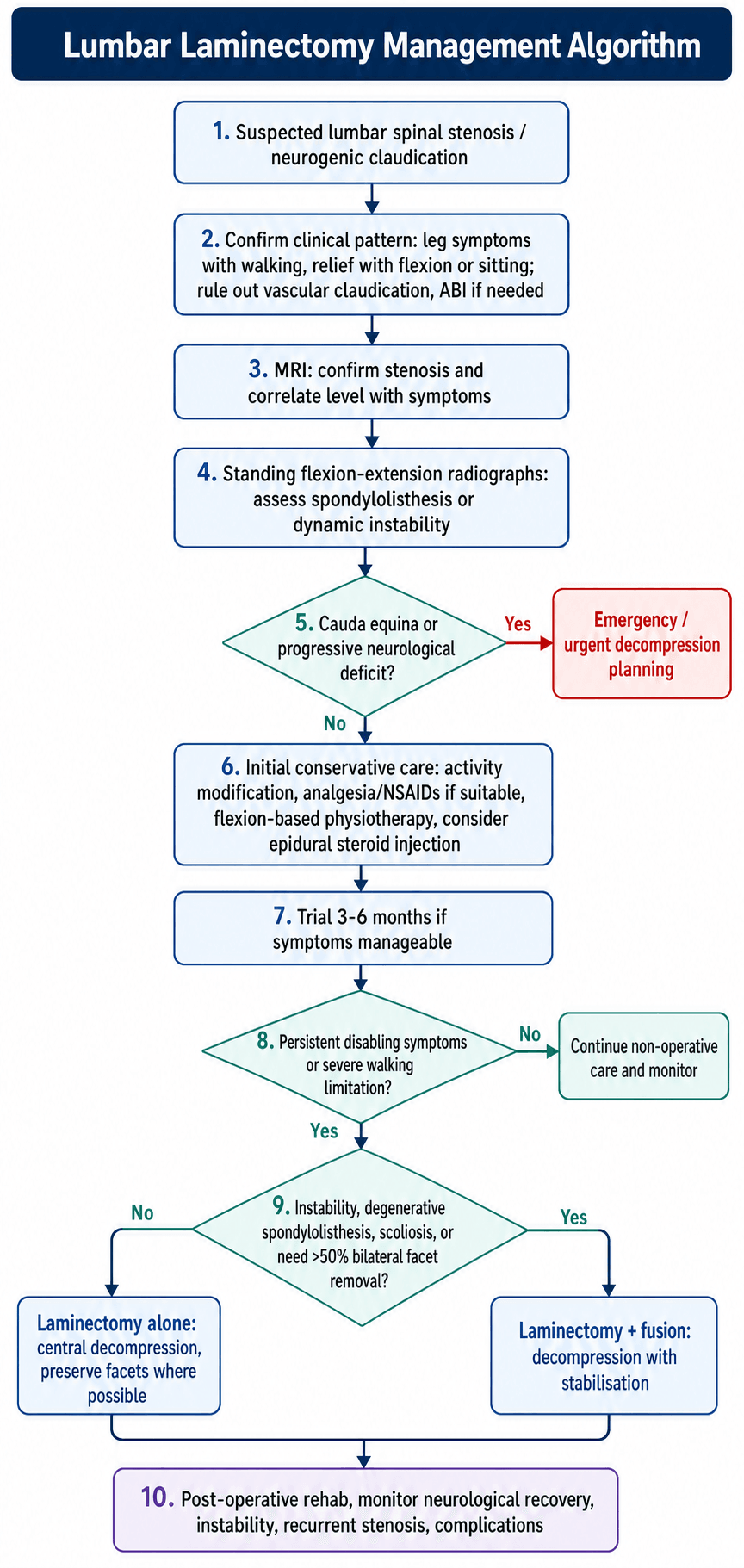
Conservative treatment first. Most patients start here, and 60-70% may improve: activity modification, NSAIDs and paracetamol, flexion-based physiotherapy, and epidural steroid injection for transient relief of leg pain. The LESS trial found the steroid added minimal or no benefit over local anaesthetic alone, so the injection is symptomatic, not disease-modifying. The trial typically runs 3-6 months and may continue if the symptoms are manageable; there is no evidence that conservative treatment prevents progression.
When to operate. Surgery is indicated when conservative measures fail or neurological progression occurs, and the urgency depends on which.
- Urgency
- Emergency
- Rationale
- Prevent permanent deficit
- Urgency
- Urgent
- Rationale
- Preserve function
- Urgency
- Elective
- Rationale
- Quality of life
- Urgency
- Elective
- Rationale
- Walking tolerance severely limited
Laminectomy alone or with fusion. Laminectomy alone is appropriate when there is no spondylolisthesis and no scoliosis, the segment is stable on flexion-extension views, more than 50% of the facets can be preserved bilaterally, and central stenosis predominates. Fusion is added when any of the following is present, a decision that rests on the stability analysis and on the extent of decompression required:
- Degenerative spondylolisthesis
- Significant degenerative scoliosis
- Dynamic instability on imaging
- More than 50% bilateral facet removal required
- Extensive multi-level decompression
Spondylolisthesis is the entry on that list the evidence has qualified: the two 2016 trials in the warning below disagree, and what they teach is that instability, rather than the presence of a slip, drives the decision.
FUSEFUSE - When to Add Fusion
Hook:FUSE when stability is at risk - don't just decompress
The single most examinable evidence point in this topic is whether to add fusion when decompressing a stenosis with grade I degenerative spondylolisthesis - and two landmark RCTs published in the same 2016 NEJM issue reached opposite conclusions:
- SLIP (Ghogawala et al., 2016): decompression plus instrumented fusion gave better physical health-related quality of life (SF-36) than decompression alone, at the cost of more blood loss and a longer stay, and with a lower reoperation rate (14% vs 34%).
- Forsth / Swedish Spinal Stenosis Study (SSSS) (Forsth et al., 2016): adding fusion gave no clinical benefit over decompression alone (whether or not spondylolisthesis was present), with higher cost and morbidity.
How to reconcile them in the viva (and what trials since, e.g. analyses favouring selective fusion, suggest): the answer is not "always fuse" or "never fuse" - it is selective. Add fusion when there is demonstrable dynamic instability (mobile slip on flexion-extension, translation over about 4 mm), deformity (scoliosis), a predominantly mechanical back-pain component, or when the decompression itself destabilises the segment (over ~50% bilateral facet resection, pars removal). A stable, non-mobile grade I slip with predominantly leg/claudication symptoms can often be treated by decompression alone - increasingly with a facet-sparing/MIS technique.
Exam point: cite SLIP (fusion better) vs Forsth/SSSS (no benefit) as the contradictory 2016 RCTs, then argue for selective fusion driven by instability, deformity, mechanical back pain and the destabilising effect of the decompression - not by the presence of a slip alone.
Multi-level stenosis means a longer operation, more blood loss, a higher dural tear risk and greater instability potential, and may need fusion if the decompression is extensive. Prioritise the symptomatic levels, weigh a limited laminotomy against a full laminectomy, and preserve the facets at each level.
Stenosis with degenerative spondylolisthesis classically occurs at L4-5, with a female predominance, associated facet arthropathy and, commonly, dynamic instability. The decision is individualised on slip stability, facet integrity and patient factors: a mobile slip on flexion-extension imaging strengthens the case for fusion, while a stable slip with preserved facets supports decompression alone.
Stenosis with degenerative scoliosis. The curve may progress after decompression, and decompression alone may worsen the deformity. Coronal and sagittal balance matter, multi-level disease is common, and the patient may need fusion and possibly correction, which makes the surgical planning complex.
The elderly and medically complex patient. Perioperative risk is higher, so select carefully, optimise the medical status preoperatively, limit the decompression and avoid fusion if possible. The patient may accept limited improvement, and conservative treatment remains an option.
Surgical Technique
Positioning and level. Prone on a Wilson frame or Jackson table with the abdomen free, the hips flexed to flatten the lordosis and the eyes protected. Mark the levels preoperatively; fluoroscopic confirmation of the level is mandatory.
Approach and exposure. The exposure in five steps:
- Midline incision over affected levels
- Subperiosteal dissection to expose laminae
- Retract paraspinal muscles laterally
- Identify interlaminar spaces
- Confirm level with fluoroscopy
Decompression. The goal is an adequate decompression that preserves as much stabilising structure as possible:
- Remove spinous process (optional, depends on technique)
- Use Kerrison rongeurs to remove lamina
- Remove hypertrophied ligamentum flavum
- Undercut medial facet for lateral recess
- Preserve more than 50% of facet joint
- Decompress to visualise normal dura proximally and distally
- Probe foramen for exiting root
Intraoperative hazards. Anticipate adherent dura in severe stenosis, use the Kerrison carefully and start at a less stenotic area if possible. Epidural veins can bleed significantly, so use bipolar cautery and haemostatic agents and avoid leaving an epidural haematoma. Identify and protect the nerve roots throughout, and retract them gently.
Incidental durotomy is the commonest intraoperative complication of laminectomy (a few percent, higher in revision, MIS/tubular, and ossified/calcified ligamentum flavum), and "you tear the dura - what now?" is a guaranteed viva question. Give a stepwise answer:
- Recognise and expose the full extent of the tear; protect any herniating rootlets/cauda and gently reduce them with a patty.
- Primary watertight repair where the defect is accessible - fine non-absorbable suture (e.g. 6-0) in a running or interrupted/locking fashion, with or without a dural graft/patch.
- If not directly repairable (small, ventral, or friable), augment with a patch (muscle/fat/fascia or a dural substitute) plus a fibrin/dural sealant.
- Test the repair with a Valsalva manoeuvre to confirm it is watertight before closing; close the deep fascia meticulously (the fascial layer is the second barrier).
- Postoperative: a period of flat bed rest is traditional for larger/ventral repairs; a subfascial/lumbar subarachnoid drain is reserved for large, ventral or refractory leaks.
Know the sequelae of a missed/failed repair: persistent CSF leak, positional (low-pressure) headache, pseudomeningocele, wound dehiscence and meningitis. A well-managed dural tear recognised and repaired at the time generally has no effect on long-term outcome - the danger is the unrecognised one.
Complications
- Incidence
- 3-5%
- Prevention
- Careful technique, anticipate adhesions
- Management
- Primary repair, sealant, bed rest
- Incidence
- Less than 1%
- Prevention
- Identify roots, gentle retraction
- Management
- Observation, steroids
- Incidence
- Rare
- Prevention
- Fluoroscopic confirmation
- Management
- Correct immediately, document
- Incidence
- Variable
- Prevention
- Meticulous haemostasis
- Management
- Bipolar, haemostatic agents
Early. Wound infection (1-3%), epidural haematoma (rare, and an emergency if symptomatic), CSF leak after a dural tear, and urinary retention. The dural tear rate is higher in revision surgery, the elderly and severe stenosis.
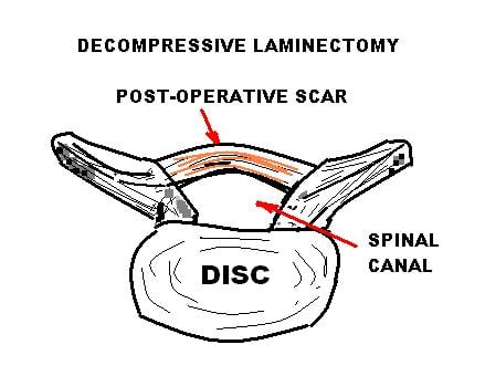
Late. Recurrent stenosis from regrowth of tissue, post-laminectomy instability, adjacent segment disease and failed back surgery syndrome. Epidural fibrosis, the post-operative scar that forms over the exposed dura, is a recognised cause of persistent or recurrent symptoms after surgery.
Post-laminectomy instability. It is more likely after:
- Excessive facet removal (more than 50%)
- Pre-existing spondylolisthesis not fused
- Degenerative scoliosis
- Multi-level decompression
- Young, active patient
It presents as recurrent symptoms after initial improvement, progressive back pain and leg symptoms, and slip progression on imaging. Prevent it by limiting facet resection, preserving the posterior tension band and adding fusion when the risk factors are present; once it develops, salvage fusion is more complex.
Postoperative Care
Mobilise the day of surgery or the next day - early ambulation is the single most useful instruction after a decompression. Document a fresh neurological examination, because this is the comparator against which any later deficit is judged, and a new deficit here is the finding that changes management urgently. Pain management and DVT prophylaxis alongside.
Physiotherapy assessment and progressive ambulation. Discharge is a functional decision, not a calendar one: ambulatory, pain controlled, voiding, and no new deficit. Watch for the two early problems that masquerade as ordinary post-operative pain - a CSF leak (positional headache, boggy wound) and cauda equina compression from haematoma (worsening rather than improving leg symptoms, retention).
Expect a longer stay (2-5 days), bracing in selected cases, more restricted early activity, and fusion precautions for 3-6 months. Note the source of the length-of-stay difference: in the Swedish Spinal Stenosis Study mean hospitalisation was 7.4 days with fusion against 4.1 without - so the added morbidity of fusion is measurable, and it bought no clinical advantage in that trial.
- Laminectomy Alone
- 1-3 days
- With Fusion
- 2-5 days
- Laminectomy Alone
- Immediate
- With Fusion
- Immediate with precautions
- Laminectomy Alone
- 2-4 weeks
- With Fusion
- 4-6 weeks
- Laminectomy Alone
- 2-4 weeks
- With Fusion
- 4-6 weeks
- Laminectomy Alone
- 6-12 weeks
- With Fusion
- 3-6 months
- Laminectomy Alone
- 3 months
- With Fusion
- 6-12 months
Rehabilitation. Physiotherapy covers flexibility, aerobic conditioning and posture and body mechanics, with core strengthening delayed 4-6 weeks. Alongside it, optimise weight, stop smoking (especially if fused) and modify activity as needed.
Outcomes and Prognosis
What to promise. Claudication improves in 70-80% of patients and 70-80% are satisfied; walking distance improves significantly, and back pain improvement is variable (50-60%). In the SPORT stenosis cohort walking capacity improved significantly with surgery.
Durability. Most patients maintain their improvement for 5-10 years. Some recur through regrowth of tissue, adjacent segment disease may develop, and the reoperation rate was approximately 18% at 8 years in the SPORT surgical stenosis cohort, with recurrent stenosis and progressive spondylolisthesis the leading indications (Gerling et al., 2016).
Prognostic factors. The patient who does well has predominant leg symptoms, severe imaging stenosis that correlates with those symptoms, a short symptom duration, no major comorbidity and does not smoke. The patient who does badly is more likely to have:
- Predominant back pain
- Mild imaging stenosis
- Long symptom duration
- Major comorbidities
- Obesity, smoking
- Psychiatric comorbidity
Guidelines, Registries & Global Practice
Global Epidemiology
Degenerative lumbar spinal stenosis is the leading indication for spinal surgery in adults over 65, and its prevalence rises steeply with age as the world's population ages. Decompression for stenosis is one of the highest-volume elective spinal procedures worldwide. Cross-country comparisons consistently show wide variation in rates of decompression and, in particular, in the proportion of decompressions accompanied by fusion - reflecting genuine clinical equipoise on the fusion question rather than differences in disease burden.
Guideline and Registry Comparison
- Position on Decompression and Fusion
- Decompression recommended for symptomatic stenosis failing non-operative care; fusion reserved for instability/deformity, not routine for stenosis alone
- Evidence Basis
- Level I-II RCT evidence (SPORT, SLIP, Swedish study)
- Position on Decompression and Fusion
- Consider decompression for stenosis when non-surgical care fails and symptoms warrant; does not endorse routine fusion for uncomplicated stenosis
- Evidence Basis
- Systematic review, Grade based
- Position on Decompression and Fusion
- Endorse decompression for refractory neurogenic claudication; individualised fusion decision based on stability
- Evidence Basis
- RCT and registry-informed
- Position on Decompression and Fusion
- Decompression first-line surgical option; fusion when documented instability or significant deformity
- Evidence Basis
- Consensus plus RCT evidence
The two pivotal fusion trials published together in 2016 - the Swedish Spinal Stenosis Study (no fusion benefit) and the SLIP trial (fusion benefit in Grade I slip) - reached discordant conclusions, which is why guideline bodies converge on an individualised, stability-driven decision rather than a blanket recommendation. Registry data (for example large national spine registries in Sweden, Norway and the United Kingdom) broadly corroborate that decompression alone produces durable patient-reported improvement and that the addition of fusion increases cost, length of stay and morbidity without a consistent outcome advantage in stable stenosis.
Current Practice and Workforce
Lumbar decompression is delivered by both orthopaedic spine surgeons and neurosurgeons across public and private healthcare systems internationally. Practice patterns track the international evidence, with emphasis on appropriate patient selection, an adequate trial of non-operative management, and an individualised approach to fusion based on stability assessment. Consistent with the trial and registry data above, most stable stenosis is managed by decompression alone, with fusion reserved for documented instability or deformity.
Documentation and Medicolegal Considerations
Key documentation requirements include confirmation of neurogenic versus vascular claudication, imaging correlation with symptoms, a documented trial of conservative treatment, explicit discussion of whether fusion is required, and informed consent for potential complications including dural tear. Standing (and where indicated flexion-extension) radiographs should form part of the preoperative workup, because spondylolisthesis and dynamic instability may not be apparent on supine MRI.
MCQ Practice Points
Q: What feature best distinguishes neurogenic from vascular claudication?
A: Relief with flexion (shopping cart sign). Neurogenic claudication is relieved by sitting or bending forward because flexion increases spinal canal diameter. Vascular claudication is relieved by rest in any position because the issue is cardiovascular, not spinal.
Q: When should instrumented fusion be added to lumbar laminectomy for stenosis?
A: Fusion should be added in the presence of:
- Degenerative spondylolisthesis
- Pre-existing spinal instability (greater than 4mm translation on flexion-extension views)
- Removal of more than 50% of facet joints (iatrogenic instability)
- Significant degenerative scoliosis Laminectomy alone is appropriate for stable stenosis without spondylolisthesis.
Q: What did the SPORT trial show for lumbar spinal stenosis treatment?
A: The SPORT trial demonstrated that surgery was superior to conservative treatment at 2-4 years for symptomatic lumbar stenosis. Patients had significant improvement in walking capacity, pain, and function. Unlike disc herniation, the stenosis cohort showed sustained surgical benefit. Intent-to-treat analysis was limited by high crossover rate.
Q: What is the most commonly affected level in lumbar spinal stenosis?
A: L4-5 is the most commonly affected level (approximately 80% of cases), followed by L3-4. The L4-5 level is susceptible due to its high mobility and degenerative stress concentration.
Q: What percentage of facet joint should be preserved during laminectomy to maintain spinal stability?
A: More than 50% of the facet joint should be preserved. Removing more than 50% of the facet bilaterally risks iatrogenic instability and post-laminectomy spondylolisthesis, which may require subsequent fusion.
Clinical Decision Scenarios
Practise clinical reasoning and management decisions out loud
“A 70-year-old woman presents with 2 years of bilateral leg pain and numbness when walking. She can walk 100 meters before needing to stop. She finds relief when pushing a shopping trolley. Peripheral pulses are normal. MRI shows L4-5 central stenosis. How would you manage her?”
“During a planned L4-5 laminectomy for stenosis, you find the patient has degenerative spondylolisthesis that was not clearly appreciated on supine MRI. What do you do?”
“A 65-year-old man presents 2 years after L4-5 laminectomy with recurrent bilateral leg symptoms. He had excellent relief initially. What is your approach?”
“How do you distinguish neurogenic claudication from vascular claudication in a patient with leg pain on walking?”
Neurogenic vs Vascular Claudication
- Neurogenic: flexion relieves (shopping cart sign)
- Vascular: rest alone relieves
- Neurogenic: better uphill, worse downhill
- Vascular: worse uphill, better downhill
- Check pulses, ABI if uncertain
Fusion Decision (FUSE)
- Fifty percent facet removal (more than 50%)
- Unstable (spondylolisthesis, dynamic instability)
- Scoliosis (degenerative)
- Extensive multi-level decompression
SPORT Trial - Stenosis
- Surgery SUPERIOR to conservative (unlike disc)
- Benefits at 2-4 years, durable
- Walking capacity, function improved
- Level I evidence supports surgery
Surgical Technique
- Prone positioning, fluoroscopy for level
- Preserve more than 50% facet bilaterally
- Remove ligamentum flavum (main contributor)
- Dural tear 3-5%
Complications
- Dural tear: 3-5%
- Post-laminectomy instability if too much facet removed
- Recurrent stenosis over time
- Adjacent segment disease
Outcomes
- 70-80% improvement in claudication
- Duration of benefit: 5-10 years
- Reoperation rate: ~18% at 8 years (SPORT)
- Surgical advantage durable to 4 years (SPORT)
Evidence-Based Practice
SPORT Trial - Stenosis (Weinstein et al., 2008)
- Multicentre randomised and observational cohorts (n=289 randomised, 365 observational) of decompressive surgery vs usual non-operative care for stenosis WITHOUT spondylolisthesis
- Intention-to-treat analysis (randomised cohort): significant benefit favouring surgery on SF-36 bodily pain (mean difference 7.8, 95% CI 1.5 to 14.1), but not physical function or Oswestry at 2 years
- As-treated combined analysis: significant advantage for surgery across all primary outcomes from 3 months, maintained at 2 years
- Establishes surgery as more effective than non-operative care for symptomatic stenosis
- Defining Level I evidence underpinning surgical decompression for stenosis
SPORT Stenosis - 4-Year Outcomes (Weinstein et al., 2010)
- Four-year follow-up of the SPORT stenosis cohort (decompressive laminectomy vs non-operative care)
- As-treated analysis: clinically significant advantages for surgery maintained through 4 years
- Treatment effects - bodily pain 12.6 (95% CI 8.5-16.7), physical function 8.6 (95% CI 4.6-12.6), Oswestry -9.4 (95% CI -12.6 to -6.2)
- Secondary measures (bothersomeness, satisfaction, self-rated progress) also favoured surgery
- Demonstrates durability of surgical benefit over the medium term