Recognition and management of neurovascular complications following total knee arthroplasty including common peroneal nerve injury and vascular injuries
- Common peroneal nerve most vulnerable due to superficial course at fibular head
- Vascular injury rare but limb-threatening - requires emergency intervention
- Risk factors: valgus deformity, flexion contracture, RA, previous surgery
- Foot drop most common presentation - check ankle dorsiflexion immediately post-op
- Prevention: careful retraction, avoid excessive correction, knee flexion post-op
- “Viva scenario: Post-TKA foot drop - systematic approach to differentiation (peroneal nerve vs sciatic vs central cause) and management algorithm is essential. Know the anatomy of the popliteal fossa cold.
Epidemiology
Nerve injury. Common peroneal nerve palsy is the most frequent motor nerve injury after TKA. [1,2] Its incidence increases substantially in revision procedures and complex primary cases.
- Primary TKA
- 0.3-1.3%
- Revision TKA
- 2-3%
- High-Risk Cases
- Up to 9.5%
- Primary TKA
- 0.05-0.1%
- Revision TKA
- 0.1-0.3%
- High-Risk Cases
- Rare
- Primary TKA
- Very rare
- Revision TKA
- 0.1-0.2%
- High-Risk Cases
- Associated with hip pathology
- Primary TKA
- Very rare
- Revision TKA
- Case reports
- High-Risk Cases
- Often epidural-related
Deformity. The palsy rate climbs with the deformity being corrected [1,3]:
- Valgus greater than 10°: 3.3% peroneal nerve palsy
- Valgus greater than 15°: 6-9.5% peroneal nerve palsy
- Flexion contracture greater than 20°: 4-5% nerve palsy
Vascular injury. Rare, at 0.03-0.17% of TKA procedures [4,5]. The popliteal artery is the vessel most often injured, by laceration, thrombosis or intimal damage, and posterior osteophyte removal and tibial preparation increase the risk. When it occurs the mortality rate is 5-7%, a historical figure that modern series report lower, and the amputation rate is 10-42% when recognition is delayed beyond 6-8 hours. [4]
Anatomy
The popliteal fossa. From superficial to deep (posterior to anterior), the fossa holds:
- Tibial nerve - most posterior, crossing the popliteal vessels from lateral to medial
- Popliteal vein - intermediate
- Popliteal artery - deepest, closest to the posterior capsule
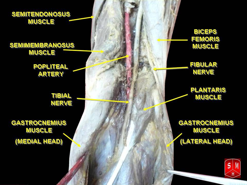
The artery's margin. With the knee in extension the popliteal artery lies about 5-10mm behind the posterior tibial cortex [6], and the distance decreases with knee flexion beyond 90°. Genicular branches tether it, limiting its mobility. Lying immediately behind the posterior capsule and the back of the tibial plateau, it is most vulnerable during posterior capsule release and tibial cutting: a posterior saw cut, an over-driven retractor, a release for a fixed deformity.
The genicular branches. Each is at risk from a particular step of the operation:
- Origin
- Popliteal, above joint
- Risk in TKA
- Medial release, posterior capsule
- Origin
- Popliteal, above joint
- Risk in TKA
- Lateral release
- Origin
- Posterior popliteal
- Risk in TKA
- Posterior cruciate ligament
- Origin
- Popliteal, below joint
- Risk in TKA
- Tibial preparation
- Origin
- Popliteal, below joint
- Risk in TKA
- Lateral tibial exposure
High-risk manoeuvres for the popliteal artery are these:
- Posterior capsule release (closest proximity)
- Posterior tibial osteophyte removal
- Oscillating saw during the tibial cut (over-penetration)
- Anterior tibial retractor placement (impingement)
- Tibia extraction after cementation
The common peroneal nerve. It originates from the sciatic nerve at the apex of the popliteal fossa, at a variable level, and then:
- Descends along the medial border of the biceps femoris tendon
- Wraps around the fibular neck, a superficial, vulnerable location
- Is separated from bone there by only 2-3mm of connective tissue
- Divides into the superficial and deep peroneal nerves
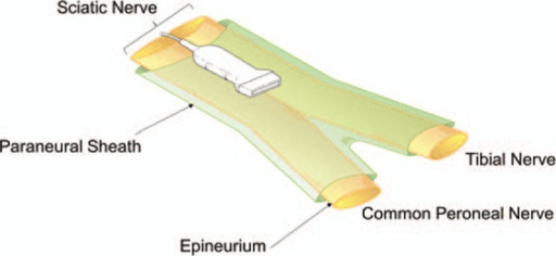
Why it is vulnerable. Its superficial course at the fibular head leaves it open to traction, compression and direct injury. Fixed at two points, the sciatic notch and the fibular tunnel, it has limited excursion when the limb lengthens, and the tether at the fibular tunnel makes it susceptible to traction during valgus correction.
Little protects it at the fibular neck. There is minimal soft tissue over it, so positioning, retractors and dressings can compress it, and its poor blood supply there makes a watershed zone vulnerable to ischaemia. Release of the iliotibial band and lateral structures places it at risk, and arthritis or osteophytes at the proximal tibiofibular joint may alter the anatomy.
Classification
Classifying neurovascular injury after TKA serves two purposes in the viva: it communicates severity and prognosis, which drives whether you observe, decompress or repair, and it directs the timing of intervention.
Nerve injury. The peroneal palsy that follows TKA is graded with the same framework used for any peripheral nerve injury, and the grade determines recovery potential.
Neurapraxia is the commonest after TKA. A conduction block from compression or ischaemia leaves axon and connective tissue intact, so there is no Wallerian degeneration, and sensation is often spared more than motor function. Recovery is expected over days to weeks (up to about 12 weeks); it is the basis of most traction and compression palsies that recover with dressing release and knee flexion.
Axonotmesis disrupts the axon but leaves the endoneurial tubes intact, and Wallerian degeneration occurs distally. It becomes detectable on EMG/NCS once degeneration is established, at 3-4 weeks. Recovery depends on axonal regrowth at roughly 1mm/day, so it takes months and is often incomplete.
Neurotmesis is complete transection or dense intraneural scar, with no spontaneous recovery. It is rare in TKA, and a transection recognised intra-operatively is an indication for repair. Otherwise it is distinguished from axonotmesis only by failure of recovery or by operative findings.
Vascular injury. Lesions are grouped by whether they occlude, bleed or present late:
- Lesion
- Thrombosis / intimal flap
- Mechanism in TKA
- Tourniquet on diseased vessel, traction, hyperextension kinking
- Typical Timing
- Intra-op to hours
- Lesion
- Laceration / transection
- Mechanism in TKA
- Saw over-penetration, posterior retractor, osteophyte removal
- Typical Timing
- Intra-op
- Lesion
- Transection with ischaemia + bleeding
- Mechanism in TKA
- Direct sharp injury to popliteal artery
- Typical Timing
- Intra-op
- Lesion
- Pseudoaneurysm
- Mechanism in TKA
- Partial wall injury, contained leak
- Typical Timing
- Days to weeks
- Lesion
- Arteriovenous fistula
- Mechanism in TKA
- Concurrent artery + vein injury
- Typical Timing
- Weeks to months
In the Pennsylvania Hospital series (Calligaro et al.) acute arterial complications were grouped by mechanism (ischaemia, bleeding, ischaemia plus bleeding, pseudoaneurysm) and by timing. Roughly half were recognised on the day of surgery and half over the first to fifth post-operative day, which is why delayed recognition drives morbidity.
Contributing Factors
Preoperative valgus is the best-supported risk factor, but resist quantifying it. The largest series (10,361 TKAs) found the association highly significant (p less than 0.0001), with 10 of its 30 palsy patients having valgus of 12 degrees or more, yet it never published a relative risk, and no study cited on this page supports a numerical multiplier such as "3-4 times" or "8-10 times". Say the risk rises steeply with the magnitude of valgus and that a large correction should change your plan; do not attach a number the literature does not contain.
And it is contested. A smaller series (1,476 TKAs, 19 events) failed to reproduce the association at all. That is a study underpowered to detect it rather than one disproving it, but it is why "valgus increases risk" is the defensible claim and any figure attached to it is not.
Why correction matters. A lateral soft-tissue contracture requires extensive release, and correction "lengthens" the lateral structures, the peroneal nerve among them. Valgus correction greater than 10-15° significantly increases nerve palsy risk through lateral soft-tissue stretching; staged correction or a constrained implant are the alternatives to consider.
Flexion contracture. Greater than 20° of fixed flexion does have a real number behind it: an odds ratio of 3.9 (p = 0.002) in 1,166 knees, from the tourniquet study cited below, where the valgus multipliers have none. The posterior capsule release it requires is close to the popliteal vessels, and restoring extension elongates the neurovascular structures.
Fixed valgus with a flexion contracture is the highest-risk combination for peroneal palsy: the lateral release lengthens the nerve.
Varus. The nerve palsy risk is lower than with valgus and the vascular injury risk similar; medial release does not tension the peroneal nerve.
Clinical Presentation
Nerve injury
Motor. Foot drop, the inability to dorsiflex the ankle, with weakness of the toe extensors (EHL, EDL) and of eversion (peroneus longus and brevis). The gait shows foot slap and a high-stepping steppage pattern to clear the foot.
Sensory. Numbness over the dorsum of the foot, in the first web space (deep peroneal territory) and over the lateral leg (superficial peroneal territory). The lateral foot (sural nerve) may be spared.
When it appears. Regional anaesthesia may mask it initially, and it is typically recognised when the block wears off, at 12-24 hours. An outpatient must be examined before discharge, and ankle dorsiflexion power documented systematically.
The examination grades and maps the deficit:
- Ankle dorsiflexion (L4-5), graded 0-5
- Great toe extension (L5), graded 0-5
- Ankle eversion (L5-S1), graded 0-5
- Sensation in the first web space (deep peroneal)
- Sensation over the dorsum of the foot (superficial peroneal)
Although the common peroneal nerve is the most important and most-tested motor nerve injury after TKA, the most frequently injured nerve of all is the infrapatellar branch of the saphenous nerve, which crosses the front of the knee from medial to lateral and is divided by the standard medial parapatellar (or midline) approach in a large proportion of TKAs. The result is an area of numbness on the antero-lateral side of the incision and, in a minority, a painful neuroma at the medial end of the scar with pain on kneeling and a positive Tinel sign. It is usually a benign, well-tolerated sensory deficit that recovers or is ignored, but it is a genuine source of patient dissatisfaction and litigation, so it belongs in the consent discussion ("a patch of numbness beside the scar is expected"). A symptomatic neuroma is managed with desensitisation first, then injection, and excision/proximal transposition for the refractory case. The exam point: distinguish this common benign sensory injury from the rare but serious peroneal motor palsy.
Vascular injury
Acute limb ischaemia. Time is critical: irreversible muscle necrosis begins at 6 hours of warm ischaemia.
PPPPPP6 Ps
Hook:The same six Ps used in compartment syndrome and acute arterial occlusion teaching
Acute arterial injury. The presentations differ in urgency:
- Clinical Features
- Acute haemorrhage, hypotension, haematoma
- Urgency
- Immediate surgery
- Clinical Features
- 6 Ps, cool limb, absent pulses
- Urgency
- Emergency - within 6 hours
- Clinical Features
- Delayed ischaemia (hours), thrombus propagation
- Urgency
- Urgent - monitor closely
- Clinical Features
- Tense compartments, pain with passive stretch
- Urgency
- Emergent fasciotomies
Compartment syndrome may occur without direct vascular injury; the arterial tourniquet and post-operative bleeding can precipitate it.
Delayed lesions. A pseudoaneurysm presents with a pulsatile mass, delayed bleeding and pain, and may rupture; duplex ultrasound is diagnostic. An arteriovenous fistula presents with a bruit and swelling, and with high-output cardiac failure in chronic cases. Deep vein thrombosis, over days to weeks, gives calf swelling, pain and Homan's sign.
Differential diagnosis of post-TKA foot drop
- Clinical Features
- Ankle dorsiflexion weak, eversion weak, sensation dorsum foot
- Investigation
- Clinical, EMG at 3-4 weeks
- Clinical Features
- All below-knee motor/sensory loss
- Investigation
- Clinical, EMG, consider MRI spine/hip
- Clinical Features
- Back pain, bladder dysfunction, bilateral weakness
- Investigation
- Urgent MRI spine - neurosurgical emergency
- Clinical Features
- Back pain, dermatomal distribution, may have reflex changes
- Investigation
- MRI lumbar spine
- Clinical Features
- Upper motor neuron signs, other neurological findings
- Investigation
- CT/MRI brain
- Clinical Features
- Severe pain, tense compartments, late paralysis
- Investigation
- Clinical diagnosis, measure pressures if uncertain
Investigations
Imaging answers the vascular question and localises compression:
- Indication
- Suspected vascular injury, pseudoaneurysm, DVT
- Findings
- Flow abnormalities, aneurysm, thrombus
- Indication
- Arterial injury, planning for intervention
- Findings
- Laceration, thrombosis, pseudoaneurysm
- Indication
- Nerve compression, haematoma localisation
- Findings
- Haematoma, nerve oedema, compression
- Indication
- Non-invasive vascular assessment
- Findings
- Arterial anatomy, occlusion
- Indication
- Diagnostic and therapeutic (embolisation)
- Findings
- Gold standard for vascular injury
Nerve conduction studies and EMG are not useful in the acute setting, because Wallerian degeneration takes 2-3 weeks. A baseline study at 3-4 weeks if there is no recovery, repeated at 3 months, assesses recovery. The studies differentiate neurapraxia from axonotmesis and can localise the level of the lesion.
Management Algorithm
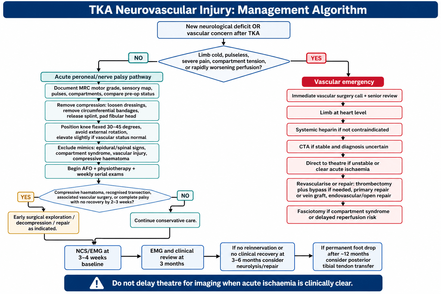
Acute nerve palsy
- Recognise and document: motor power (MRC grade 0-5) and the sensory examination, compared with pre-operative status if available, and a photograph of any skin changes.
- Remove compression: loosen all dressings immediately, remove circumferential bandages, release any splint, and make sure nothing presses on the fibular head.
- Position: flex the knee to 30-45° to reduce nerve tension, avoid external rotation of the leg, pad the fibular head, and elevate the leg slightly.
- Rule out other causes: epidural haematoma or abscess (back pain, bladder, bilateral signs), compartment syndrome (compartment tension) and vascular injury (pulses, perfusion).
- Document and communicate: inform the patient and family, record it in the notes, refer early to physiotherapy, and consider early surgical exploration if a compressive haematoma is a concern.
Acute vascular injury
- Recognise: a cold, pulseless, painful limb.
- Call for help: immediate vascular surgery consultation.
- Position: the limb at heart level, not elevated.
- Heparinise: systemic heparin if not contraindicated.
- Image: CTA if stable, direct to theatre if unstable.
- Do not delay: if the clinical diagnosis is clear, do not wait for imaging; proceed to the operating room.
By injury type, the immediate and definitive steps differ:
- Immediate Action
- Direct pressure, call vascular
- Definitive Treatment
- Primary repair or vein graft
- Immediate Action
- Heparinise, urgent vascular consult
- Definitive Treatment
- Thrombectomy ± bypass
- Immediate Action
- Emergency fasciotomies
- Definitive Treatment
- All 4 compartments, leave open
- Immediate Action
- Monitor if small, intervention if expanding
- Definitive Treatment
- Endovascular or open repair
- Immediate Action
- Usually not urgent
- Definitive Treatment
- Elective surgical or endovascular repair
Fasciotomy. All four compartments must be released. A single lateral incision can access the anterior, lateral and superficial posterior compartments, and a separate posteromedial incision the deep posterior. Leave the wounds open for delayed primary closure or split-skin grafting.
Surgical Technique
Most peroneal palsies are managed non-operatively. Surgery is reserved for (1) intra-operative recognition of nerve transection, (2) a compressive haematoma, (3) failure to recover by 3-6 months (decompression/neurolysis), (4) established permanent foot drop (tendon transfer), and (5) any vascular injury.
Indication. No clinical or electrical recovery by about 3 months, or a documented compressive lesion.
Approach. Supine or lateral, with a tourniquet optional and used judiciously. A lazy-S or curvilinear incision over the fibular neck, behind the biceps femoris tendon, finds the common peroneal nerve posterior to biceps femoris, and it is traced distally around the fibular neck.
Key steps at the fibular neck:
- Release the fibrous arch at the origin of peroneus longus, the classic constriction point
- External neurolysis of epineurial fibrosis, decompressing the superficial and deep branches
- Intraneural neurolysis only if a discrete fascicular constriction is found
- Preserve the nerve's vascular supply and avoid devascularising mobilisation
Rationale. Mont and colleagues found 30/31 patients (97%) improved and discontinued the AFO after decompression, against 3/9 (33%) managed non-operatively, supporting decompression when non-operative measures fail.
Pitfalls. Operating too early (most Sunderland I-II palsies recover spontaneously), missing a more proximal sciatic lesion, and iatrogenic injury during dissection at the fibular neck.
Complications
Neurovascular injury after TKA is itself a complication, but it generates its own cascade of secondary complications that the examiner expects you to anticipate, recognise and manage.
- Context
- Palsies that fail to recover fully (varies widely by series)
- Prevention
- Avoid over-correction of valgus; protect nerve; release dressings early
- Recognition & Management
- AFO; tendon transfer after ~12 months if no recovery
- Context
- After prolonged ischaemia/reperfusion or bleeding
- Prevention
- Limit tourniquet time; meticulous haemostasis; high index of suspicion
- Recognition & Management
- Pain on passive stretch, tense compartments; emergency 4-compartment fasciotomy
- Context
- Delayed recognition of vascular injury
- Prevention
- Recognise ischaemia early; do not delay for imaging when clear
- Recognition & Management
- Urgent revascularisation within the ischaemic window
- Context
- Following late revascularisation
- Prevention
- Timely revascularisation; consider fasciotomy
- Recognition & Management
- Monitor CK, renal function, potassium; fluids, renal support
- Context
- Delayed (days to months)
- Prevention
- Careful posterior dissection; recognise partial wall injury
- Recognition & Management
- Duplex/CTA; endovascular or open repair
- Context
- After significant nerve injury
- Prevention
- Early mobilisation, analgesia, nerve care
- Recognition & Management
- Multidisciplinary pain management
- Context
- Vascular injury (historical rates; lower in modern series)
- Prevention
- Early recognition, haemodynamic resuscitation
- Recognition & Management
- Resuscitation, haemorrhage control, ITU support
Postoperative Care
After a vascular repair. Care is shared with the vascular team. Distal pulses, Doppler signals, capillary refill and compartment status are monitored, hourly at first, with vigilance for compartment syndrome after reperfusion and a low threshold for fasciotomy. Vascular surgery directs the anticoagulant or antiplatelet regimen, and reperfusion injury is monitored with CK, renal function and electrolytes.
Rehabilitation milestones for both injuries:
- Nerve Injury Focus
- Dressing release, knee flexion, document exam
- Vascular Injury Focus
- Perfusion monitoring, fasciotomy if needed
- Nerve Injury Focus
- AFO, physiotherapy, baseline EMG at 3-4 weeks
- Vascular Injury Focus
- Wound care, anticoagulation, graft surveillance
- Nerve Injury Focus
- Repeat EMG; assess reinnervation
- Vascular Injury Focus
- Duplex surveillance of repair/graft
- Nerve Injury Focus
- Decide on neurolysis if no recovery
- Vascular Injury Focus
- Manage claudication, late stenosis
- Nerve Injury Focus
- Tendon transfer for permanent deficit
- Vascular Injury Focus
- Long-term vascular follow-up
Outcomes
Peroneal palsy recovers in most patients. Complete recovery is reported in 50-90% after primary TKA (the literature varies widely) and 40-70% after revision, and is variable in high-risk cases; partial recovery occurs in 10-30%, and no recovery in 5-20%. Recovery, when it comes, typically spans 12-24 months (mean 12-18 months) and may be incomplete; maximum recovery is typically reached by 2-3 years.
- Better Prognosis
- Delayed onset (dressings)
- Worse Prognosis
- Immediate post-operative
- Better Prognosis
- Partial palsy
- Worse Prognosis
- Complete palsy
- Better Prognosis
- Evidence of reinnervation
- Worse Prognosis
- No motor unit recruitment
- Better Prognosis
- Traction/compression
- Worse Prognosis
- Direct transection
- Better Prognosis
- No diabetes or neuropathy
- Worse Prognosis
- Diabetic neuropathy present
- Better Prognosis
- Early dressing release, AFO
- Worse Prognosis
- Delayed recognition
Function. Most patients with complete recovery have normal function, and those left with an AFO-dependent foot drop can still walk. Satisfaction is lower than after an uncomplicated TKA; consider revision or tendon transfer for a permanent deficit.
Vascular injury. Outcome depends on how quickly the injury is recognised:
- Limb Salvage Rate
- 90-95%
- Amputation Rate
- 5-10%
- Limb Salvage Rate
- 70-80%
- Amputation Rate
- 20-30%
- Limb Salvage Rate
- 40-60%
- Amputation Rate
- 40-60%
After limb salvage. Patients may have claudication, repeat intervention may be required, and chronic pain and disability are common.
Medicolegal. Neurovascular injury is a recognised complication of TKA, the consent discussion must be documented, and the litigation risk is significant.
The dominant theme examiners probe is that delayed recognition - not the injury itself - is the driver of catastrophic outcome and the most common source of litigation. A documented pre-operative neurovascular status and a documented timely post-operative re-examination are your protection both clinically and medicolegally.
Prevention Strategies
Before surgery. Document the pre-operative neurological status (ankle dorsiflexion, sensation) and assess the peripheral pulses, with an ABI if vascular disease is suspected. Consider a vascular surgery consultation for calcified vessels or prior vascular surgery, review previous surgical records for anatomical variations, and inform the patient of the increased risk if risk factors are present.
Planning the correction. Template to predict the correction required. Consider a constrained implant for severe deformity, which reduces the soft-tissue release, plan staged correction for valgus greater than 15°, and have vascular surgery backup in a high-risk case.
Retractors. A lateral retractor on the fibular head compresses the peroneal nerve directly, so place it on the proximal tibia, and do so with the knee flexed. Retract gently and intermittently, avoiding prolonged pressure, and consider visualising the nerve in high-risk cases.
Correcting the deformity. Excessive valgus correction is a traction injury to the peroneal nerve. Limit valgus correction to 10-15° in a single stage; for severe valgus consider a constrained implant to reduce the release, stage the correction for extreme deformity, and accept slight under-correction rather than a nerve palsy.
Tourniquet. Prolonged tourniquet time causes ischaemic nerve injury. A wider cuff (10cm minimum) distributes the pressure, the inflation pressure should be lower (limb occlusion pressure + 100mmHg), duration is limited to less than 90-120 minutes, and tourniquet-free TKA is worth considering in high-risk patients.
Closure and dressings. Tight closure and dressings cause compression and compartment syndrome. Avoid excessive tension on closure and circumferential tight bandages, use loose, well-padded dressings, and monitor post-operatively.
In the highest-risk knee — a severe fixed valgus deformity with a flexion contracture that needs extensive lateral release and significant lengthening of the lateral side — some surgeons perform a prophylactic decompression of the common peroneal nerve at the time of TKA, releasing it at the fibular neck (the constricting peroneus-longus arch) so the corrected, lengthened nerve is not tethered. The rationale is to pre-empt the traction palsy rather than treat it afterwards, as an adjunct to the standard preventive measures. It remains debated and selective — the evidence is limited and it adds operative time and a second wound — so it is not routine, but it is a legitimate, examinable option for the extreme valgus / flexion-contracture knee, while the mainstay is still correction strategy and postoperative knee-flexion positioning.
After surgery. Document pulses and ankle dorsiflexion in recovery, and repeat the examination when the regional block wears off. Monitor for compartment syndrome, especially if a tourniquet was used, and keep a low threshold for loosening dressings if there is concern.
The high-risk knee. Splint the knee in flexion: slight flexion (20-30°) at closure in high-risk cases, and 30-45° for 48-72 hours in the high-risk post-operative protocol. Add serial neurological examinations and early physiotherapy input, and consider routine Doppler assessment if there is vascular concern.
Guidelines, Registries & Global Practice
Global Epidemiology, Guidelines and Registry Evidence
Global Burden and Practice Variation
Total knee arthroplasty is among the highest-volume elective orthopaedic procedures worldwide, with over a million performed annually in the United States alone and rising volumes across Europe, the United Kingdom, Australia and Asia. Because neurovascular injury is rare, single-centre series under-represent it; the most reliable incidence figures come from large institutional cohorts and prospectively collected datasets. The verified pooled picture is consistent across geographies: peroneal palsy in roughly 0.3-1.3% of primary TKA (higher with valgus deformity and revision), and arterial injury in roughly 0.03-0.17%.
Practice variation centres on three modifiable areas: tourniquet use (duration and whether used at all), deformity correction strategy (single-stage versus staged, constrained implants), and pre-operative vascular screening of patients with peripheral arterial disease. Tourniquet-free and limited-tourniquet TKA has gained traction in Europe and Australasia partly to reduce ischaemic nerve risk, whereas tourniquet use remains common in North America.
Guidance Comparison
- Relevant Position / Guidance
- No dedicated neurovascular-injury guideline; the AAOS surgical management of osteoarthritis of the knee CPG informs patient selection and shared decision-making, and emphasises documented informed consent for recognised complications
- Evidence Level / Status
- Consensus/CPG-based
- Relevant Position / Guidance
- Covers patient selection, optimisation and information-giving for primary hip/knee replacement; complications such as nerve and vascular injury fall under the consent and shared-decision recommendations rather than a specific algorithm
- Evidence Level / Status
- Guideline (GRADE-informed)
- Relevant Position / Guidance
- Best-practice principles for knee arthroplasty stress pre-operative neurovascular documentation, vascular assessment in PVD, and prompt escalation of suspected ischaemia; align with general vascular-emergency standards
- Evidence Level / Status
- Professional consensus
- Relevant Position / Guidance
- Educational principles emphasise protecting the popliteal vessels (retractor medial to PCL, avoid hyperextension) and the peroneal nerve during lateral release, mirroring the Ninomiya anatomical findings
- Evidence Level / Status
- Expert/educational consensus
- Relevant Position / Guidance
- Promotes registry-based outcome monitoring and risk-factor awareness (valgus deformity, flexion contracture, RA, PVD); supports tourniquet stewardship
- Evidence Level / Status
- Consensus/registry-informed
- Relevant Position / Guidance
- Acute limb ischaemia pathways apply directly: urgent revascularisation within the ischaemic window, heparinisation, and consideration of endovascular first-line at experienced centres
- Evidence Level / Status
- Guideline
Registry Evidence
- What it captures
- Implant survival, revision rates, indications - not specific neurovascular complications
- Relevance to this topic
- Confirms procedural volume and revision burden (a higher-risk group for nerve injury); complication-specific data require linked datasets
- What it captures
- Outcomes and revision; complication coding limited
- Relevance to this topic
- Large denominators contextualise rarity of vascular injury; mortality and revision tracked
- What it captures
- Long-running outcome and revision data
- Relevance to this topic
- Benchmark for outcomes; reinforces that neurovascular injury is rare relative to aseptic loosening/infection
- What it captures
- Prospectively collected systemic and local complications
- Relevance to this topic
- The primary source of the verified incidence figures used throughout
- Neurovascular complication rates remain consistent across international registries and institutional series; high-volume centres with fellowship-trained surgeons report lower overall complication rates.
MCQ Practice Points
High-Yield MCQ and Exam Points
Q: A patient develops foot drop after TKA. Which muscle action is the most sensitive bedside test of common peroneal nerve function? A: Ankle dorsiflexion (tibialis anterior). Loss of dorsiflexion is the hallmark; eversion (peroneus longus/brevis) and great-toe extension are also affected.
Q: Where is the popliteal artery in relation to the joint line during TKA, and where should a posterior retractor sit? A: It is a lateral structure at the joint line (Ninomiya); the retractor should be placed medial to the tibial plateau midline, and inserted no more than 1cm, avoiding placement lateral to the PCL.
Q: When should nerve conduction studies/EMG be performed after a post-TKA peroneal palsy? A: At 3-4 weeks for a baseline (Wallerian degeneration is not detectable acutely), repeated at ~3 months for prognosis. Ordering EMG on day 1 is a classic wrong answer.
Q: What is the single most important prognostic factor for recovery of a post-TKA peroneal palsy? A: Whether the palsy is incomplete (partial) versus complete at onset - partial palsies are far more likely to recover fully (Asp/Rand).
Discriminating facts examiners test:
- The popliteal artery is a lateral structure at the joint line; a posterior retractor placed lateral to the PCL or inserted more than 1cm endangers it (Ninomiya). Place retractors medial to the tibial plateau midline.
- Hyperextension tents and kinks the popliteal artery - dangerous during patellar preparation, especially in atherosclerotic vessels.
- Recovery is best when the palsy is incomplete (partial) at onset and when there is delayed onset attributable to dressings (Asp/Rand; Idusuyi/Morrey).
- Tourniquet time over 120 minutes carries a 7.7% neurological complication rate (Horlocker); flexion contracture over 20 degrees gives OR 3.9.
- Operative decompression after failed non-operative treatment improved 97% of patients versus 33% (Mont).
- Indirect (traction/thrombotic) injury is the commonest vascular mechanism in TKA, in contrast to direct injury in THA (Parvizi/Pulido).
Exam Viva Scenarios
Practise clinical reasoning and management decisions out loud
“You are asked to see a 68-year-old woman on the ward day 1 after a left TKA for severe valgus osteoarthritis. The nurses are concerned because she cannot lift her left foot off the bed. How do you approach this patient?”
“Two hours after a primary TKA, the recovery nurse reports the foot is cold, mottled and the patient has severe calf pain. The dorsalis pedis and posterior tibial pulses are absent. Walk me through your management.”
“A patient has a complete peroneal nerve palsy after TKA for valgus deformity. There is no clinical or electrical recovery at 4 months. The patient asks what can be done. How do you counsel and manage them?”
Incidence
- Peroneal nerve palsy: 0.3-1.3% primary TKA
- Valgus greater than 15°: 6-9.5% peroneal palsy rate
- Vascular injury: 0.03-0.17%
- Amputation rate with delayed vascular recognition: 10-42%
Risk Factors
- Valgus greater than 10-15° correction
- Atherosclerosis / PVD
- Stiff knee / flexion contracture greater than 20°
- Calcified vessels
- Underlying RA
- Limb lengthening (excessive correction)
- Anatomical variants / previous surgery
- Revision surgery
Anatomy
- Peroneal nerve wraps around fibular neck - only 2-3mm protection
- Popliteal artery 5-10mm from posterior tibial cortex
- Nerve fixed at sciatic notch and fibular tunnel - limited excursion
- Vulnerable to traction with valgus correction
Clinical Features
- Foot drop = ankle dorsiflexion weakness (L4-5)
- Cannot extend toes or evert ankle
- Sensory loss: dorsum of foot, first web space
- Vascular: 6 Ps - Pain, Pallor, Pulseless, Paresthesia, Paralysis, Poikilothermia
Prevention
- Retractor on tibia not fibular head
- Limit valgus correction to 10-15° single stage
- Flexed knee during posterior work
- Loose dressings, slight knee flexion post-op
- Tourniquet: wider cuff, lower pressure, limited duration
Management - Nerve
- Immediate: loosen dressings, flex knee 30-45°
- AFO for ambulation
- EMG at 3-4 weeks baseline
- 50-90% complete recovery over 12-24 months
- Posterior tibial tendon transfer if no recovery at 12 months
Management - Vascular Emergency
- Immediate vascular surgery consultation
- Heparinize if not contraindicated
- CTA if stable, direct to OR if unstable
- Irreversible ischemia at 6 hours - do NOT delay
- Fasciotomies for compartment syndrome - all 4 compartments
Viva Answers
- Systematic exam: motor, sensory, vascular, compartments
- Exclude emergencies: compartment syndrome, vascular injury, epidural pathology
- Immediate: loosen dressings, flex knee, document, counsel
- EMG too early acutely - need 3-4 weeks for Wallerian degeneration
- Prognosis: 70-90% recover, 12-24 months timeline
Evidence Base
Landmark Evidence
Nerve Injury After Primary TKA - Incidence and Recovery (Columbia Series)
- 19 neurological complications among 1,476 primary TKAs (1970-1998) - overall incidence 1.3%
- A larger-than-expected proportion of rheumatoid knees sustained neurological injury
- In this series valgus deformity, flexion contracture, epidural analgesia and prolonged tourniquet were not statistically associated with palsy - illustrating genuine controversy over risk factors
- All patients showed at least partial recovery with conservative treatment, most recovering completely
Peroneal Nerve Palsy After TKA - Predisposing and Prognostic Factors (Mayo Clinic)
- 32 peroneal palsies among 10,361 TKAs (1979-1992)
- Preoperative valgus deformity (p less than 0.0001), epidural analgesia for postoperative pain (p less than 0.03) and previous lumbar laminectomy (p less than 0.04) were significantly associated with palsy
- High frequency of DELAYED presentation - postulated to relate to impaired proprioception/sensation under epidural analgesia and unprotected limb positioning
- The epidural signal is strongest in a dedicated subgroup the card previously omitted: among 4,388 arthroplasties from 1988 to 1992 there were 25 palsies, of which 18 followed epidural anaesthesia against 5 general and 2 spinal (p less than 0.03)
- A NEGATIVE WORTH KNOWING: previous proximal tibial osteotomy doubled the relative risk but did not reach significance (p less than 0.4), so it is not in the same evidential class as valgus, epidural or laminectomy
- Ten of the thirty patients had preoperative valgus alignment of 12 degrees or more; the control group was 100 patients computer-matched for age, sex and operating surgeon
- Double-crush phenomenon proposed for patients with prior laminectomy or asymptomatic neuropathy
Peroneal Palsy After TKA - Prognosis for Recovery (Mayo Clinic)
- 26 peroneal palsies after 8,998 TKAs (1972-1985); 18 complete and 8 incomplete
- At mean 5.1 years, recovery was complete in 13 and partial in 12 (only one had no recovery)
- Complete recovery was significantly more likely when the initial palsy was incomplete (partial)
- Patients with partial palsy and with complete recovery had significantly higher knee scores - severity at onset is the key prognostic factor
Popliteal Artery Location and Risk During TKA (Cadaveric + MRI Anatomy)
- Intra-operative arteriograms and 50 transverse MRI scans defined the popliteal artery as a LATERAL structure at the joint line
- A posterior retractor placed the artery at risk when positioned lateral to the PCL or inserted more than 1cm into soft tissue
- Hyperextension produced dramatic tenting/kinking of the artery; both hyperflexion and hyperextension are dangerous, especially in atherosclerotic vessels
- Recommendation: place posterior retractors MEDIAL to the tibial plateau midline and avoid extremes of flexion/extension
Tourniquet Time and Neurological Complications After TKA
- 1,166 knee replacements with tourniquet time over 120 min; 129 nerve palsies in 90 patients (overall 7.7%) - the counts differ because 39 patients had BOTH peroneal and tibial deficits (85 peroneal and 44 tibial lesions in total)
- Mean total tourniquet time was 145 minutes (range 120 to 308)
- Risk rose with longer tourniquet time (OR 2.8 per 30-min increase, p less than 0.001) and preoperative flexion contracture over 20 degrees (OR 3.9, p = 0.002); YOUNGER age was also associated, the odds ratio being 0.7 per 10-year increase in age (p less than 0.001)
- Complete recovery in 76/85 (89%) peroneal and 44/44 (100%) tibial palsies
- A reperfusion (deflation) interval only modestly reduced risk - total tourniquet time is the dominant driver
Operative Decompression for Peroneal Nerve Palsy
- 31 patients undergoing decompression after at least 2 months of non-operative management
- Epineurial fibrosis and fibrous bands constricting the nerve at the fibular head and proximal peroneus longus origin were found intra-operatively
- 30/31 (97%) improved and discontinued the AFO versus only 3/9 (33%) managed non-operatively (p less than 0.01)
- Supports operative decompression when non-operative measures fail to improve within ~2 months
References
References
-
Schinsky MF, Macaulay W, Parks ML, Kiernan H, Nercessian OA. Nerve injury after primary total knee arthroplasty. J Arthroplasty. 2001;16(8):1048-1054. PMID: 11740762. DOI
-
Idusuyi OB, Morrey BF. Peroneal nerve palsy after total knee arthroplasty. Assessment of predisposing and prognostic factors. J Bone Joint Surg Am. 1996;78(2):177-184. PMID: 8609107. DOI
-
Nercessian OA, Ugwonali OF, Park S. Peroneal nerve palsy after total knee arthroplasty. J Arthroplasty. 2005;20(8):1068-1073. PMID: 16376265. DOI
-
Calligaro KD, Dougherty MJ, Ryan S, Booth RE. Acute arterial complications associated with total hip and knee arthroplasty. J Vasc Surg. 2003;38(6):1170-1177. PMID: 14681604. DOI
-
Rand JA. Vascular complications of total knee arthroplasty. Report of three cases. J Arthroplasty. 1987;2(2):89-93. PMID: 3612144. DOI
-
Ninomiya JT, Dean JC, Goldberg VM. Injury to the popliteal artery and its anatomic location in total knee arthroplasty. J Arthroplasty. 1999;14(7):803-809. PMID: 10537254. DOI
-
Asp JP, Rand JA. Peroneal nerve palsy after total knee arthroplasty. Clin Orthop Relat Res. 1990;(261):233-237. PMID: 2245551.
-
Rose HA, Hood RW, Otis JC, Ranawat CS, Insall JN. Peroneal nerve palsy following total knee arthroplasty. A review of The Hospital for Special Surgery experience. J Bone Joint Surg Am. 1982;64(3):347-351. PMID: 7061551.
-
Horlocker TT, Hebl JR, Gali B, Jankowski CJ, Burkle CM, Berry DJ, et al. Anesthetic, patient, and surgical risk factors for neurologic complications after prolonged total tourniquet time during total knee arthroplasty. Anesth Analg. 2006;102(3):950-955. PMID: 16492857. DOI
-
Mont MA, Dellon AL, Chen F, Hungerford MW, Krackow KA, Hungerford DS. The operative treatment of peroneal nerve palsy. J Bone Joint Surg Am. 1996;78(6):863-869. PMID: 8666604.
-
Parvizi J, Pulido L, Slenker N, Macgibeny M, Purtill JJ, Rothman RH. Vascular injuries after total joint arthroplasty. J Arthroplasty. 2008;23(8):1115-1121. PMID: 18676115. DOI
-
Pulido L, Parvizi J, Macgibeny M, Sharkey PF, Purtill JJ, Rothman RH, Hozack WJ. In hospital complications after total joint arthroplasty. J Arthroplasty. 2008;23(6 Suppl 1):139-145. PMID: 18722311. DOI
-
Troutman DA, Dougherty MJ, Spivack AI, Calligaro KD. Updated strategies to treat acute arterial complications associated with total knee and hip arthroplasty. J Vasc Surg. 2013;58(4):1037-1042. PMID: 23747133. DOI
Note on sources: a previously cited reference (attributed to Abularrage et al., "Arterial injury during primary total joint arthroplasty: a 13-year review", J Vasc Surg 2013) could not be located in PubMed during verification and has been removed; the same epidemiological points are covered by the verified Calligaro/Troutman series above.