Catastrophic Complication | Quadriceps or Patellar Tendon
- Devastating complication with poor functional outcomes
- Prevention is critical - protect extensor mechanism during surgery
- Patellar tendon ruptures have worse outcomes than quadriceps
- Allograft reconstruction is treatment of choice for chronic ruptures
- Many patients cannot achieve active extension despite treatment
- “Risk factors: previous surgery, diabetes, steroid use, MUA
- “Acute: primary repair with augmentation
- “Chronic: allograft (Achilles, whole extensor mechanism)
- “Extension lag is functional measure of success
Overview and Epidemiology
Extensor mechanism disruption after TKA is uncommon but devastating. It covers rupture of the patellar tendon, rupture of the quadriceps tendon, and patellar fracture extending into the tendon insertion. Outcomes are often poor, and many patients lose the ability to actively extend the knee.
Why it matters. Without active extension the patient cannot climb stairs, rise from a chair or walk normally. Even after a successful reconstruction many are left with a permanent extension lag and reduced functional capacity, which makes this a quality-of-life altering complication.
Who is at risk. Previous knee surgery is the strongest factor, and a revision TKA carries a 3-4x higher risk; multiple operations leave scarred, less vascular tissue. The other factors:
- Tibial tubercle osteotomy
- Diabetes, smoking and vascular disease, through poor vascularity
- Steroid use, systemic or local; steroid injection weakens collagen
- Inflammatory arthritis, including RA, through impaired tissue quality
- Forceful manipulation, including MUA
- A pre-existing extension lag, the sign of an already weak extensor mechanism
How it happens. Failure may occur during surgery, in the early postoperative period, late and spontaneously in weakened tissue, or during an MUA. The classification by cause below sets these out with how each is prevented.
Pathophysiology and Mechanisms
The chain. The extensor mechanism runs from the quadriceps muscle through the quadriceps tendon, patella and patellar tendon to the tibial tubercle. Quadriceps force reaches up to 7x body weight and patellofemoral contact 3x body weight on stairs, with tendon stress concentrated at the insertions. After TKA the weakest point is the patellar tendon.
Blood supply decides healing. The quadriceps tendon has a good supply from muscle, heals better, and primary repair is often possible. The patellar tendon depends on the inferior pole of the patella and the fat pad; fat pad resection, previous surgery, diabetes and lateral release all compromise it, and loss of these sources leaves poor healing potential. That is why patellar tendon injuries have worse outcomes and often need allograft. The tibial tubercle has moderate vascularity and heals as bone, and a TTO is fixed with screws.
What the arthroplasty itself does. Three technical factors weaken the mechanism:
- Fat pad resection - compromises the patellar tendon's blood supply
- Lateral release - destabilises the patella
- Component malposition - abnormal stress on the mechanism
Classification Systems
Three descriptions are used together: where the mechanism failed, how long ago, and why. Location determines prognosis and treatment options; timing determines whether the ends can still be repaired; cause points to prevention.
- Incidence
- 30-40%
- Prognosis
- Better
- Primary Treatment
- Primary repair with augmentation
- Incidence
- 40-50%
- Prognosis
- Worst
- Primary Treatment
- Allograft reconstruction
- Incidence
- 20-30%
- Prognosis
- Variable
- Primary Treatment
- ORIF vs revision patella
Clinical Assessment
History. The answers that shape management:
- Mechanism - a fall, a sudden pop, or an intraoperative event
- Timing relative to the TKA
- Symptoms - inability to extend, giving way
- Previous surgeries and comorbidities (diabetes, steroids, RA), for the risk profile
Examination. Look for swelling and ecchymosis around the knee and note the previous surgical scars. Feel for a gap at the rupture site and note where the patella sits. Then test active extension, which decides whether the rupture is complete.
Inability to actively extend the knee is pathognomonic for complete extensor mechanism rupture. Test with patient supine, leg straight - ask to lift leg off bed. If unable to maintain straight leg raise = complete disruption.
- Patella Position
- Patella ALTA (high-riding)
- Palpable Defect
- Below patella
- Extension Test
- Complete loss of extension
- Patella Position
- Patella BAJA (low)
- Palpable Defect
- Above patella
- Extension Test
- Complete loss of extension
- Patella Position
- May be normal
- Palpable Defect
- Tenderness only
- Extension Test
- Weak extension, may have lag
Extension lag. The knee can hold extension if placed there but cannot actively achieve full extension. A lag indicates a partial disruption or a weak or reconstructed mechanism, and it is measured in degrees (a 15-degree extension lag, for example).
Differential. A new active extension deficit after TKA has causes other than rupture, infection among them.
- Distinguishing Feature
- Total loss of active extension, palpable gap, patella alta/baja
- Key Investigation
- Lateral XR (Insall-Salvati), USS/MRI
- Management Direction
- Repair or reconstruction
- Distinguishing Feature
- Weak extension with lag but some active power
- Key Investigation
- USS/MRI for tendon continuity
- Management Direction
- Often non-operative, brace and monitor
- Distinguishing Feature
- Defect at superior pole, patella baja
- Key Investigation
- XR plus MRI
- Management Direction
- Repair to patella bone tunnels
- Distinguishing Feature
- Crepitus, fracture line on XR, extension deficit
- Key Investigation
- Skyline plus lateral XR
- Management Direction
- ORIF, partial patellectomy or revision
- Distinguishing Feature
- Pain, effusion, instability rather than true lag
- Key Investigation
- Serial XR, exam under anaesthesia
- Management Direction
- Revision arthroplasty
- Distinguishing Feature
- Pain, warmth, raised CRP/ESR; may cause tissue failure
- Key Investigation
- Aspiration, CRP/ESR, alpha-defensin
- Management Direction
- Treat infection BEFORE any reconstruction
- Distinguishing Feature
- Intact tendon, lag resolves with effort/rehab
- Key Investigation
- Clinical, normal imaging
- Management Direction
- Physiotherapy, no surgery
A new extensor lag in a previously functioning TKA may be the first sign of periprosthetic joint infection eroding the tendon. Reconstructing over an unrecognised infection guarantees failure. Check CRP/ESR and aspirate the joint before planning any extensor mechanism surgery.
Investigations
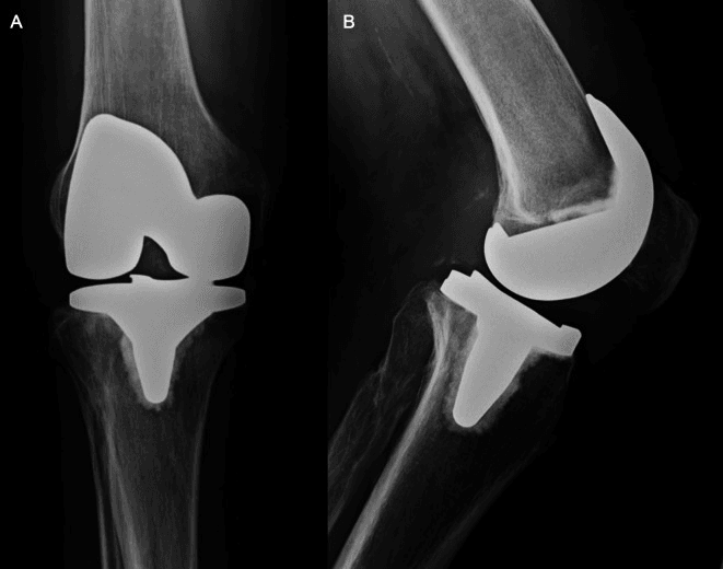
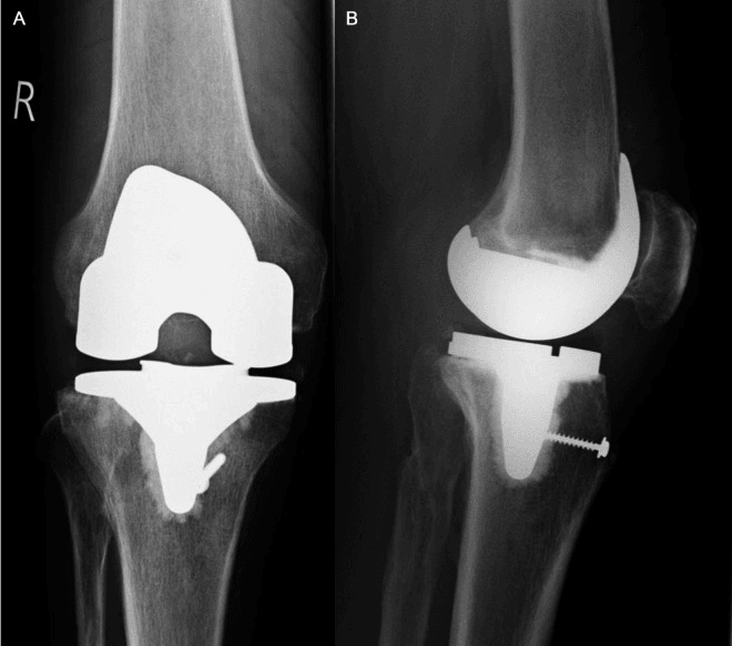
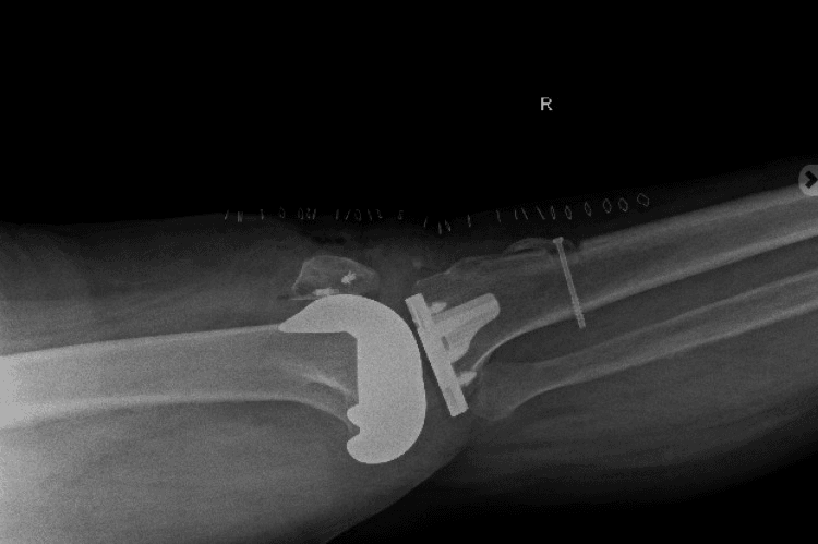
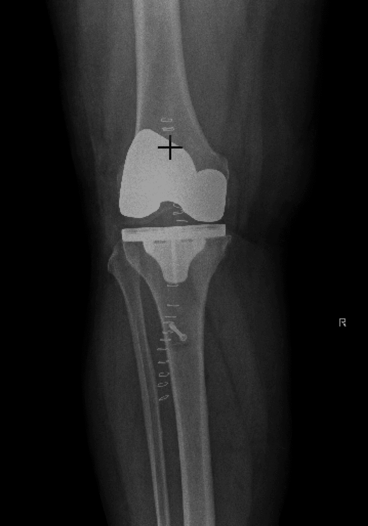
Radiographs first. The lateral view shows where the patella sits, alta or baja, and any patellar fracture or avulsion fragment. The films also show component position, which still matters, and a tibial tubercle fragment if a TTO has failed. Compare with previous films.
Insall-Salvati ratio (patellar tendon length / patella length): Normal 0.8-1.2. Patella alta (greater than 1.2) suggests patellar tendon rupture. Patella baja (less than 0.8) suggests quadriceps rupture. Compare to contralateral.
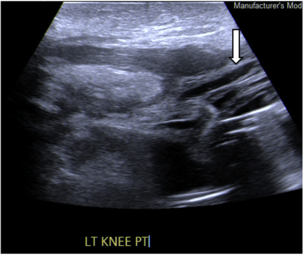
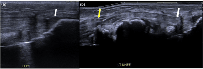
Ultrasound is the bedside option. It shows tendon continuity, gap size and retraction, and although operator-dependent it is useful for confirming the clinical diagnosis.
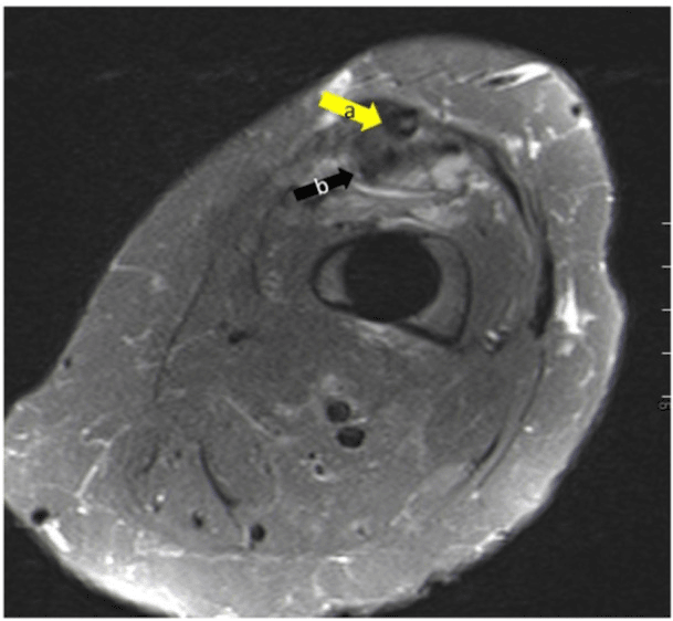
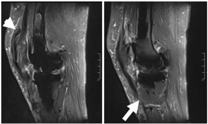
MRI is for when the diagnosis is unclear. It defines the rupture site, gap size and tissue quality, and shows associated pathology such as muscle atrophy, which makes it useful for planning a reconstruction. The prosthesis may cause artefact.
Management Algorithm
The decision. Site, timing and tissue quality decide the operation. The quadriceps tendon has better tissue, and an acute quadriceps rupture with good tissue can often be repaired primarily with augmentation. Acute patellar tendon tissue is usually poor and primary repair often fails, so early allograft is the alternative. Once a rupture is chronic, retracted and scarred, allograft reconstruction is the treatment of choice at either site.
- Tissue Quality
- Good tissue
- Management
- Primary repair with augmentation
- Tissue Quality
- Poor/friable
- Management
- Primary repair with allograft augmentation; prognosis guarded
- Tissue Quality
- Usually poor
- Management
- Primary repair (often fails) or early allograft; prognosis poor
- Tissue Quality
- Retracted, scarred
- Management
- Allograft reconstruction
- Tissue Quality
- Retracted, scarred
- Management
- Allograft extensor mechanism
Repair and augment. Primary repair with augmentation is preferred when the tissue allows, the augment being hamstring autograft, synthetic mesh or allograft. The repair is tensioned with the knee in extension and protected for 6-8 weeks in an extension splint. The technique and the reason for augmenting are under Surgical Technique.
Surgical Technique
Why augment. Primary repair alone often fails after TKA because the tissue quality and vascularity are poor. Augmentation provides mechanical reinforcement, a scaffold for healing, and protection during rehabilitation.
Surgical Steps
A midline incision through the previous scar. Identify the rupture site and tendon ends, debride non-viable tissue and assess tissue quality.
Freshen the tendon ends and create bone tunnels in the patella or tibial tubercle as needed. A retracted tendon may need mobilising.
Heavy non-absorbable suture (Krackow or Bunnell technique) passed through the bone tunnels and tensioned with the knee in full extension.
Reinforce the repair with semitendinosus autograft, synthetic mesh (e.g. polypropylene) or allograft tissue for additional strength.
Medial Gastrocnemius Flap Reconstruction
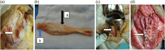
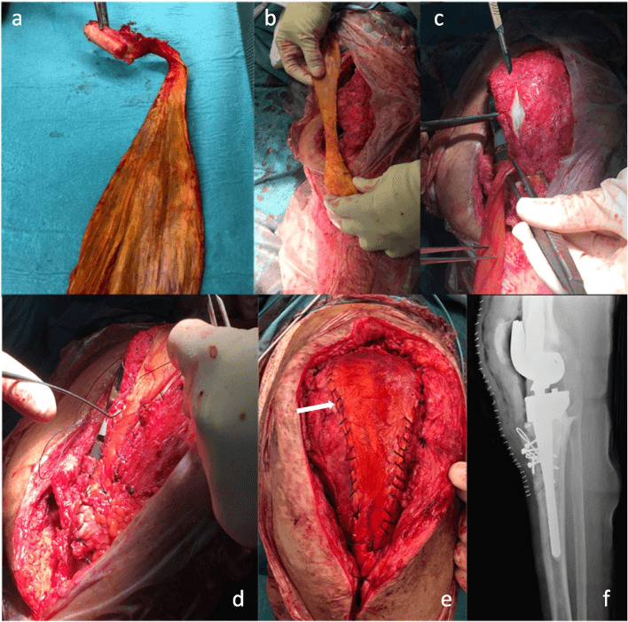
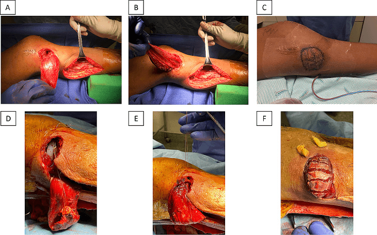
Why the medial head. The medial head of gastrocnemius is longer, has a wider arc of rotation, and reaches the anterior knee and patella more reliably than the lateral head, which is shorter and endangers the common peroneal nerve.
The technique. The flap is raised on its proximal pedicle, detached distally and rotated anteriorly over the extensor defect. Its tendinous or fascial portion is woven into or sutured to the quadriceps remnant and patella under tension in full extension, and the muscle belly is then skin-grafted or covered with local tissue.
Indications. The flap is used for:
- Extensor mechanism loss with an anterior soft-tissue defect or exposed implant
- Salvage after a failed allograft or synthetic mesh
- Thin, previously irradiated or multiply-operated skin over the tubercle
- Staged reconstruction where coverage must precede or accompany the graft
The flap is frequently combined with mesh or allograft, which reinforce the reconstruction while the flap solves the coverage problem, so it is often an adjunct rather than a stand-alone construct. Its reported results are under Controversies.
The gastrocnemius flap is the answer to the viva stem "the extensor mechanism is gone and the skin will not hold a graft." It simultaneously provides soft-tissue coverage and extensor continuity.
Synthetic Mesh (Marlex) Reconstruction Technique
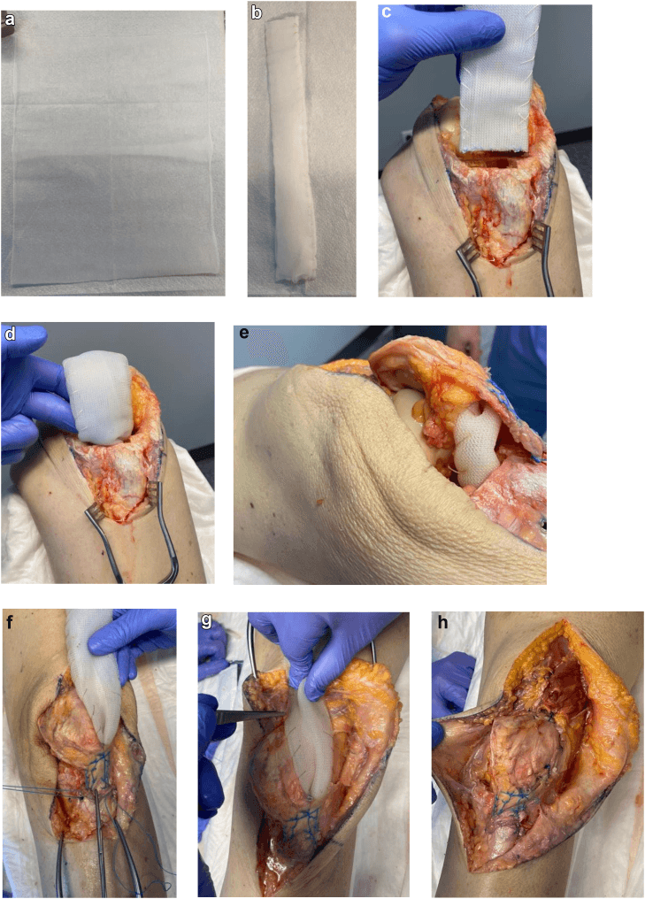
The construct popularised by Browne and Hanssen uses a knitted monofilament polypropylene (Marlex-type) mesh that host tissue can grow into. It can reconstruct the extensor mechanism at the time of component revision when required.
Marlex Mesh Reconstruction Steps
Create a bone trough or slot in the proximal tibia at the tubercle. Fashion the mesh into a tube, folded on itself for strength, and bury its distal end in the trough.
Secure the distal mesh with heavy non-absorbable sutures, staples or screws, and fixation may be reinforced with cement, particularly in poor bone or at revision. This distal anchor is the construct's weak link if it pulls out.
Pass the mesh beneath the residual patellar tendon and patella, then weave and imbricate the proximal end into the quadriceps tendon and the medial and lateral retinaculum, capturing all viable host tissue.
Tension the whole construct with the knee in full extension (the Burnett principle) and close the retinaculum over the mesh for a soft-tissue envelope.
Where mesh fails, and where it helps. Failures are usually early, within 6 months, and driven by infection rather than by the mesh itself. Mesh is especially useful when the components are being revised at the same sitting or when a previous allograft has already failed. How it compares with allograft is set out under Controversies.
Complications
- Incidence
- 10-30%
- Risk Factors
- Poor tissue, non-compliance
- Management
- Repeat reconstruction or accept
- Incidence
- 50-70%
- Risk Factors
- Expected outcome
- Management
- PT, accept residual lag
- Incidence
- 5-15%
- Risk Factors
- Multiple surgeries, diabetes
- Management
- Debridement, antibiotics
- Incidence
- 10-20%
- Risk Factors
- Immune response, poor incorporation
- Management
- Revision reconstruction
- Incidence
- Variable
- Risk Factors
- Immobilisation, fibrosis
- Management
- PT, possible MUA (cautious)
Re-rupture. Inadequate protection, early aggressive range of motion, poor tissue quality and allograft failure all raise the risk. The repair is always protected for the full 6-8 weeks in an extension splint.
Postoperative Care
Principles. Protect the repair, and an extension splint is mandatory. Delay flexion so the repair is not stressed, progress gradually without rushing, and accept limitations, because an extension lag is likely.
Rehabilitation Protocol
Extension splint or cylinder cast full-time, with no active flexion. Weight bearing as tolerated with the brace locked, preventing flexion forces on the repair.
Begin gentle passive flexion, 0-30 degrees progressing to 0-60. Continue the brace for walking and start isometric quadriceps work, progressing gradually.
Progressive active range of motion, closed-chain quadriceps strengthening and gait training. Wean from the brace gradually as strength allows.
Continue strengthening, assess the final range and extension lag, and adapt to residual deficits. A long-term brace may be needed for some activities.
Red flags. Watch for:
- Sudden loss of the ability to extend
- A new palpable gap
- Significant swelling
- Inability to maintain extension
Outcomes and Prognosis
- Primary Repair Success
- 60-70%
- Allograft Survival (5yr)
- N/A if repair holds
- Functional Outcome
- Fair-Good
- Primary Repair Success
- Low
- Allograft Survival (5yr)
- 60-70%
- Functional Outcome
- Fair
- Primary Repair Success
- Low
- Allograft Survival (5yr)
- 50-60%
- Functional Outcome
- Poor-Fair
Extension lag is the measure. Even after a successful reconstruction most patients have a lag of 10-30 degrees, reduced stair climbing and, for some, a need for assistive devices; complete active extension is rarely achieved. Patients with a lag under 15 degrees can usually manage stairs and transfers, while a lag over 30 degrees often requires assistive devices. Set realistic expectations before surgery.
Reading the series. Some small series in the evidence below report smaller mean lags: 4.3 degrees in Burnett's 13 grafts tensioned tightly in full extension, and 3 degrees in Crossett's 9 Achilles reconstructions at short-term follow-up.
Guidelines, Registries & Global Practice
Extensor mechanism disruption after TKA has an incidence of 0.1-2.5%, rising several-fold in the revision setting. No single society publishes a dedicated guideline; practice is shaped by registry data, expert consensus and the systematic reviews above.
- Region
- USA
- Relevant Position
- Tracks revision TKA and complications; emphasises infection workup before reoperation; no graft-specific mandate
- Region
- UK
- Relevant Position
- NJR captures revision TKA volume and indications; BOA stresses MDT management of failed extensor mechanism at high-volume revision units
- Region
- Australia
- Relevant Position
- Registry data on revision burden and patella-related revisions informs implant and technique choice
- Region
- Europe
- Relevant Position
- Supports reconstruction (allograft or mesh) tensioned in full extension; favours staged management when infection present
- Region
- Global
- Relevant Position
- Tibial tubercle osteotomy principles: long osteotomy, secure multi-point fixation to prevent iatrogenic disruption
- Tissue-bank allograft (Achilles, whole mechanism) readily available
- Synthetic mesh used increasingly for cost and availability
- Two-stage protocols when PJI present
- MRI/USS and metal-artefact-reduction imaging accessible
- Local autograft (semitendinosus, gastrocnemius flap) when allograft/mesh unavailable
- Greater reliance on clinical exam and plain radiographs
- Bracing/KAFO and non-operative pathways more commonly definitive
- Tissue banking and revision expertise concentrated in referral centres
Examiners worldwide expect: recognition of risk factors (revision, diabetes, TTO, steroids), the patellar-tendon blood-supply problem, exclusion of infection first, knowledge of the reconstruction ladder (repair to autograft to allograft/mesh) tensioned in full extension, and honest counselling about residual extension lag and approximately 25-32% failure.
Related pages: Extensor Mechanism Ruptures is the native-knee counterpart, and the contrast is the whole point — a healthy extensor mechanism repairs reliably, while after arthroplasty the tissue is scarred, devascularised by prior approaches and often previously infected, which is why direct repair fails in up to 63 percent of patellar tendons here; Patellar Tendon Rupture and Quadriceps Tendon Rupture carry the primary injuries and their repair techniques; TKA Patellar Complications for maltracking, patellar fracture and component loosening, which share risk factors and often coexist; Patella Fractures and Patellar Height Abnormalities for the patella alta that signals a patellar tendon rupture and the baja that follows a tightly tensioned reconstruction; Periprosthetic Joint Infection and TKA Complications: Infection are the dominant reason these reconstructions fail — infection was present in 39 to 44 percent of failures in the largest synthesis, so exclude it before reconstructing and expect it afterwards; Revision TKA and Revision TKA: Bone Loss, Cones and Sleeves because extensor failure is usually one problem in a failing knee rather than an isolated event; TKA Complications: Instability and TKA Stiffness complete the differential of the poorly functioning knee, and stiffness matters twice here since manipulation is a recognised cause of iatrogenic rupture; Knee Arthrofibrosis for that manipulation decision; Gastrocnemius Anatomy for the medial head flap described above; Tibial Tubercle Fractures for the avulsion pattern encountered during exposure; and Megaprosthesis for Non-Oncologic Salvage for the endpoint when reconstruction has failed repeatedly and the alternatives are arthrodesis or amputation.
Controversies and Areas of Uncertainty
Allograft or mesh. The Shau 2018 meta-analysis found equivalent success, approximately 76% for allograft and 74% for mesh, with similar failure rates. Mesh is cheaper, off-the-shelf, and carries no disease transmission and no immune resorption; allograft offers biological incorporation. No high-level trial settles the debate.
Augment every acute repair? Direct repair fails in roughly 34% of quadriceps and up to 63% of patellar tendon repairs. Whether to reinforce every acute repair with mesh or allograft, or reserve augmentation for poor tissue, remains unresolved.
Autograft. Small series of semitendinosus autograft and gastrocnemius flap report low failure and the least lag; in the Rajgopal 2025 meta-analysis the autograft and medial gastrocnemius flap cohorts had the least extensor lag (approximately 5 degrees for autografts) and no failures. The cohorts are tiny, and whether autograft truly outperforms allograft or mesh, or the apparent superiority reflects selection of better hosts, is uncertain.
Operate at all? In a low-demand or high-comorbidity patient a braced, lagging knee may be safer than a complex reconstruction with approximately 25-32% failure and a high infection risk. The threshold to operate is individualised, not evidence-defined.
Across all reconstruction techniques, periprosthetic joint infection is present in 39-44% of failures (Rajgopal 2025). Many "failed reconstructions" are in fact unrecognised or recurrent infections. This is the dominant area of uncertainty and the key to improving outcomes.
MCQ Practice Points
Q: Which extensor mechanism component has the worst prognosis when ruptured after TKA? A: Patellar tendon - Has poorest blood supply (depends on fat pad) and worst healing potential. Primary repair often fails, requiring allograft reconstruction with guarded outcomes.
Q: What patella position is expected with patellar tendon rupture? A: Patella alta (high-riding patella) - The patella migrates proximally when patellar tendon is disrupted. Insall-Salvati ratio greater than 1.2 indicates patella alta.
Q: What is the most commonly used allograft for extensor mechanism reconstruction? A: Achilles tendon allograft with calcaneal bone block - The bone block is fixed to the patella and the tendon portion is sutured to the quadriceps, bridging the defect.
Q: How long should the knee be protected in extension after extensor mechanism repair/reconstruction? A: 6-8 weeks - This period allows healing of repair or incorporation of allograft. Active flexion is avoided during this time. Premature mobilization risks re-rupture.
Q: What residual extension lag is commonly seen after extensor mechanism reconstruction? A: 10-30 degrees - Most patients have permanent extension lag even with successful reconstruction. Full active extension is rarely achieved after allograft reconstruction.
Exam Viva Scenarios
Practise clinical reasoning and management decisions out loud
“A 68-year-old man falls 3 weeks after revision TKA and cannot extend his knee. Examination shows a palpable gap below the patella and patella alta on XR. He has diabetes.”
“During revision TKA with difficult exposure, you perform a tibial tubercle osteotomy. When you flex the knee to deliver the tibia, the tubercle avulses completely from the remaining tibia. What do you do?”
“A 70-year-old woman presents 4 months after primary TKA. She has had progressive difficulty extending her knee. She now has a 40-degree extension lag and cannot climb stairs. MRI confirms chronic quadriceps tendon rupture with 4cm retraction.”
“You are about to perform MUA for stiffness at 10 weeks post-TKA. The patient has diabetes and had a previous TKA revision. How do you minimize risk of extensor mechanism rupture?”
Key Facts
- Patellar tendon rupture = WORST prognosis
- Quadriceps tendon = better tissue quality, better outcomes
- Primary repair often fails in TKA setting
- Allograft reconstruction is treatment of choice for chronic
Risk Factors
- Revision surgery (3-4x higher risk)
- Tibial tubercle osteotomy (TTO)
- Poor vascularity (diabetes, smoking)
- Excessive tension (MUA, forceful manipulation)
Clinical Signs
- Patellar tendon rupture = patella ALTA
- Quadriceps rupture = patella BAJA
- Inability to actively extend = complete rupture
- Extension LAG = partial or reconstructed
Management
- Acute: Primary repair + AUGMENTATION
- Chronic: ALLOGRAFT reconstruction (Achilles or whole mechanism)
- Protection: 6-8 weeks extension splint
- Non-op if unfit for surgery
Outcomes
- 50-70% 5-year allograft survival
- 10-30 degree extension lag EXPECTED
- Full extension rarely achieved
- PREVENTION is the best treatment
Evidence Base
Burnett RSJ et al. — Tensioning of Extensor Mechanism Allograft
- 20 consecutive whole extensor mechanism allograft (tubercle-patellar tendon-patella-quadriceps tendon) reconstructions after TKA.
- All 7 loosely-tensioned grafts FAILED (mean residual lag 59 degrees, HSS score 52). All 13 grafts tensioned tightly in FULL EXTENSION succeeded (mean lag 4.3 degrees, HSS 88; p less than 0.0001).
- Flexion was not significantly compromised by full-extension tensioning.
Nazarian DG, Booth RE — Extensor Mechanism Allografts
- 40 knees with chronic extensor mechanism disruption after TKA treated with whole extensor mechanism allograft; 13 previously infected.
- At mean 3.6 years, 34 of 36 evaluable patients (94%) had a successful clinical result; mean Knee Society function score improved 37 to 68.
- 8 allografts ruptured and required re-grafting; mean extensor lag 13 degrees.
Crossett LS et al. — Achilles Allograft for Patellar Tendon Rupture
- 9 patellar tendon ruptures after TKA reconstructed with fresh-frozen Achilles tendon allograft + calcaneal bone block.
- Mean extensor lag improved from 44 degrees to 3 degrees; mean ROM 88 to 107 degrees; Knee/function scores 26/14 to 81/53.
- 2 early graft failures, both successfully re-repaired; mean proximal patellar migration 17.8 mm did not impair function.
Browne JA, Hanssen AD — Synthetic Mesh Reconstruction
- 13 subacute/chronic patellar tendon disruptions after TKA reconstructed with knitted monofilament polypropylene mesh (5 had failed prior allograft).
- 9 of 13 succeeded with extensor lag 10 degrees or less and maintained flexion; 3 failed within 6 months, 1 recurrent infection.
- Mesh was significantly cheaper than allograft and avoids disease transmission.
Cadambi A, Engh GA — Semitendinosus Autograft
- 7 patellar ligament ruptures after TKA reconstructed with autogenous semitendinosus tendon woven through patella and tibial tubercle.
- Restored active extension in this small series — established autograft as a viable option when allograft/mesh are unavailable.
- Avoids disease transmission and immune/incorporation issues of allograft.
Shau D et al. — Synthetic Mesh vs Allograft (Meta-analysis)
- Systematic review and meta-analysis of 14 studies, 204 knees, comparing allograft vs synthetic mesh extensor mechanism reconstruction.
- No significant difference in success (76% allograft vs 74% mesh), Knee Society Scores, extensor lag, or complications — approximately 25% failure in both groups.
- Periprosthetic joint infection is a leading cause of failure in both groups; mesh is cheaper and universally available.
Rajgopal A et al. — EMD After TKA (Systematic Review & Meta-analysis)
- 32 studies (655 reconstructions, 119 repairs); incidence of extensor mechanism disruption 0.1-2.5%.
- Direct repair failure: quadriceps 33.9% vs patellar tendon 63% — confirming patellar tendon repair is markedly less reliable.
- Allograft and mesh reconstruction failure approximately 32%; PJI present in 39-44% of failures; autograft/gastrocnemius flap had the least lag in small cohorts.