Osteolysis | Polyethylene Wear | Particle Disease
- Polyethylene wear debris triggers osteolysis (particle disease)
- Tibial component most commonly affected
- Rule out infection in ALL cases before proceeding to revision
- Revision requires addressing bone loss, component stability, and alignment
- Stems and augments often needed for revision
- “Progressive radiolucent lines greater than 2mm = loosening
- “Component migration on serial XR is diagnostic
- “CRP/ESR + aspiration mandatory to exclude infection
- “AORI classification for bone loss
Overview and Epidemiology
Aseptic loosening is the mechanical failure of the bone-implant interface without infection. It remains a leading cause of TKA revision, second only to infection in many series; in the multicentre series of Dalury and colleagues (Evidence Base) it was the single most common reason for revision.
The trend. Loosening rates have decreased with cross-linked polyethylene, improved cementing technique, better alignment targets and modern implant designs, which have reduced the problem but not eliminated it. With increasing TKA volume and younger patients, the absolute numbers requiring revision continue to rise.
Patient risk factors. High BMI increases wear and loosening, and a high activity level increases wear. Young age brings higher activity and a longer exposure. Poor bone quality (osteoporosis, RA) and smoking, which impairs bone healing, complete the list.
Technical risk factors. Varus or valgus malalignment increases tibial stress, and malrotation is an error of component rotation. Poor cementing leaves cement mantle defects, undersizing concentrates stress, and the polyethylene type matters: conventional versus cross-linked (XLPE).
Early Versus Late Aseptic Loosening
The timing of loosening is not incidental. It points to the underlying mechanism and changes how you investigate the knee, and the biological-versus-mechanical split in the summary maps onto a temporal dichotomy that examiners expect you to articulate.
Early loosening, within about 2 years, is predominantly mechanical or technical:
- Inadequate primary fixation
- Cement mantle defects or tibial baseplate debonding
- Malalignment or malrotation concentrating interface stress
- Failure of osseointegration in cementless implants
In the Insall Award series (Sharkey), over half of all revisions occurred within 2 years, and these early failures were driven by instability, malalignment and failure of fixation.
Early means technical error or infection first. The particle-osteolysis cascade has not had time to mature, so a component that loosens within the first 2 years should make you think of technical error or infection, not particle disease. Early "aseptic" loosening is a diagnosis of exclusion, accepted only after a rigorous negative workup has ruled out occult low-grade infection.
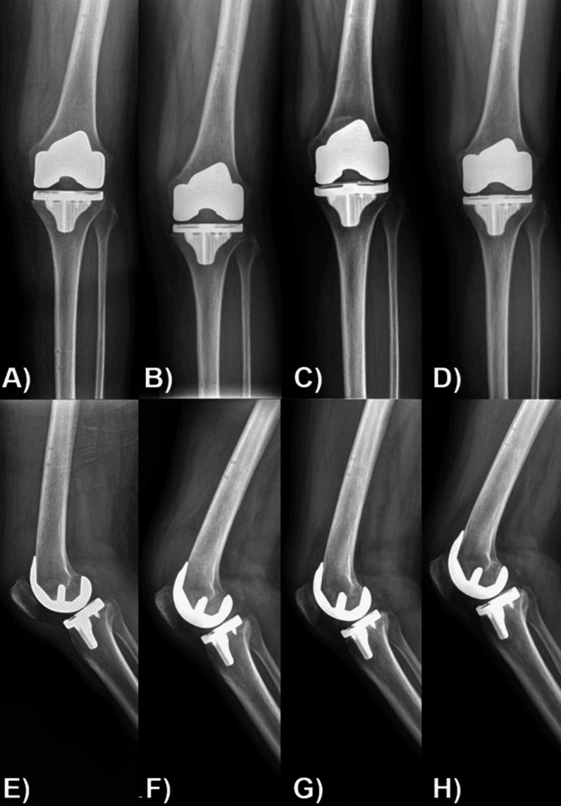
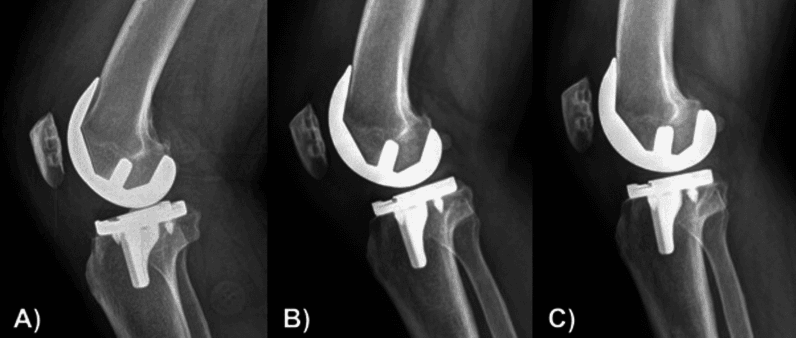
Late loosening, after about 5-10 years, is predominantly biological. Cumulative polyethylene wear generates a rising particle burden, driving macrophage-mediated osteolysis and progressive interface failure, and this is the mechanism most reduced by cross-linked polyethylene. It is the pattern that follows a symptom-free interval with insidious start-up pain. Osteolysis is typically more advanced by then, so reconstruction more often needs augments, cones or sleeves.
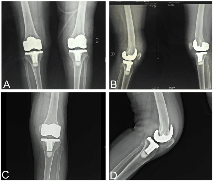
Pathophysiology and Mechanisms
Particle disease. Polyethylene wear debris, particles of 0.3-10μm, is phagocytosed by macrophages, which release cytokines (IL-1, IL-6, TNF-α, RANKL) that activate osteoclasts. The result is periprosthetic bone resorption, osteolysis and component loosening. It is a dose-dependent biological response to the particle load.
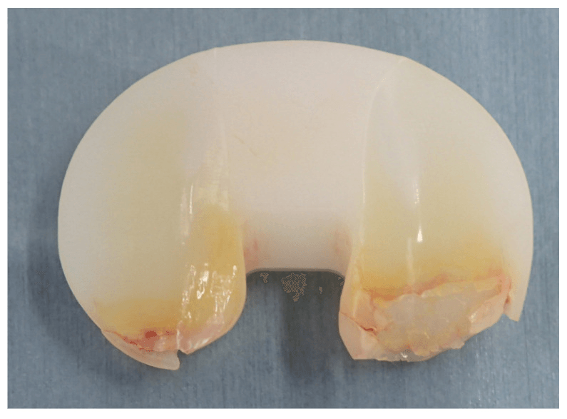
Why the tibia. The tibial component is the one most commonly affected, because of higher wear at the tibial polyethylene surface and varus malalignment forces. In a varus knee the highest polyethylene contact stress falls on the medial compartment, and malalignment increases edge loading and accelerates wear. Load passes through the polyethylene and the tibial baseplate to bone, so any gap or poor support beneath the baseplate leads to micromotion and loosening.
Tibial fixation. The cement mantle is critical, the proximal tibial bone supports the load, and varus malalignment concentrates stress medially.
Femoral fixation. The posterior condyles bear the flexion load and the anterior flange the extension load, and the femur is generally more tolerant of slight malalignment. Femoral loosening is less common, but may indicate rotational malalignment. Rotation also affects patellofemoral tracking, and an internally rotated component brings patellofemoral problems.
- Loading Pattern
- Axial compression, shear
- Common Failure Mode
- Medial subsidence, varus collapse
- Revision Consideration
- Stems, metal augments, cones
- Loading Pattern
- Contact stress, wear
- Common Failure Mode
- Delamination, oxidative degradation
- Revision Consideration
- XLPE, adequate thickness
- Loading Pattern
- Flexion/extension loading
- Common Failure Mode
- Posterior condylar loosening
- Revision Consideration
- Stems, augments if needed
- Loading Pattern
- Shear, eccentric loading
- Common Failure Mode
- Loosening, fracture
- Revision Consideration
- Address alignment/rotation
Classification Systems
Bone loss: the AORI classification. The Anderson Orthopaedic Research Institute classification grades the integrity of the metaphyseal bone, is applied separately to the tibia and the femur, and matches each grade to a reconstruction. Grade it on imaging and confirm it at surgery, because imaging underestimates defect size in 50% of cases.
- Description
- Intact metaphyseal bone, minor defects: less than 5mm, contained
- Reconstruction Options
- Standard revision, cement alone, no augments
- Description
- Damaged metaphyseal bone, one condyle/plateau
- Reconstruction Options
- Metal augments, particulate graft
- Description
- Damaged bone, both condyles/plateaus
- Reconstruction Options
- Larger augments, stems, structural graft
- Description
- Deficient metaphyseal bone (metaphyseal segment deficient)
- Reconstruction Options
- Cones, sleeves, structural allograft, megaprosthesis, allograft-prosthetic composite (APC)
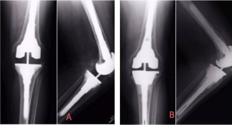
Femoral defects. Femoral bone loss is typically posterior, so assess the flexion gap carefully. Engh's femoral defect classification grades them from F1 to F4:
- Description
- Intact metaphysis, minor damage
- Reconstruction
- Standard revision, augments if needed
- Description
- Damaged metaphysis, intact condyles
- Reconstruction
- Augments, possible stems
- Description
- Damaged metaphysis and one condyle
- Reconstruction
- Augments, stems, structural graft
- Description
- Damaged metaphysis and both condyles
- Reconstruction
- Megaprosthesis, allograft-prosthetic composite
Clinical Assessment
The story. A symptom-free interval after primary TKA followed by gradual onset of pain is classic for aseptic loosening. Pain from the start suggests an initial fixation problem, infection or component malposition.
The history. Characterise the pain as start-up, activity-related or rest pain, and locate it: anterior pain points to the patella, medial or lateral pain to a component. Take the details of the index TKA and any revision, and measure the decline in function by walking distance, stairs and activities of daily living.
Examination. Watch the gait, look at the alignment, compare the range of motion with previous, and test stability and patellar tracking:
- Gait - antalgic, varus or valgus thrust
- Alignment - obvious deformity
- Range of motion - compared with previous
- Stability - varus/valgus stress, AP drawer
- Patellar tracking - J-sign, subluxation
- Typical Presentation
- Medial pain, start-up pain
- Examination Findings
- Tenderness over tibial component, varus thrust
- Typical Presentation
- Global knee pain, posterior pain
- Examination Findings
- Less localisable, may have flexion instability
- Typical Presentation
- Anterior knee pain
- Examination Findings
- Patellar crepitus, tenderness, tracking issues
- Typical Presentation
- Severe pain, instability
- Examination Findings
- Obvious deformity, gross instability
- Discriminating Features
- Symptom-free interval then start-up pain; RLL/migration; normal markers
- Key Investigation
- Serial radiographs; aspiration to exclude PJI
- Discriminating Features
- No pain-free interval, rest pain, warmth/effusion; raised CRP/ESR
- Key Investigation
- Aspiration (synovial WCC, PMN%, culture, alpha-defensin)
- Discriminating Features
- Giving way, recurrent effusions, often well-fixed components
- Key Investigation
- Stress views, examination, fluoroscopy
- Discriminating Features
- Anterior knee/patellofemoral pain, maltracking
- Key Investigation
- CT rotational profile
- Discriminating Features
- Hip OA, lumbar radiculopathy, vascular, CRPS
- Key Investigation
- Hip/spine exam and imaging; vascular assessment
- Discriminating Features
- Mechanical catching, focal tenderness
- Key Investigation
- Clinical exam; ultrasound if needed
NEVER proceed to revision for presumed aseptic loosening without ruling out infection. A painful TKA is periprosthetic joint infection until proven otherwise, and aseptic loosening can only be diagnosed after a negative infection workup: at minimum CRP, ESR and aspiration with synovial WCC, differential (PMN%) and culture, judged against the MSIS criteria. Missed infection is a disaster.
Investigations
Bloods. CRP and ESR are first line and must be done. Elevated values suggest infection, and if they are raised the knee is aspirated before any surgical planning.
Aspiration is mandatory, and the synovial fluid is sent for:
- WCC - greater than 1100/μL in chronic, greater than 3000 in acute infection
- PMN% - greater than 64%
- Culture - held for 14 days
- Alpha-defensin, if available
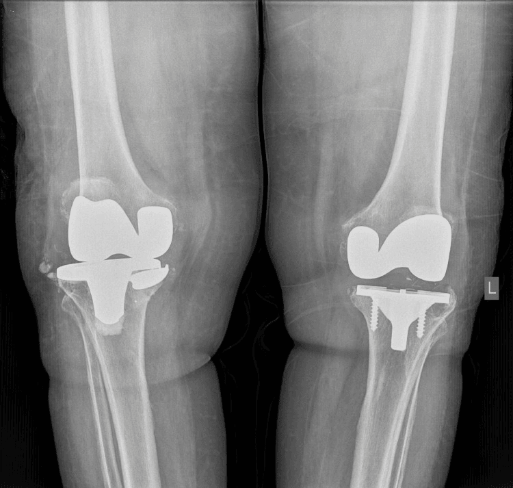
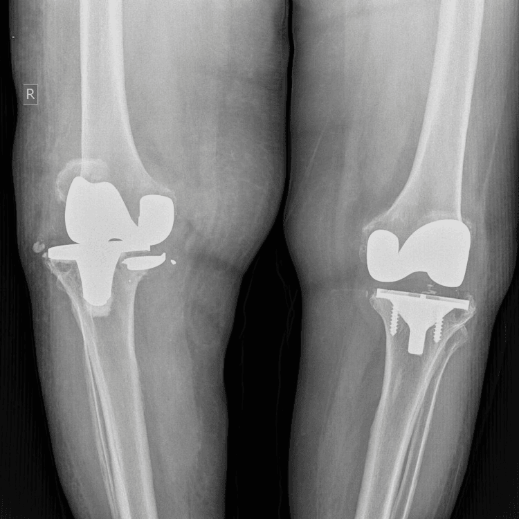
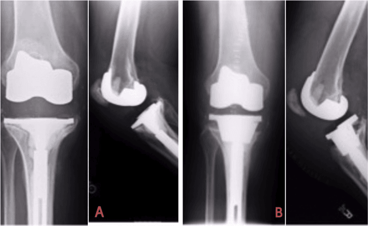
Radiographs. Weight-bearing AP, lateral and skyline views, always compared with the immediate post-operative films; serial films are critical. Look for radiolucent lines (RLL), migration, osteolysis and component position.
Reading the radiolucent line. A new or enlarging line is significant, and lucency at the bone-cement interface is more concerning than at the cement-implant interface. Width and behaviour on serial films grade the finding:
- Significance
- Normal/expected
- Action
- Monitor
- Significance
- Possible early loosening
- Action
- Close monitoring, serial XR
- Significance
- Probable loosening
- Action
- Workup for revision
- Significance
- Definitive loosening
- Action
- Plan revision
- Significance
- Failed fixation
- Action
- Revision indicated
The Knee Society divides the tibial component into 7 zones (AP and lateral views) and the femoral component into 7 zones, and a complete radiolucency around a component means it is definitely loose. Beyond the line itself, migration or subsidence of the component, periprosthetic osteolysis and cement mantle fracture are the other radiographic signs of loosening.
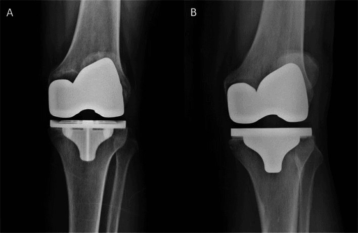
Advanced imaging is used where needed:
- CT - bone defect quantification (AORI) and planning for augments, component rotation, occult osteolysis
- MRI MARS - soft tissue, occult osteolysis
- Nuclear medicine or PET - if the diagnosis is unclear, but of limited utility in differentiating a loose from a well-fixed component
Metal artefact reduction protocols (MARS) on CT or MRI are considered for metal components.
Implant Migration and Radiostereometric Analysis (RSA)
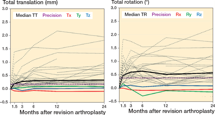
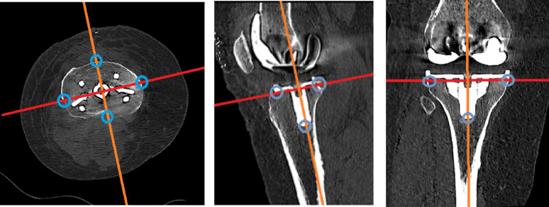
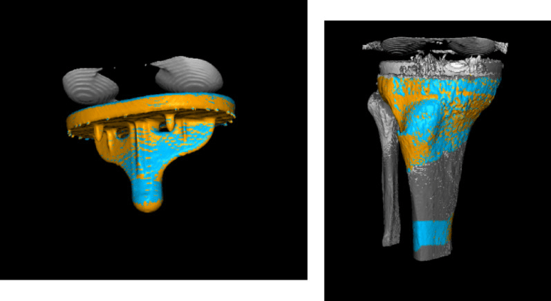
What RSA measures. Radiostereometric analysis measures in-vivo micromotion of the implant relative to bone, using implanted tantalum bead markers and paired (stereo) radiographs. Its accuracy of roughly 0.1 to 0.5 mm is far below the plain-film detection threshold. Maximum total point motion (MTPM) is the standard reported parameter, and RSA is the reference standard for quantifying early fixation and comparing new implant designs.
Stabilising versus continuous migration. This is the single discriminator RSA teaches. Components that migrate initially then stabilise within the first year rarely go on to loosen; components that migrate continuously beyond the first year are at high risk of late mechanical loosening. Continuous migration, broadly ongoing motion greater than roughly 0.2 mm per year, is the danger signal, and late plain-film migration is the visible endpoint of a process that started early.
What it is used for. It underpins the stepwise (phased) introduction of new implants: early RSA migration is used as a surrogate for long-term revision risk before a design is released widely. It also reinforces the clinical rule to compare every follow-up radiograph against the immediate post-operative baseline.
Ryd L et al. RSA as a Predictor of Mechanical Loosening of Knee Prostheses
- RSA follow-up of tibial components showed most migrated about 1mm in the first year; roughly one-third migrated continuously thereafter while two-thirds ceased migrating after 1-2 years. In the group later revised for mechanical loosening, 14 of 15 components had migrated continuously and had already migrated significantly more at one year than the stable group - yet clinical symptoms prompting revision could be delayed by up to 10 years. Mechanical loosening therefore begins early even when it presents late.
Management Algorithm
Confirm the diagnosis first. Rule out infection, which is mandatory, confirm mechanical loosening, and identify the contributing factors such as alignment and rotation. The findings then place the knee in one of these scenarios:
- Investigation Findings
- Normal XR, elevated markers
- Management
- Exclude infection: aspirate before revision
- Investigation Findings
- Awaiting aspiration
- Management
- Do NOT proceed until infection ruled out
- Investigation Findings
- Normal markers, equivocal XR
- Management
- Repeat XR at 3-6 months, trial conservative
- Investigation Findings
- Greater than 2mm RLL, no migration
- Management
- Close monitoring vs revision
- Investigation Findings
- Component migration or subsidence on serial XR; negative infection workup
- Management
- Plan revision TKA with a bone defect strategy
- Investigation Findings
- Progressive on serial XR
- Management
- Consider early revision to preserve bone
- Investigation Findings
- Large cavitary defects
- Management
- Classify bone loss; cones, sleeves, augments
Conservative treatment has a limited role in true loosening. Activity modification, analgesia and bracing may temporise but cannot reverse the problem, so consider it in patients unfit for surgery or those with limited symptoms. The timing of revision balances bone loss progression against patient factors and symptoms.
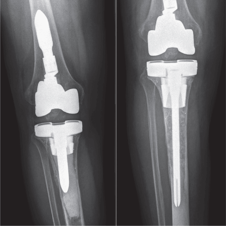
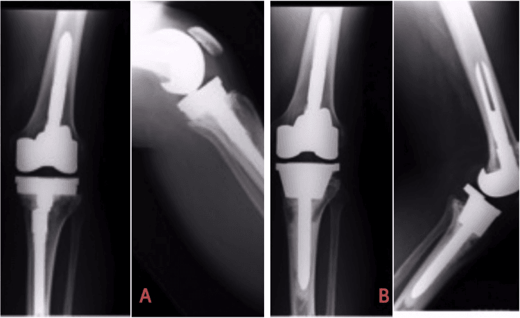
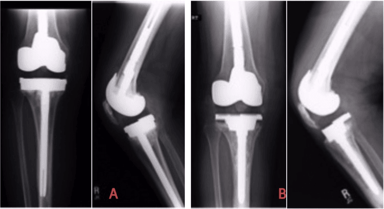
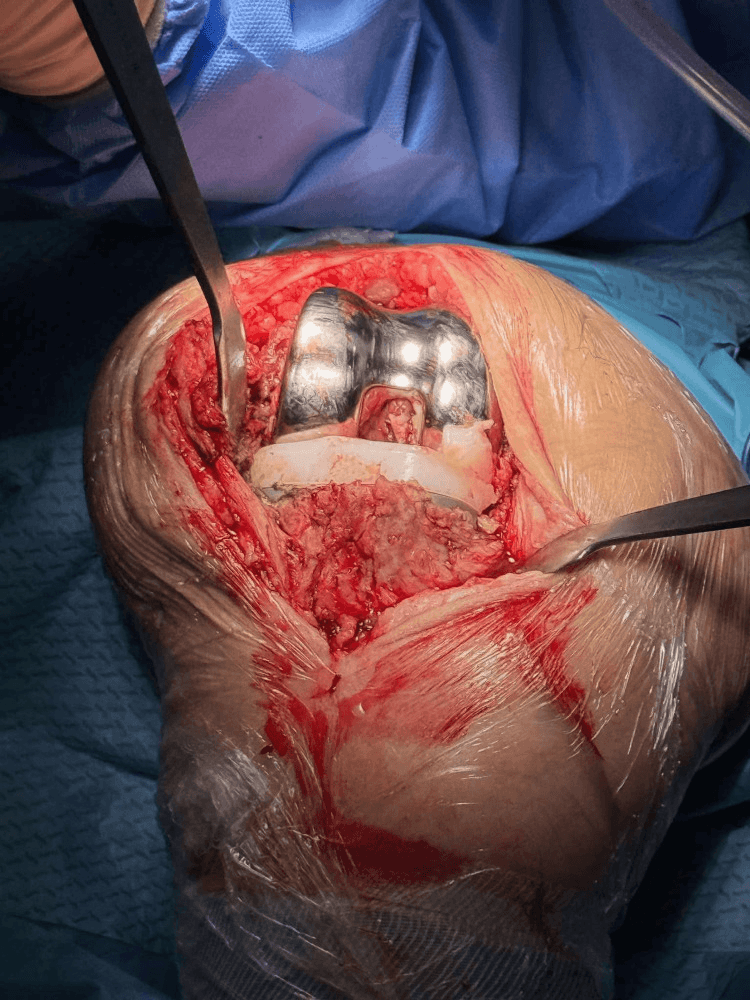
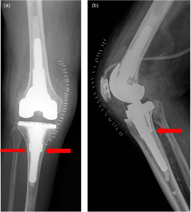
What a revision must do. Remove the components, debride the membrane, manage the bone loss (AORI), restore alignment and use appropriate constraint; stems and augments are typically required. Plan augments, grafts and stems from the AORI grade and order the appropriate revision system. Then choose the implant:
- Constraint - based on ligament competence
- Fixation - stems, including offset stems, for metaphyseal bypass
- Augments - metal blocks, wedges, cones, sleeves
Zonal fixation. Revision fixation is organised in three zones, and revision relies on Zone 3 press-fit or cemented stems:
- Zone 1, epiphysis - minimal support after revision
- Zone 2, metaphysis - damaged in loosening, augments needed
- Zone 3, diaphysis - healthy bone for stem fixation
Final implant selection depends on intraoperative findings, so plan for extensile exposure and have backup options available.
Surgical Technique
Surgical Steps
Use the previous incision, the lateral-most if there are several, and typically a medial parapatellar arthrotomy. Be prepared for extensile exposure.
Mobilise the patella carefully; a stiff knee may need a quad snip. Protect the patellar tendon throughout.
Debride the hypertrophic synovium and the particle-laden membrane (pseudomembrane). It contains wear particles and osteolytic cytokines, so remove it thoroughly.
Extensile options. Adequate exposure is critical, and rushing leads to complications:
- Quad snip - 45-degree proximal extension of the arthrotomy
- Rectus snip - the rectus tendon is cut
- VY turndown - V-shaped turndown of the quadriceps tendon
- Tibial tubercle osteotomy (TTO) - for severe stiffness or difficult exposure; best for severe stiffness, and allows direct proximal retraction
Complications
- Risk Factors
- Longer surgery, revision setting
- Prevention
- Antibiotics, laminar flow, technique
- Management
- DAIR vs 2-stage revision
- Risk Factors
- Poor exposure, inadequate rehab
- Prevention
- Adequate exposure, early ROM
- Management
- MUA, possible re-revision
- Risk Factors
- Inadequate constraint, ligament damage
- Prevention
- Proper constraint selection
- Management
- Poly exchange vs revision
- Risk Factors
- TTO, aggressive mobilisation
- Prevention
- Careful technique, protect tendon
- Management
- Repair, reconstruction, allograft
- Risk Factors
- Osteoporosis, cortical windows
- Prevention
- Careful cement removal, stems
- Management
- ORIF, revision with long stems
Protect the extensor mechanism throughout surgery. Patellar tendon avulsion is a devastating complication. If TTO performed, secure fixation and protect postoperatively.
Re-revision. Revision TKA has higher failure rates than primary TKA (see Outcomes and Prognosis). Re-revision is more complex with progressive bone loss, so consider referral to a high-volume revision centre.
Postoperative Care
Rehabilitation Protocol
Pain management, DVT prophylaxis and wound monitoring; CPM if available. Begin range-of-motion exercises.
Progressive range of motion, quadriceps strengthening, gait training with aids and wound care.
Progressive resistance and functional exercises; wean the walking aids. Monitor for stiffness requiring MUA (typically by 6-8 weeks if needed).
Full activities as tolerated, a final range-of-motion assessment, and serial XR to confirm stability.
Weight bearing. After a standard revision the patient typically bears weight as tolerated with aids. Bone graft may restrict weight bearing for 6 weeks. After a TTO, weight bearing is partial and active extension is limited for 6-8 weeks, and after a megaprosthesis the surgeon's protocol applies.
Follow-up. Reviews fall at fixed points:
- 2 weeks - wound check, staple removal
- 6 weeks - XR, range-of-motion assessment
- 3 months - progress check; consider MUA if stiff
- Annual - long-term surveillance
Outcomes and Prognosis
- Revision for Aseptic Loosening
- 80-85%
- Primary TKA (Comparison)
- 95%+
- Revision for Aseptic Loosening
- 70-80
- Primary TKA (Comparison)
- 85-95
- Revision for Aseptic Loosening
- 75-85%
- Primary TKA (Comparison)
- 90%
- Revision for Aseptic Loosening
- 10-15%
- Primary TKA (Comparison)
- 2-5%
- Revision for Aseptic Loosening
- 5-10%
- Primary TKA (Comparison)
- 1-2%
Prognosis. Outcomes are better with single-component loosening, adequate bone stock, younger age and a good soft-tissue envelope. They are worse with global loosening, massive bone loss, multiple previous revisions and extensor mechanism compromise.
Guidelines, Registries & Global Practice
Aseptic loosening is a leading cause of revision worldwide. Across major national registries (NJR England/Wales, AJRR USA, AOANJRR Australia, SHAR Sweden, NZJR New Zealand), loosening and infection consistently top the list of indications for TKA revision, while polyethylene wear and osteolysis have declined in the cross-linked-polyethylene era. Cumulative percent revision at 10 years for primary TKA is broadly 3-6% across registries, with implant- and bearing-specific differences (see below).
- Region
- Australia, Sweden, UK, USA
- Key Message for Loosening
- Loosening and infection are the dominant revision indications; cross-linked polyethylene reduces wear/osteolysis-driven revision
- Region
- USA
- Key Message for Loosening
- Validated implant/bearing selection, accurate alignment, exclude PJI before revising for presumed aseptic loosening
- Region
- UK
- Key Message for Loosening
- Routine post-op surveillance radiographs; refer complex revision to higher-volume centres
- Region
- Europe (global)
- Key Message for Loosening
- Zonal-fixation principle and metaphyseal cones/sleeves for AORI Type 2B/3 defects; least constraint compatible with stability
- Modular revision systems: cones, sleeves, augments, offset stems
- Cross-linked polyethylene and validated bearings as standard
- Two-stage PJI pathways and arthroplasty MDT for bone loss
- Registry-linked surveillance and outcome benchmarking
- Restricted access to cones/sleeves and megaprostheses
- Greater reliance on cement, bone graft and basic stems/augments
- Conventional polyethylene still in use, raising wear/osteolysis risk
- Earlier referral and selective revision where implant inventory is constrained
Examiners worldwide expect: particle-disease pathophysiology, a systematic infection-exclusion algorithm before revision, AORI classification, the zonal-fixation concept, and reconstruction options (augments, cones, sleeves, megaprosthesis) calibrated to bone loss and resource setting.
Related pages: Revision TKA is the operation this page leads to and carries exposure, component removal and constraint selection in full; Revision TKA: Bone Loss, Cones and Sleeves holds the AORI grading and the reconstruction menu in depth, including the metaphyseal fixation devices that postdate the 1999 classification carded above; Periprosthetic Joint Infection and TKA Complications: Infection are the diagnosis that must be excluded before any knee is called aseptic - the single commonest error on this page is revising a low-grade infection as loosening, and the Hadley series is a reminder that infection, not loosening, drove 80 percent of cone removals; TKA Complications: Instability and TKA Stiffness are the other two members of the painful-TKA differential and the commonest reasons an early revision is not for loosening at all; Polyethylene UHMWPE and XLPE and Highly Cross-Linked and Vitamin E Polyethylene for the bearing material behind particle disease and behind the registry advantage in the Vertullo analysis; Wear Mechanisms and THA Wear and Osteolysis for the particle-macrophage-osteoclast cascade that drives late loosening; PMMA Bone Cement for the cement-bone interface whose failure defines the problem, and Tantalum for the porous metal used in the cones; TKA Alignment Philosophies because malalignment is the modifiable mechanical driver of early loosening; TKA Periprosthetic Fractures and TKA Extensor Mechanism Failure for the complications of the revision itself; and Megaprosthesis for Non-Oncologic Salvage for the endpoint when metaphyseal fixation is no longer achievable.
Controversies & Areas of Uncertainty
Both tantalum cones and titanium sleeves provide durable metaphyseal fixation for AORI Type 2B/3 defects, but no high-quality randomised data establish superiority of one over the other. Choice is driven by defect geometry, system availability and surgeon familiarity.
The hybrid (cemented metaphysis, press-fit diaphyseal stem) versus fully cemented stem debate persists. Registry and cohort data show comparable survivorship; end-of-stem pain is more associated with long press-fit stems.
Isolated polyethylene exchange for wear/osteolysis with well-fixed components is tempting but carries high re-revision rates; most authors reserve it for highly selected cases with secure fixation and accurate component position.
Whether kinematic or functional alignment reduces long-term loosening compared with mechanical alignment remains unresolved; current evidence shows equivalent short- to mid-term survivorship.
There is no validated threshold that perfectly separates a painful-but-well-fixed knee from early loosening on a single radiograph. Serial imaging, component migration and a rigorous infection workup - not any one test - drive the decision to revise.
MCQ Practice Points
Q: Which component is most commonly affected by aseptic loosening in TKA? A: Tibial component - Due to higher contact stress at the polyethylene surface, varus malalignment forces, and the smaller fixation surface compared to femoral component.
Q: What is the primary biological mechanism of osteolysis in TKA? A: Macrophage activation by polyethylene wear debris - Wear particles (0.3-10μm) are phagocytosed by macrophages, which release cytokines (IL-1, IL-6, TNF-α, RANKL) that stimulate osteoclast-mediated bone resorption.
Q: What radiolucent line width at the bone-cement interface indicates probable loosening? A: Greater than 2mm - RLL greater than 2mm or any progressive radiolucent line on serial radiographs indicates loosening. Complete radiolucency around a component is definitive.
Q: What does AORI Type 2B bone defect signify? A: Damaged metaphyseal bone involving both condyles/plateaus - Type 2A involves one condyle/plateau, Type 2B involves both. Type 2 defects require metal augments or graft for reconstruction.
Q: In revision TKA, which zone provides the most reliable fixation? A: Zone 3 (diaphysis) - The metaphysis (Zone 2) is typically damaged in loosening. Stems that engage the healthy diaphyseal bone (Zone 3) provide reliable fixation for revision components.
Exam Viva Scenarios
Practise clinical reasoning and management decisions out loud
“A 72-year-old woman presents 8 years after primary TKA with increasing medial knee pain. She had been pain-free for 6 years. Radiographs show 3mm radiolucent line around the tibial component with medial tibial subsidence. CRP and ESR are normal.”
“A 65-year-old man has a 15-year-old TKA with massive osteolysis. CT shows AORI Type 3 defects in both tibia and femur. He is moderately symptomatic but mobile. What are your options?”
“You are planning revision TKA for apparent aseptic loosening. Preoperative CRP is 15 mg/L (normal less than 5). The surgeon wants to proceed. What do you do?”
“During revision TKA for tibial loosening, you remove the component and find the medial tibial plateau is completely absent with a large cavitary defect extending into the diaphysis. You had planned for AORI Type 2 but this is Type 3. What do you do?”
Pathophysiology
- Polyethylene wear → macrophage activation → cytokines → osteolysis
- TIBIAL component most commonly affected
- Varus malalignment increases medial stress
- Dose-dependent biological response to particles
Diagnosis
- RLL greater than 2mm = probable loosening
- Progressive RLL or migration = definitive
- ALWAYS rule out infection: CRP, ESR, ASPIRATION
- CT for bone loss quantification
Classification (AORI)
- Type 1: Intact metaphysis, minor defects → cement alone
- Type 2A: One condyle/plateau → augments
- Type 2B: Both condyles/plateaus → larger augments, stems
- Type 3: Metaphyseal deficient → cones, sleeves, megaprosthesis
Revision Principles
- Zone 3 (diaphysis) fixation with STEMS
- Metal AUGMENTS for bone defects
- CONES and SLEEVES for metaphyseal fixation
- Higher CONSTRAINT if ligaments compromised
Exam Pearls
- Never revise without ruling out infection
- Imaging underestimates bone loss in 50%
- Symptom-free interval then pain = classic presentation
- 10-year survival 80-85% (vs 95% primary)
Evidence Base
Sharkey PF et al. Insall Award: Why Are Total Knee Arthroplasties Failing Today?
- Retrospective review of 212 revision TKAs at one institution. Leading causes ranked by prevalence: polyethylene wear, aseptic loosening, instability, infection, arthrofibrosis. Notably, over half were revised within 2 years of the index operation, with early failures driven by instability, malalignment and failure of fixation.
Engh GA, Ammeen DJ. Bone Loss with Revision TKA: Defect Classification and Reconstruction (AORI)
- Defined the standardized AORI classification of femoral and tibial bone defects (Types 1-3) based on metaphyseal bone integrity, and described reconstruction options - cement, augments, allograft - matched to defect severity. Emphasised selecting the least constraint compatible with stability and using long stems when major structural allografts are needed.
Morgan-Jones R et al. Zonal Fixation in Revision Total Knee Arthroplasty
- Describes three anatomical fixation zones in both femur and tibia - epiphysis (Zone 1), metaphysis (Zone 2) and diaphysis (Zone 3). Recommends obtaining solid fixation in at least two of the three zones, since the metaphysis is typically compromised in loosening.
Dalury DF et al. Why Are Total Knee Arthroplasties Being Revised?
- Multicentre retrospective series of 820 consecutive revision TKAs (2000-2012). Aseptic loosening was the single most common reason for revision (23.1%), ahead of infection (18.4%), polyethylene wear (18.1%) and instability (17.7%). Compared with earlier eras there were fewer revisions for poly wear, osteolysis, instability and malalignment, attributed to improved implants and technique.
Hadley ML, Abdel MP et al. Porous Tantalum Tibial Metaphyseal Cones in Revision TKA: Excellent 10-Year Survivorship
- Single-institution review of 228 revision TKAs using porous tantalum tibial metaphyseal cones with stemmed components. Ten-year survivorship free of aseptic loosening leading to cone removal was 97%, and free of any cone removal was 88%. Periprosthetic joint infection - not aseptic loosening - accounted for 80% of cone removals. Knee Society scores improved from 38 to 69.
Vertullo CJ et al. Optimum Prosthesis Combination Reduces Revision Risk by 60%: A Registry Analysis
- National registry analysis of 482,373 primary TKAs. The lowest-risk combination across five design categories (minimally stabilised, fixed bearing, cross-linked polyethylene, patella resurfaced) had a 10-year cumulative percent revision of 2.4% versus 5.5% for alternative combinations - a 60% reduction. Loosening/lysis was the most common cause of revision in the higher-risk cohort (1.1%) but only the second most common, and far less frequent, with the optimum combination (0.3%).
AAOS Clinical Practice Guideline: Surgical Management of Osteoarthritis of the Knee
- Evidence-based recommendations for surgical management of knee osteoarthritis and TKA. Reinforces accurate alignment, validated implant/bearing selection and systematic exclusion of periprosthetic joint infection before revision for presumed aseptic loosening.