Complex Reconstruction | Bone Loss Management | Registry Data Critical
- Aseptic loosening is the most common indication for revision THA (60-70%)
- Paprosky classification guides acetabular reconstruction strategy
- Component removal must preserve bone stock - avoid excessive reaming
- Infection must be ruled out - ESR and CRP in all cases, aspiration with culture if raised or clinically suspicious
- AOANJRR data: Cemented acetabular components have lower revision rate than uncemented in revision setting
- “Type IIIB acetabular defects require structural support (cage, triflange, or reconstruction ring)
- “Extended trochanteric osteotomy (ETO) provides excellent femoral stem exposure with low complication rate
- “Re-revision risk increases with each subsequent surgery - counsel patients accordingly
- “Australian registry shows higher failure rates for cementless acetabular components in revision
Overview and Epidemiology
Revision THA is one of the most technically demanding procedures in adult reconstruction. With rising primary THA volumes and ageing populations, the revision burden is projected to increase by 137% by 2030, and revision volume keeps rising even as implant survival improves.
The numbers to counsel with. The Australian registry (AOANJRR) records an 18% cumulative revision rate at 15 years after primary THA, with aseptic loosening the leading indication.
Re-revision. After a first revision the re-revision rate is 35% at 10 years, which is why getting the first revision right matters so much. Re-revision risk climbs with each subsequent operation, and younger patients carry a higher cumulative risk because of their longer lifespan.
Who is revised. The mean age at revision is 65-70 years, younger than at primary, and revision is slightly more common in men. The median interval from the primary operation is 10-12 years.
What a revision costs the patient. Against a primary THA the complication rate is 2-3 times higher and the operation takes 2-4 hours rather than 1-1.5. Average blood loss is 600-800ml, more with bone loss, and the hospital stay is 5-7 days against 2-3 days.
Indications for Revision THA
Aseptic loosening leads, but infection has to be excluded in every case before a failure is called aseptic. The approximate share of revisions by indication:
- Aseptic loosening - 60-70%
- Periprosthetic joint infection - 10-15%
- Periprosthetic fracture - 5-15%
- Recurrent dislocation - 5-10%
- Polyethylene wear - 5-10%
- Adverse reaction to metal debris - 2-5%
- Component failure - under 2%
These proportions are not universal. US administrative data (Bozic, in the Evidence Base) rank instability marginally ahead of mechanical loosening, whereas in some national-registry breakdowns aseptic loosening predominates. A failed previous revision is an indication in its own right, with re-revision for recurrent loosening or instability.
Aseptic loosening. Cup loosening presents as groin pain with activity and start-up pain; stem loosening as thigh pain on weight-bearing and a limp. The radiographic signs, and the point at which each justifies revision:
- Radiographic Signs
- Radiolucent lines over 2mm, cup migration, screw breakage
- Clinical Presentation
- Groin pain with activity, start-up pain
- Threshold for Revision
- Symptomatic loosening with function limitation
- Radiographic Signs
- Subsidence over 5mm, progressive radiolucency, cement mantle fracture
- Clinical Presentation
- Thigh pain with weight-bearing, limp
- Threshold for Revision
- Painful subsidence or osteolysis threatening bone stock
- Radiographic Signs
- Dual loosening with osteolysis
- Clinical Presentation
- Severe hip pain, reduced function
- Threshold for Revision
- Symptomatic with deteriorating bone stock
Infection. The diagnosis is made against the MSIS criteria (see Investigations). Confirmed chronic periprosthetic joint infection is treated by two-stage revision, which controls infection in 85-90% at 5 years:
- Stage 1 - component removal, debridement, an antibiotic spacer and 6 weeks of intravenous antibiotics
- Stage 2 - reimplantation once the infection markers have normalised and a repeat aspiration is negative
Instability. Revision is for dislocation that recurs despite conservative measures, and the cause selects the operation (the instability algorithm is under Management):
- Component malposition - the most common
- Abductor insufficiency
- Impingement, prosthetic or bony
- Polyethylene wear
- Insufficient femoral offset
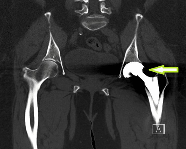
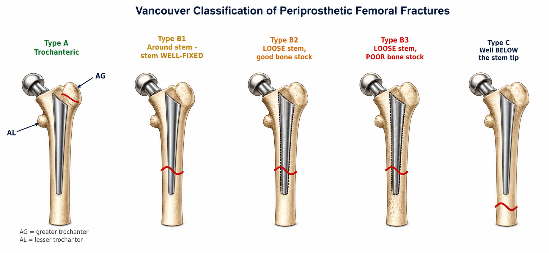
Periprosthetic fracture. The Vancouver classification guides treatment: B2 and B3 fractures need stem revision, and an acetabular fracture with a loose component needs revision.
Polyethylene wear. Severe liner wear causes osteolysis. An isolated liner exchange is enough if the components are well fixed and there is no bone loss; otherwise the cup is revised.
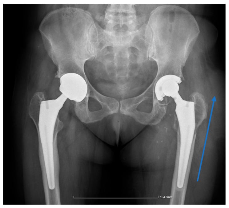
Adverse reaction to metal debris. Metal-on-metal tribocorrosion and metallosis raise serum cobalt and chromium ions and can form a pseudotumour. The bearing is revised to ceramic or polyethylene.
Component failure. Ceramic liner fracture, modular neck fracture and dissociation are rare but catastrophic, and require complete revision.
Anatomy and Biomechanics
The acetabulum. The anterior (iliopubic) column supports the anterior rim and dome, and the posterior (ilioischial) column supports the posterior wall and dome; the quadrilateral surface is the medial wall between them. On the radiograph the teardrop marks medial wall integrity and Kohler's line shows medial migration of the cup.
The femur. Standard stems fit the metaphysis; revision stems take distal fixation in the diaphysis. The isthmus, the narrowest point, is critical for canal-filling stems. Cortical thickness decides the risk of perforation during revision, and canal geometry ranges from champagne-flute to stovepipe.
The hip centre. Restore the anatomic hip centre whenever possible; the AAOS guideline asks for it within 2cm of the anatomic position. A high hip centre raises joint reaction forces by 30-50%, shortens the abductor lever arm, increases the demand on the abductors, and leads to a Trendelenburg gait and instability.
The trade-off. Accepting a high centre trades short-term stability for long-term abductor insufficiency: a construct that is stable but malpositioned trades early fixation for later instability and wear.
- Implication for Revision
- Bone loss reduces load-bearing area
- Technical Solution
- Maximise host bone contact, use augments to restore rim support
- Implication for Revision
- Metaphyseal deficiency prevents proximal fixation
- Technical Solution
- Bypass defect, achieve distal diaphyseal fixation over 4cm
- Implication for Revision
- Insufficient offset causes impingement and instability
- Technical Solution
- Use modular stems, adjust neck length, plan templating
- Implication for Revision
- Excessive lengthening causes nerve injury
- Technical Solution
- Limit to under 4cm, accept slight shortness if needed
Classification Systems
Paprosky grades bone loss on each side of the joint, and the grade tells you what can still hold an implant. Vancouver classifies periprosthetic femoral fractures by where the fracture lies and whether the stem is still fixed.
What it grades. How much supportive host bone is left, and so whether a hemispherical cup can be made stable. Rim preservation decides whether a hemispherical cup is achievable, posterior column integrity is critical to implant stability, and host bone quality affects the fixation strategy. Medial migration is judged against Kohler's line on the AP radiograph.
- Bone Loss Pattern
- Minimal bone loss, intact hemisphere
- Rim Integrity
- Rim intact, supportive
- Bone Loss Pattern
- Superior or medial bone loss, distorted hemisphere
- Rim Integrity
- Rim partially supportive
- Bone Loss Pattern
- Superior migration under 3cm, teardrop intact
- Rim Integrity
- Rim supportive
- Bone Loss Pattern
- Medial wall deficiency, migration into pelvis
- Rim Integrity
- Rim intact
- Bone Loss Pattern
- Superior migration under 3cm, teardrop obscured
- Rim Integrity
- Rim partially supportive (over 50%)
- Bone Loss Pattern
- Superior migration over 3cm, medial wall deficient
- Rim Integrity
- Rim minimally supportive (under 50%)
The differentiator is the supportive rim. A Type IIIA defect leaves over 50% host bone contact potential for a jumbo cup; a Type IIIB leaves under 40% and needs structural support. Superior migration over 3cm with medial wall deficiency is IIIB.
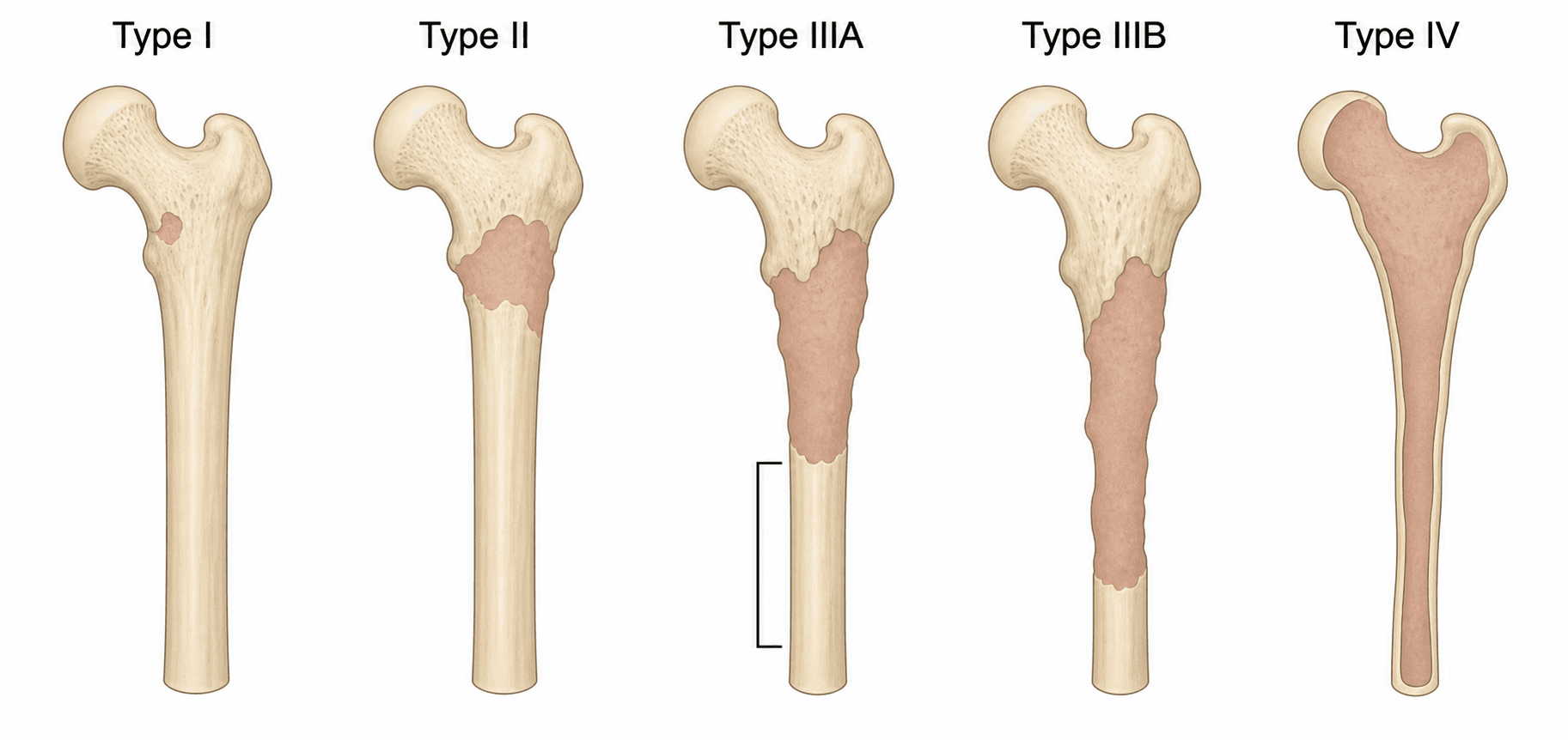
Clinical Assessment
History. The site of pain points to the failing component: groin for the acetabulum, thigh for the femur. Separate start-up pain from pain with activity, then establish:
- Function - walking distance, stairs, activities of daily living
- Previous surgery - number of operations, approaches, complications, implant types
- Infection - prolonged wound drainage, fevers, systemic illness
- Instability - number of dislocations, mechanism, whether closed reduction succeeded
Examination.
- Gait - Trendelenburg (abductor insufficiency), antalgic, limb length
- Scars - previous incisions, their quality, any sinus
- Range of motion - terminal flexion pain (loosening), a restricted arc
- Abductor strength - gluteus medius integrity
- Leg length - measured from ASIS to medial malleolus
- Neurovascular baseline - femoral pulse, sciatic function, documented
Any of these warrants aspiration before revision:
- Wound drainage persisting over 6 weeks from primary surgery
- Recurrent atraumatic dislocations (infection destabilises soft tissues)
- Early failure (under 5 years) without mechanical explanation
- Constitutional symptoms (fevers, night sweats, weight loss)
- Elevated inflammatory markers (CRP over 10, ESR over 30)
The painful THA. Pain after THA has a differential, and the discriminators below separate the causes that revision can cure from those it cannot.
- Pain Pattern
- Rest and night pain, constant, early failure
- Key Discriminator
- Raised CRP/ESR, sinus, wound issues
- Confirmatory Test
- Aspiration: cell count, culture, alpha-defensin
- Pain Pattern
- Start-up pain; groin (cup) or thigh (stem)
- Key Discriminator
- Progressive radiolucency, migration, subsidence
- Confirmatory Test
- Serial radiographs; normal inflammatory markers
- Pain Pattern
- Episodic giving-way, mechanical
- Key Discriminator
- Documented dislocation, component malposition
- Confirmatory Test
- Radiographs of cup version/inclination, exam
- Pain Pattern
- Deep ache, swelling, clunk
- Key Discriminator
- Metal-on-metal or modular taper bearing
- Confirmatory Test
- Serum cobalt/chromium, MARS MRI for pseudotumour
- Pain Pattern
- Acute pain after fall, inability to weight-bear
- Key Discriminator
- Trauma history, deformity
- Confirmatory Test
- Radiographs (Vancouver classification)
- Pain Pattern
- Variable, may not relate to hip activity
- Key Discriminator
- Normal hip imaging, positive spine or peripheral signs
- Confirmatory Test
- Diagnostic intra-articular local anaesthetic, spine imaging
Outcome scores. Three instruments measure the same hip in different ways, and the one chosen determines what can be claimed. What matters is who scores it: surgeon-scored instruments tend to read more favourably than the patient's own assessment of the same hip, which is one reason older revision series look better than modern registry data. Harris Hip Score. Surgeon-administered, and includes objective range of motion and deformity. It is a 100-point scale weighted heavily towards pain (44) and function (47), with range of motion (5) and deformity (4) contributing little: under 70 poor, 70-79 fair, 80-89 good, 90-100 excellent.
Its limitation. It is widely used, which makes historical comparison easy, but it ceilings out in a patient who is comfortable and walking, so it discriminates poorly at the good end.
WOMAC. Patient-reported, with 24 items across pain, stiffness and function. It is the most sensitive to change, so it detects the incremental improvement that matters when the starting point is a failed arthroplasty rather than a native arthritic hip.
Oxford Hip Score. A 12-item patient questionnaire scored 0-48, with 48 best. It is short enough to be completed reliably in clinic or by post and is validated in revision populations, which is why it dominates registry and large-cohort reporting, where completeness of follow-up matters more than granularity.
The Decision to Revise: Never Operate Without a Diagnosis
The governing principle. Offer revision only when a specific, correctable diagnosis that matches the symptoms has been established. Revising a painful THA for unexplained pain, without a defined mechanical or infective cause, gives poor and unpredictable results and may leave the patient worse off. It is a classic trap: no diagnosis means no revision.
The work-up, in order.
- Exclude infection first. It is the single most important diagnosis to exclude before attributing symptoms to a mechanical cause. Check CRP and ESR, then aspirate (cell count, differential, culture, with or without alpha-defensin) if they are raised or there is any clinical suspicion. This is non-negotiable: revising an unrecognised infected joint as aseptic is catastrophic.
- Exclude extrinsic and referred sources, because none of them is cured by revising the hip: the lumbar spine (radiculopathy or stenosis causing groin or thigh pain), vascular claudication, inguinal or femoral hernia, and greater trochanteric pain syndrome or abductor pathology.
- Confirm an intra-articular source. A diagnostic intra-articular local anaesthetic injection that relieves the pain supports a true intra-articular cause; failure to relieve it points away from the prosthetic hip.
- Define the failure mode with the imaging and tests described here: aseptic loosening, instability, adverse local tissue reaction or taper corrosion (serum cobalt-chromium, MARS MRI), periprosthetic fracture, or wear and osteolysis.
Only then operate, with the procedure matched to the diagnosis. If a thorough work-up yields no correctable diagnosis, do not revise: continue investigating, optimise non-operative measures, and observe.
Investigations
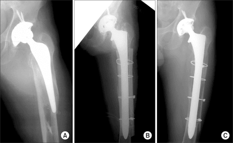
Radiographs. An AP pelvis and lateral hip are mandatory, with a full-length AP femur including the knee. Together they classify the defect, measure migration and bone loss, and show the femoral canal for planning stem length.
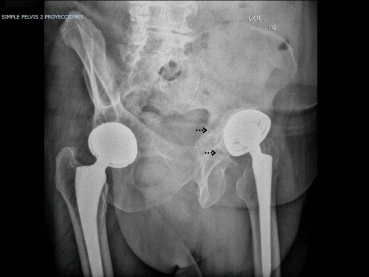
Judet views, when the acetabulum is deficient. The 45° obturator and iliac obliques assess anterior and posterior column integrity and are essential for detecting pelvic discontinuity. The iliac oblique shows the posterior column; the obturator oblique shows the anterior column.
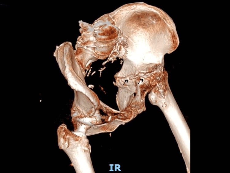
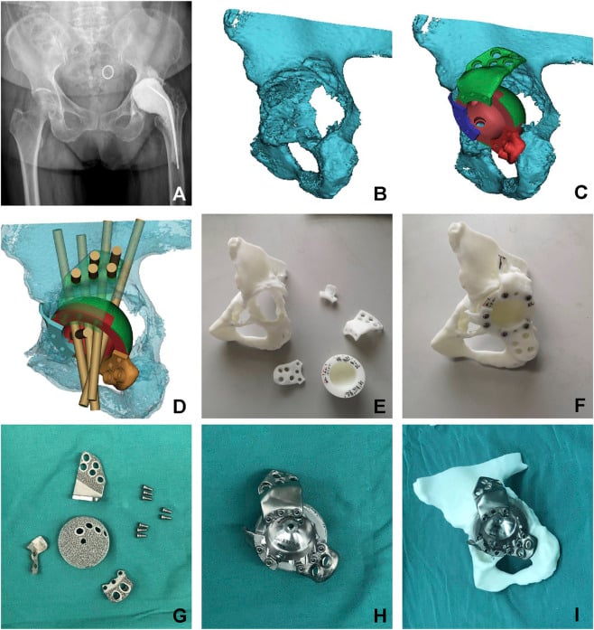
CT for complex bone loss. Three-dimensional reconstruction of the pelvis and femur measures the bone loss precisely and plans custom (triflange) implants. It is the gold standard for detecting pelvic discontinuity and essential in planning for a Paprosky IIIB defect.
MRI in selected cases. Metal artefact reduction sequences (MARS) look for pseudotumour around metal-on-metal bearings and assess soft tissue such as abductor tears. MRI is not routinely needed for standard aseptic loosening.
Blood and joint fluid. Every revision starts with inflammatory markers; aspiration follows when they are raised or clinical suspicion persists.
- Normal Value
- Under 10 mg/L
- Interpretation
- Elevated in infection but also loosening, metallosis
- Action
- If over 10: Aspiration recommended
- Normal Value
- Under 30 mm/hr
- Interpretation
- Less specific than CRP, elevated in many conditions
- Action
- If over 30: Aspiration recommended
- Normal Value
- WBC under 3000, PMN under 80%
- Interpretation
- Gold standard for infection diagnosis pre-revision
- Action
- Send cell count, culture, consider alpha-defensin
- Normal Value
- Negative
- Interpretation
- Biomarker with high sensitivity/specificity for PJI
- Action
- Adjunct when CRP/ESR equivocal, not first-line
MSIS criteria for PJI. Any one of the following establishes the diagnosis:
- A sinus tract communicating with the joint
- Two positive cultures with the identical organism
- Four of the six minor criteria, which include elevated CRP/ESR, elevated synovial WBC (over 3000), elevated synovial neutrophil percentage (over 80%), positive histology and a single positive culture
Planning. Revision is planned before the incision, and the plan is a checklist:
- The previous operative notes - approach, implant type, fixation method
- Templated radiographs - AP pelvis, lateral hip, full femur to the knee
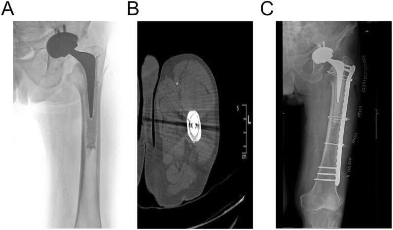
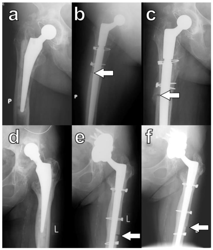
- A plan for component removal - extended trochanteric osteotomy or a femoral window
- Augments and graft ready - metal augments, allograft, bone graft substitutes
- Equipment - implant trials, extraction tools, a cement removal kit
Acetabular templating. Measure superior migration as the distance from the teardrop to the cup dome, assess the medial wall against Kohler's line and the teardrop, classify the defect, and choose between a hemispherical cup, a jumbo cup, a cage and a triflange. Reconstruction of the hip centre is planned here too, by measuring superior migration and offset on the template. The new cup is usually 2-4mm larger than the primary, and for a Paprosky I-II revision one size larger should be ready.
Femoral templating. Decide the removal strategy (ETO for a well-fixed stem, extraction for a loose one) and measure the canal diameter at the isthmus and 4cm distal to the stem. Choose the stem type (standard, extensively coated or modular tapered) and a length that bypasses defects by twice the diameter with at least 4cm of contact, then template leg length and femoral offset.
Management Algorithm
The principles. Every reconstruction answers to the same aims:
- Preserve bone stock at all costs - plan extraction before the incision, use an ETO for a well-fixed stem, and avoid aggressive reaming of the acetabulum
- Restore the hip centre and offset, and address leg length discrepancy
- Obtain stable initial fixation, biological when possible; inadequate fixation leads to early loosening
- Use modular components
- Consider a constrained liner for instability
One question. The reconstructions are alternatives matched to the defect in front of you, not stages a patient passes through, and one question selects between them: is there enough supportive host rim to achieve press-fit stability? Above roughly 50% rim support a hemispherical cup will hold; below it, the construct must obtain fixation from the ilium and ischium instead.
- Type I - standard hemispherical uncemented cup
- Type II - hemispherical cup, which may be oversized (jumbo), supplemented according to the defect: medial bone graft or a superior augment for IIA, superior support for IIB, a medial augment or mesh for IIC; screws for supplemental fixation
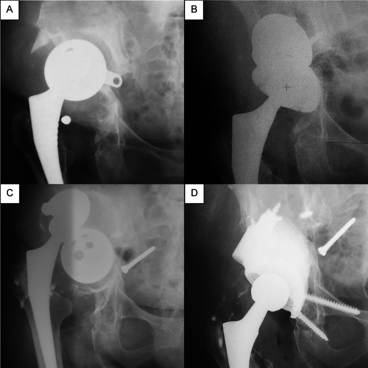
- Type IIIA - superior migration under 3cm with the posterior column intact: jumbo cup (62-66mm) with superior metal augments or structural allograft and multiple screws, and cages if needed; the superior dome augment restores the anatomic hip centre
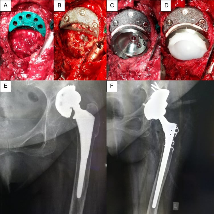
- Type IIIB - structural support: a cup-cage construct (uncemented cup plus Burch-Schneider cage), a custom triflange or a reconstruction ring; if there is a pelvic discontinuity, plate the columns first
Pelvic discontinuity. IIIB is discontinuity territory, and a discontinuity must be actively excluded before reaming. Unrecognised, it turns a cup revision into a construct that will never integrate, because the two halves of the pelvis move independently under load.
- Defect Type
- Paprosky I-II acetabular, Cortical type femoral
- Treatment Strategy
- Standard hemispherical cup, cementless stem
- Key Pearl
- Prepare for one size larger cup
- Defect Type
- Paprosky IIIA acetabular
- Treatment Strategy
- Jumbo cup with superior augment or bone graft
- Key Pearl
- Superior dome augment restores hip centre
- Defect Type
- Paprosky IIIB acetabular
- Treatment Strategy
- Cup-cage construct or custom triflange
- Key Pearl
- Requires pelvic discontinuity assessment
- Defect Type
- Paprosky III femoral
- Treatment Strategy
- Extended trochanteric osteotomy + long stem
- Key Pearl
- ETO allows controlled extraction with bone preservation
Surgical Technique
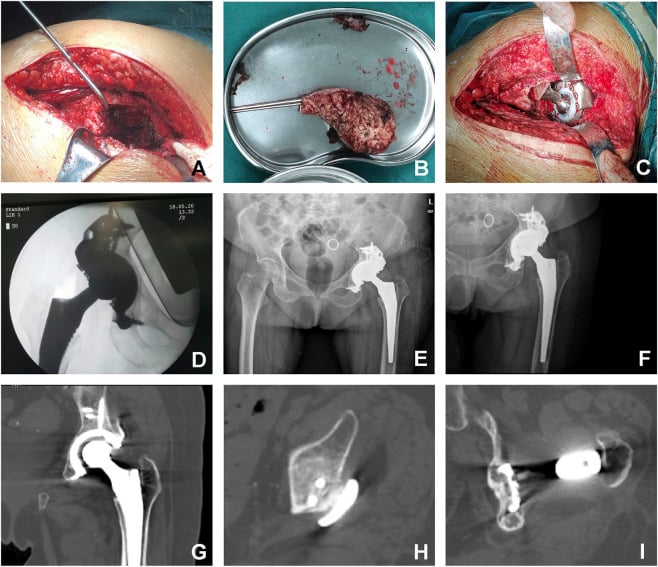
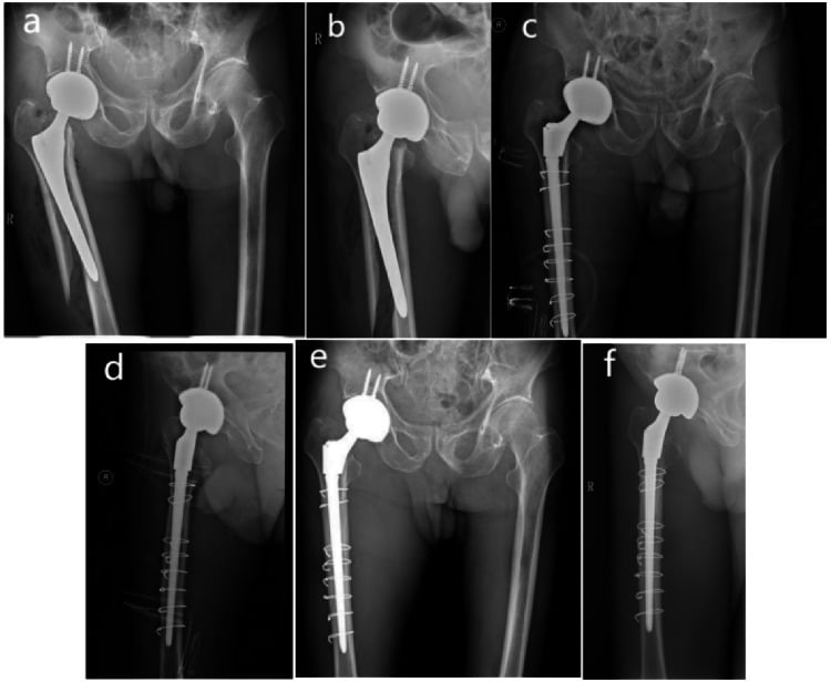
Extensile exposure. Poor visualisation leads to complications. Do not hesitate to extend the incision distally for femoral access or proximally for acetabular exposure.
- Advantages
- Extensile, excellent visualisation, can extend distally
- Disadvantages
- Abductor detachment risk if extends too superior
- Best Use
- Most revision cases, especially femoral-sided work
- Advantages
- Gluteus medius preserved, lower dislocation if primary was AL
- Disadvantages
- Limited distal extension, abductor damage possible
- Best Use
- Acetabular-only revisions, preserve abductors
- Advantages
- Stable construct, good acetabular exposure
- Disadvantages
- Abductor disruption, Trendelenburg risk
- Best Use
- Isolated acetabular revisions with stable stem
- Advantages
- Avoids additional scars, preserves blood supply
- Disadvantages
- May not be optimal for revision exposure
- Best Use
- Default unless primary approach precludes adequate exposure
Complications
Preventing the catastrophic ones. Exclude infection preoperatively, avoid lengthening the leg over 4cm, and plan component removal: an ETO for a well-fixed stem prevents perforation. Restore the hip centre, because superior migration increases dislocation and abductor insufficiency, and obtain stable initial fixation.
- Incidence
- 10-15% (vs 2-5% primary)
- Risk Factors
- Abductor insufficiency, malposition, bone loss limiting constraint
- Management
- Closed reduction, abduction brace. If recurrent: component revision to correct position or constrained liner
- Incidence
- 2-10% (higher with bone loss)
- Risk Factors
- Previous infection, wound issues, comorbidities, operative time over 3 hours
- Management
- Acute (under 3 weeks): Debridement, liner exchange, antibiotics. Chronic: Two-stage revision with spacer
- Incidence
- 5-15% at 10 years (higher for cementless cups in revision)
- Risk Factors
- Inadequate fixation, bone quality poor, infection
- Management
- Re-revision with increased bone loss
- Incidence
- 3-5%
- Risk Factors
- Osteoporosis, cortical perforation, ETO, forceful impaction
- Management
- Intraoperative: Cable fixation, extend stem. Postoperative: ORIF if stem stable, revision if loose
- Incidence
- 1-3% (sciatic most common)
- Risk Factors
- Leg lengthening over 4cm (risk rises exponentially), posterior approach, retractor placement
- Management
- Most are neurapraxia - observation for 6-12 months. EMG at 6 weeks if no recovery. Surgical exploration rarely indicated
- Incidence
- 5-7%
- Risk Factors
- Inadequate fixation, patient noncompliance, infection
- Management
- If asymptomatic: Observation. If symptomatic: Revision cable fixation, bone grafting, protected weight-bearing
Postoperative Care and Rehabilitation
Immediate Postoperative Period (Days 0-7)
DVT prophylaxis is chemical (LMWH or rivaroxaban) plus mechanical (sequential compression), continued for 35 days. Analgesia is multimodal: paracetamol, NSAIDs if not contraindicated, opioids as needed.
Mobilise with physiotherapy on a walker or crutches, with hip precautions after a posterior approach. Weight-bearing depends on fixation and bone quality:
- Standard revision (good fixation): touch to partial weight-bearing
- Structural graft or cage: touch weight-bearing for 6-12 weeks
- ETO: partial weight-bearing for 6-12 weeks until union
Check the wound for drainage, erythema and dehiscence. The goals are independent transfers and toileting and short distances with a walking aid; discharge when pain is controlled, the patient is mobilising safely and home supports are arranged.
Hip precautions after a posterior approach. Avoid flexion over 90 degrees, adduction past the midline and internal rotation for 6 weeks. They reduce the dislocation risk from 15% to under 5%.
Outcomes and Prognosis
Survival follows the defect. The figures fall as the defect worsens. That is the defect's prognosis, not the implant's failure rate, and quoting it that way is what a viva examiner is listening for.
- Implant Strategy
- Hemispherical uncemented cup
- Survival at 10 Years
- 85-90% (Type I), 80-85% (Type II)
- Notes
- Outcomes approach primary THA if good bone stock
- Implant Strategy
- Jumbo cup + augments
- Survival at 10 Years
- 75-85%
- Notes
- Augments provide better longevity than structural graft alone
- Implant Strategy
- Cup-cage or triflange
- Survival at 10 Years
- 70-80%
- Notes
- Custom triflange showing promising results in severe defects
- Implant Strategy
- Standard cementless (Type I) or extensively coated (Type II) stem
- Survival at 10 Years
- 90-95%
- Notes
- Excellent outcomes with modern cementless stems
- Implant Strategy
- Modular tapered stem
- Survival at 10 Years
- 85-90%
- Notes
- Bypassing defect with distal fixation highly successful
- Implant Strategy
- Salvage: modular tapered long stem with struts, impaction grafting or allograft-prosthetic composite
- Survival at 10 Years
- 70-80%
- Notes
- Salvage scenarios
Prognostic factors. Prognosis is good for a first, aseptic revision with minimal bone loss (Paprosky I-II) and good bone quality in a healthy host, and in a younger patient with good bone stock. It is poor with:
- Previous infection, even if eradicated
- Severe bone loss (Paprosky IIIB, IV)
- Multiple prior revisions
- Medical comorbidities (uncontrolled diabetes, immunosuppression)
- Abductor deficiency, which increases instability
What the registry adds. In the AOANJRR, infection accounts for a higher proportion of re-revisions (20%) than of first revisions (10%), and modular femoral stems have a higher revision rate than monoblock stems in the revision setting. These findings should guide implant selection and patient counselling.
Cemented vs Cementless Fixation in the Revision Setting
The AOANJRR has historically shown cemented acetabular components with a lower revision rate than cementless ones in the revision setting, the reverse of primary THA.
Why fixation behaves differently. The revision bed is sclerotic, relatively avascular, often deficient and previously instrumented, so the biological ingrowth on which cementless fixation depends is harder to achieve than in healthy primary bone. That mechanistic disadvantage is why older registry signals favoured cemented cups in revision.
Cement in sclerotic bone. Equally, cementing into a smooth, sclerotic old canal or socket gives poor cement interdigitation, so primary-style cement fixation is also less reliable. The fundamental problem is the host bone, not the fixation mode per se.
The caveat: highly porous metals. Modern highly porous cementless cups and augments (trabecular metal, tantalum) achieve ingrowth even in deficient bone and have substantially narrowed, or reversed, the historical cemented advantage. Much of the old registry signal reflects earlier, less porous cementless designs, so treat it as a registry generalisation rather than an absolute law.
Match fixation to bone: the acetabulum. Use a highly porous cementless hemispherical cup when there is at least 50% host bone contact and a stable press-fit is achievable. Where structural support is needed (cup-cage, reconstruction ring), cement a polyethylene liner into the cage. A cemented cup alone in deficient sclerotic bone loosens early and is reserved for low-demand elderly patients or cementing into a cage.
Match fixation to bone: the femur. Revision is dominated by cementless extensively porous-coated or modular fluted tapered stems with distal diaphyseal fixation. Cemented femoral revision keeps a defined role, notably impaction bone grafting into a contained canal (the Exeter, UK and Netherlands tradition, covered in the bone-loss reconstruction topic) and the elderly patient with poor bone, but not primary-style cementing into a bare sclerotic canal.
Guidelines, Registries & Global Practice
Global Epidemiology and Failure Modes
- Finding
- US total hip revisions projected to grow 137% between 2005 and 2030; demand to double by 2026
- Source
- Kurtz, JBJS Am 2007 (PMID 17403800)
- Finding
- Instability/dislocation 22.5%, mechanical loosening 19.7%, infection 14.8% (51,345 revisions)
- Source
- Bozic, JBJS Am 2009 (PMID 19122087)
- Finding
- Approximately 18% cumulative percent revision at 15 years after primary THA; re-revision risk much higher
- Source
- AOANJRR Annual Report
- Finding
- Single-dose prophylaxis as effective as multiple doses for PJI prevention
- Source
- Tan, JBJS Am 2019 (PMID 30845037)
Guideline and Registry Positions (Side-by-Side)
- Region
- USA
- Key Position
- Rule out infection before assuming aseptic failure; restore hip centre; classify bone loss to guide reconstruction
- Evidence Basis
- Clinical practice guidance / consensus
- Region
- Global
- Key Position
- Standardised PJI definition and workup (serum CRP/ESR, aspiration with cell count and culture, biomarkers); two-stage exchange for chronic PJI
- Evidence Basis
- Delphi consensus, multinational
- Region
- United Kingdom
- Key Position
- Revision arthroplasty concentrated in higher-volume units/networks; structured infection pathways and MDT decision-making
- Evidence Basis
- Guidance and best-practice standards
- Region
- Europe / Australia / USA
- Key Position
- Mandatory implant and outcome capture; survival benchmarking informs implant selection in revision
- Evidence Basis
- Observational registry data
Reconstruction philosophy varies by region: trabecular-metal/cup-cage constructs and custom triflanges are favoured in North America and Australasia, whereas impaction bone grafting (Exeter/Sloof-Ling technique) has a strong tradition in the UK and the Netherlands for contained defects. Across all systems the constants are the same: exclude infection, classify bone loss (Paprosky), restore the hip centre, and obtain stable fixation that bypasses the defect.
Hospital Systems Considerations
Revision THA Pathway in Australian Public Hospitals
Symptomatic loosening typically triaged as Category 2 (target treatment within 90 days). Severe pain or bone loss threatening viability may warrant Category 1 (urgent, within 30 days).
Mandatory: CRP, ESR, radiographs (AP pelvis, lateral hip, full femur), hip aspiration if inflammatory markers elevated. CT scan if complex bone loss. Cardiac and anesthesia clearance.
Length of stay: Average 5-7 days vs 2-3 for primary. Blood cross-match 2-4 units. DVT prophylaxis 35 days. Physiotherapy daily. Occupational therapy for home modifications.
Supports: Arrange home modifications, community physiotherapy, walking aids. Follow-up at 6 weeks, 3 months, 12 months, then annually. Registry reporting.
Key documentation requirements:
- Informed consent must include: Higher complication rates than primary (dislocation 10-15%, infection 5-10%, nerve injury 1-3%), re-revision risk 35% at 10 years, prolonged recovery 6-12 months, leg length discrepancy possibility
- Preoperative infection workup: Document CRP, ESR results. If aspiration performed, document indication and results. Failure to exclude infection before aseptic revision is major litigation risk
- Component selection rationale: Document why specific implant chosen (e.g., "Paprosky IIIA defect - jumbo cup with superior augment selected to restore hip center")
- Intraoperative complications: If perforation, fracture, or nerve injury occurs, document immediately and inform patient postoperatively
Common litigation issues:
- Unrecognized infection revised as aseptic (catastrophic outcome)
- Sciatic nerve injury from excessive leg lengthening (document leg length measured intraoperatively)
- Recurrent dislocation (document component position optimization and abductor repair)
- Early re-revision (document bone quality and fixation achieved)
Comprehensive documentation and realistic patient expectations are essential medicolegal protection.
MCQ Practice Points
Q: A 65-year-old patient has acetabular component loosening with superior migration of 3.5cm and medial wall deficiency. What Paprosky classification is this? A: Paprosky Type IIIB. Type IIIB is defined by superior migration over 3cm and typically includes medial wall deficiency. This differs from Type IIIA which has migration under 3cm. Type IIIB requires structural support (cage or triflange) whereas Type IIIA can often be managed with jumbo cup and augments.
Q: What is the minimum length an Extended Trochanteric Osteotomy should extend distal to the tip of a well-fixed femoral stem? A: The ETO should extend at least to the tip of the stem, and preferably 1-2cm beyond to allow adequate access for stem extraction. The total length is typically 10-15cm. The new revision stem must bypass the osteotomy by at least 2x the stem diameter distally to prevent stress concentration.
Q: According to AOANJRR data, what is the approximate re-revision rate at 10 years after first revision THA? A: 35% at 10 years. This highlights that revision THA has significantly worse outcomes than primary THA (18% revision rate at 15 years). Re-revision risk increases with each subsequent surgery, emphasizing importance of getting the first revision right and counseling patients appropriately.
Q: What is the most common nerve injured during revision THA and what is the primary risk factor? A: Sciatic nerve is most commonly injured (1-3% incidence). Primary risk factor is leg lengthening over 4cm, which causes traction injury to the nerve. Risk increases exponentially with lengthening beyond 4cm. Other risk factors include posterior approach and prolonged retractor placement.
Q: What is the threshold for host bone contact with a hemispherical cup below which structural support (cage) is typically required? A: Under 50% host bone contact typically requires structural support with a cage, reconstruction ring, or custom triflange. This corresponds to Paprosky Type IIIB defects. 40-60% host bone contact (Type IIIA, Kohler's line intact) can usually be managed with a jumbo cup and augments alone.
Q: According to AOANJRR, do cemented or cementless acetabular components have better survival in the revision THA setting? A: Cemented acetabular components have lower revision rates than cementless in the revision setting according to AOANJRR data. This is contrary to primary THA where cementless cups perform better. The finding suggests biological fixation is more difficult to achieve in deficient bone, and cement may provide more reliable initial stability.
Exam Viva Scenarios
Practise clinical reasoning and management decisions out loud
“A 68-year-old woman presents with progressive groin pain 12 years after primary THA. Radiographs show superior migration of the acetabular component by 2.5cm, with obscured teardrop. The femoral stem is well-fixed. CRP is 8, ESR is 25. How would you assess and manage this patient?”
“You are planning revision THA for a 72-year-old man with aseptic loosening of the acetabular component. The femoral stem is well-fixed and extensively porous-coated. Walk me through your approach to femoral component removal and reconstruction.”
“Intraoperatively during revision THA, after removing a loose acetabular component, you discover severe superior and medial bone loss with independent motion between anterior and posterior columns. How do you proceed?”
Key Classifications
- Paprosky Acetabular: Type I (intact rim), Type II (distorted, superior/medial loss), Type IIIA (migration under 3cm, over 50% rim), Type IIIB (migration over 3cm, under 50% rim)
- Paprosky Femoral: Type I (minimal loss), Type II (metaphyseal loss, diaphysis intact), Type IIIA (diaphysis over 4cm intact), Type IIIB (diaphysis damaged), Type IV (extensive loss)
- Vancouver Fracture: A (trochanteric, stem stable), B1 (around stem, stable), B2 (around stem, loose), B3 (loose with poor bone), C (distal to stem)
- Pelvic discontinuity = independent column motion, requires plating + cage/triflange
Surgical Approach
- Type I-II acetabular: Hemispherical uncemented cup, ream to bleeding bone, under-ream 1-2mm
- Type IIIA acetabular: Jumbo cup (over 62mm) + superior augments or structural graft, restore hip center
- Type IIIB acetabular: Cup-cage construct or custom triflange, requires structural support
- ETO for well-fixed femoral stem: 10-15cm long, extending distal to tip, anterior 1/3, posterior hinge, minimum 3 cables, bypass 2x diameter
Critical Steps
- ALWAYS exclude infection: CRP/ESR, aspiration if elevated or suspicious
- Plan component removal before incision: ETO for well-fixed stems prevents perforation
- Restore anatomic hip center: Superior migration leads to abductor insufficiency
- Achieve stable initial fixation: Inadequate fixation = early loosening
- Minimum 4cm diaphyseal contact for femoral stems, bypass defects by 2x diameter
Surgical Pearls
- Jumbo cups need over 50% host bone contact for success without cage
- Metal augments have lower resorption than structural allograft
- ETO union rate over 90% with cable fixation, nonunion risk 5-7%
- Constrained liners for recurrent instability have 5-8% failure at 5 years
- Leg lengthening over 4cm increases sciatic nerve injury risk exponentially
Complications
- Dislocation 10-15% (vs 2-5% primary) - malposition, abductor insufficiency
- Infection 2-10% (higher with bone loss, operative time over 3 hours)
- Nerve injury 1-3% (sciatic from lengthening over 4cm)
- Re-revision 35% at 10 years (counsel patients about high failure rate)
- ETO nonunion 5-7% (protected weight-bearing 6-12 weeks)
Evidence Base and Key Trials
Epidemiology and Projected Burden of Revision THA
- US Nationwide Inpatient Sample of 51,345 revision THAs: most common causes were instability/dislocation 22.5%, mechanical loosening 19.7% and infection 14.8%
- All-component revision was the most frequent procedure type (41.1%)
- Companion projection study (Kurtz, JBJS Am 2007, PMID 17403800, DOI 10.2106/JBJS.F.00222) forecasts US total hip revisions to grow 137% between 2005 and 2030
- Demand for hip revision projected to double by 2026
- Provides the quantitative basis for workforce and resource planning
Australian Orthopaedic Association National Joint Replacement Registry (AOANJRR)
- Approximately 18% cumulative percent revision at 15 years for primary conventional THA
- Re-revision rates after a first revision are substantially higher than after primary THA
- Loosening/lysis and dislocation are leading recorded reasons for revision
- Cemented and hybrid fixation perform well in many revision and older-patient subgroups
- Mandatory near-complete national capture of arthroplasty procedures
Paprosky Acetabular Defect Classification (Landmark)
- Original description of the acetabular defect classification (Types 1 to 3A/3B) on 147 cemented cups revised to cementless press-fit components
- Mean follow-up 5.7 years; classification based on degree of superior, medial and rim bone loss
- Only 6 of 147 components (4.0%) were unstable, all Type 3B, confirming structural support is required for the most severe defects
- Adequate remaining host bone is essential for ingrowth of hemispherical components
- Defined the framework still used to plan acetabular reconstruction strategy