Fracture | Instability | Clunk Syndrome | Aseptic Loosening
- Historically a leading cause of TKA reoperation - but contemporary revision series place aseptic loosening, infection, wear and instability ahead of patellofemoral causes
- Extensor mechanism integrity is the key determinant of management for fractures
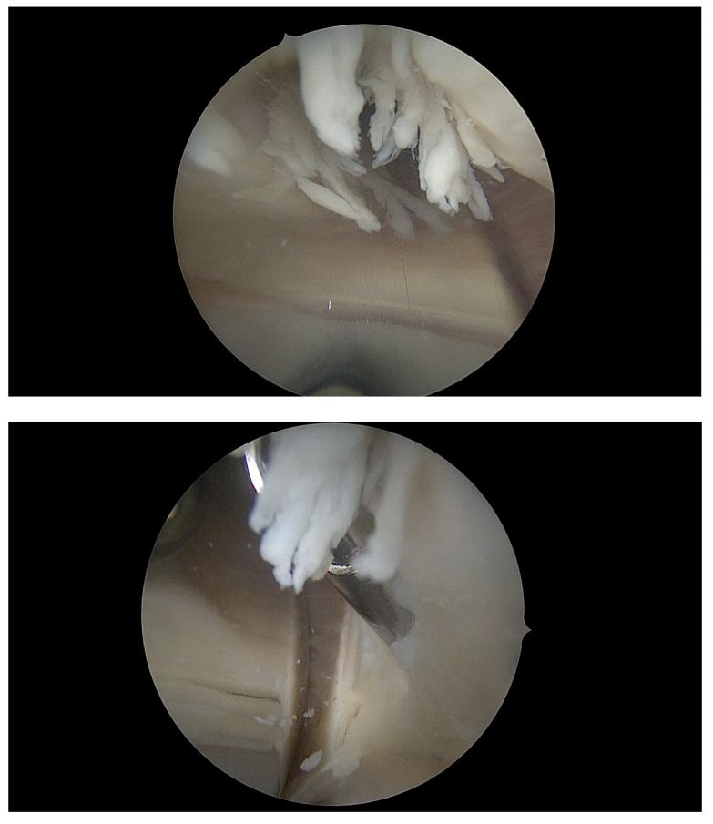
- Patellar clunk syndrome presents as a painful 'clunk' at 30-45 degrees of flexion (extension from flexion)
- Malrotation of femoral/tibial components is a major cause of patellar instability
- Avascular necrosis risk is increased by lateral retinacular release (sacrifices superior lateral genicular artery)
- “Look for component malrotation on axial CT (Berger Protocol)
- “Patellar clunk is treated with arthroscopic debridement of the fibrous nodule
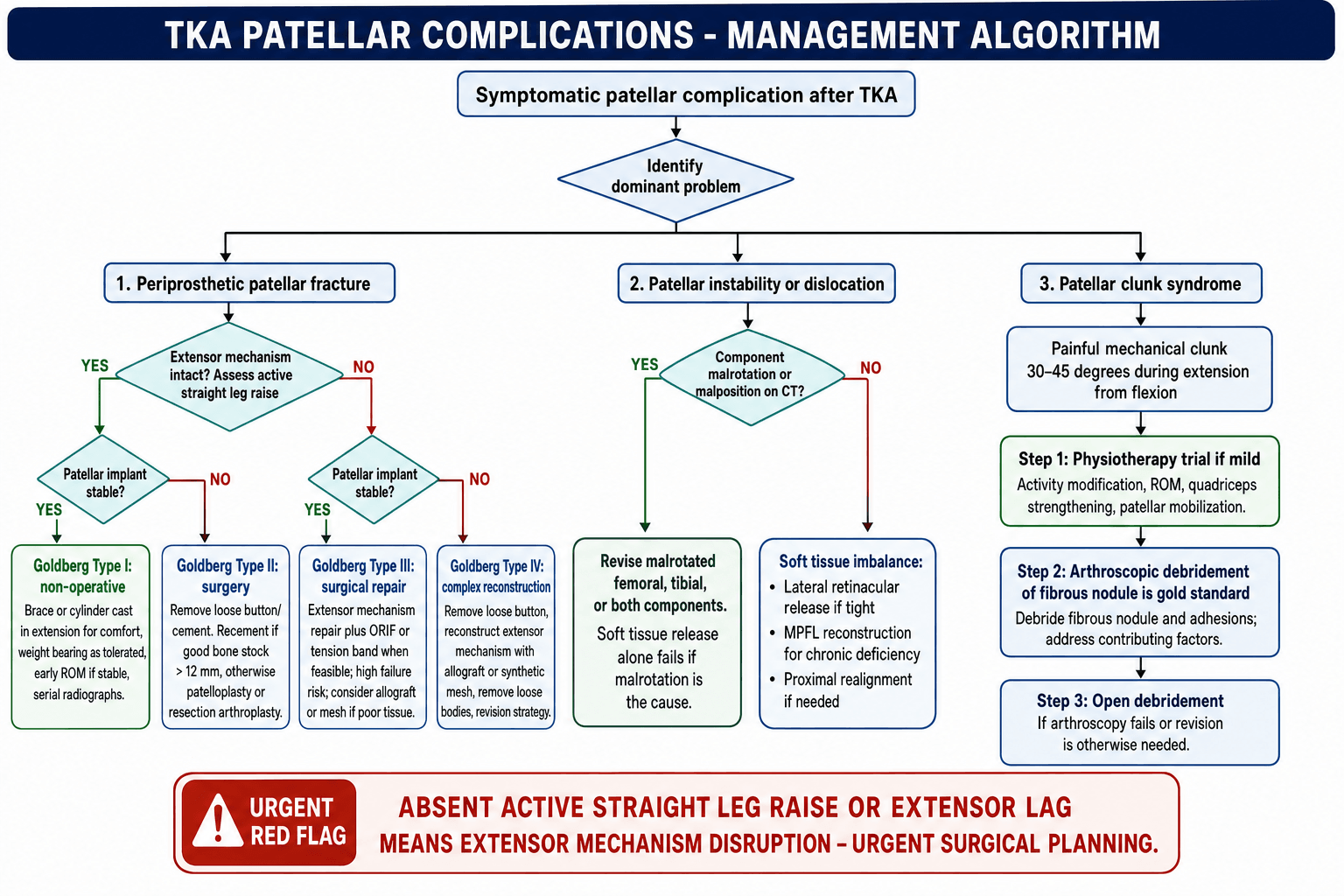
- “Goldberg classification drives treatment: Is the extensor mechanism working? Is the implant loose?
- “Avoid ORIF for patellar fractures in TKA if possible - high non-union rate (poor bone stock)
Overview and Epidemiology
Patellar complications after total knee arthroplasty are fracture, instability, clunk syndrome and aseptic loosening.
How common. In the 1980s and early 1990s patellofemoral problems were widely reported as accounting for a large share of TKA reoperations. The "up to 50%" figure still quoted is from that era and is not sourced to a single study, so quote it as history. Modern trochlear geometry, better rotational instrumentation and selective resurfacing have changed the picture, and contemporary revision series place aseptic loosening, infection, polyethylene wear and instability ahead of patellofemoral causes.
Fracture. Patellar fracture itself is uncommon, at 0.68% of 12,464 arthroplasties in the largest denominator-based series (Ortiguera and Berry, carded below). The risk factors fall into three groups:
- Vascular: lateral release (AVN)
- Technical: over-resection (residual bone less than 12 mm) and excessive thermal necrosis
- Patient: osteoporosis, high activity and male sex
Anatomy and Biomechanics
The blood supply. The patella is supplied primarily by the genicular anastomosis, in which the superior lateral genicular artery and the inferior lateral genicular artery are the most critical vessels. The superior lateral genicular artery runs in the substance of the lateral retinaculum, and a wide lateral retinacular release sacrifices it. That significantly increases the risk of patellar AVN and subsequent fragmentation or fracture, so leave a cuff of tissue if possible.
Technical errors. Each of these has an effect on the patella and a surgical counter:
- Effect on Patella
- Increases Q-angle
- Surgical Pearl
- Avoid internal rotation. Use Whiteside's line/TEA
- Effect on Patella
- Lateralises tubercle
- Surgical Pearl
- Align to medial third of tibial tubercle
- Effect on Patella
- Increases Q-angle
- Surgical Pearl
- Lateralise femoral component to improve tracking
- Effect on Patella
- Increases shear force
- Surgical Pearl
- Measure pre-op thickness and reproduce it
Overstuffing and Patellar Composite Thickness
The patellar composite. After resurfacing, the composite is the residual patellar bone plus the button, and the aim is to reproduce the native patellar thickness, measured with a calliper before cutting. Native adult patellar thickness is roughly 22-25 mm, and about 12-15 mm of residual bone should be preserved to avoid fracture and osteonecrosis.
Too thin. Over-resection, leaving residual bone under about 12 mm, weakens the patella and predisposes to fracture and AVN. The risk is compounded if the superior lateral genicular supply is also lost.
Too thick. Overstuffing is a composite thicker than native, or an oversized or proud anterior femoral flange. It increases patellofemoral contact pressure and the extensor lever-arm demand, causing reduced flexion, anterior knee pain and higher shear, which in turn drive maltracking, instability and component wear.
The femoral side. Oversizing or a proud anterior contour can overstuff the patellofemoral compartment even when the patellar composite thickness is restored. Anterior stuffing can be quantified by comparing the anterior chamfer bone resection with the corresponding implant thickness: a component thicker than the bone removed increases anterior offset and patellofemoral pressure.
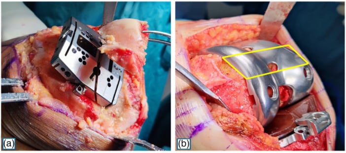
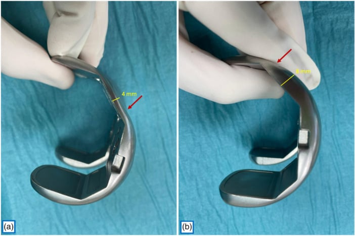
The rule. Measure the patella before resection and resurface to reproduce native thickness, and avoid an oversized or anteriorly translated femoral component. Both errors are patellofemoral: too thin risks the bone, too thick risks the joint.
Classification Systems
Goldberg's is the standard classification for periprosthetic patellar fractures. The grid below, extensor-mechanism integrity against implant stability, is the treatment-oriented framework commonly used in exams. The original 1988 paper classified fractures by location and involvement instead (Types I, II, IIIA, IIIB and lateral fracture-dislocation), but both converge on the principle that an intact mechanism with a stable implant does best.
- Mechanism Intact?
- YES
- Implant Stable?
- YES
- Treatment
- Non-operative
- Mechanism Intact?
- YES
- Implant Stable?
- NO
- Treatment
- Revision (excise loose button/cement)
- Mechanism Intact?
- NO (disrupted)
- Implant Stable?
- YES
- Treatment
- Surgery (repair/ORIF + liner exchange)
- Mechanism Intact?
- NO (disrupted)
- Implant Stable?
- NO
- Treatment
- Surgery (resection/patellectomy/allograft)
Two further factors also count: where the fracture lies relative to the implant, and the quality of the bone stock.
Patellar Crepitus vs Clunk: The Fibrosynovial Spectrum
One spectrum. Patellar crepitus and patellar clunk sit on one spectrum of peripatellar fibrosynovial hyperplasia in posterior-stabilised (PS) knees. Both arise from fibrous and synovial tissue at the quadriceps-patella junction interacting with the intercondylar box of the PS femoral component.
Crepitus is a palpable or audible grating through the mid-flexion arc, from softer peripatellar fibrosynovial hyperplasia. Clunk is a discrete, often painful catch as a mature fibrous nodule at the superior pole snaps out of the box during terminal extension, at around 30-45 degrees.
Risk factors. The two share them: PS designs, smaller or thinner patellar components, patella infera, a less accommodating trochlear box and certain implant designs. The culprit zone is the anterosuperior patella-quadriceps junction.
Telling them apart. Both preserve range of motion, which separates them from arthrofibrosis, a stiff knee. Crepitus is a grating and often incidental; clunk is a painful, reproducible catch on extension and must also be told apart from instability. The distinction matters for treatment, and naming both shows you understand the spectrum.
Clinical Assessment
History. Each complication has its own story:
- Instability: "the knee gives way", "the patella jumps out"
- Clunk: a painful catch or clunk as the knee extends from flexion
- Fracture: acute pain and inability to straight leg raise
- Pain: anterior knee pain, worse on stairs and rising
Examination. Look at the Q-angle and for valgus alignment, and palpate for tenderness and the fibrous nodule of a clunk. Assess patellar tracking for a J-sign, and the implant for gross loosening or instability.
The straight leg raise. The test that must be done is an active straight leg raise. Inability implies a Type III or IV fracture or a tendon rupture requiring surgery, and an absent raise or an extensor lag calls for urgent surgical planning.
In any painful TKA, infection must be ruled out first. Aseptic loosening and patellar complications can mimic infection. Obtain ESR/CRP and aspirate if elevated.
Investigations
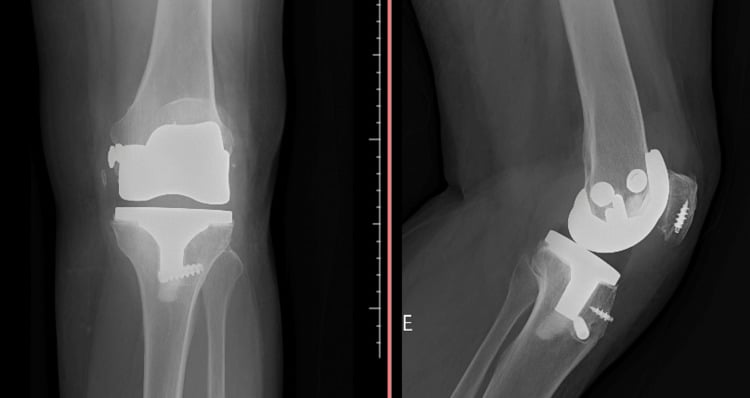
Radiographs. AP and lateral films show the fracture, implant position and bone stock. The skyline (Merchant) view is critical for patellar tilt, subluxation and the fracture profile, and it is essential when AP and lateral films understate patellofemoral failure.
What the skyline finds. Severe patellar resorption, a pattern consistent with patellar osteonecrosis, is one. Lateral-facet impingement is another: contact produced by a medialised patellar button and a prominent prosthetic trochlear margin, or facet wear in an unresurfaced patella, is a mechanical pain source that AP films can miss.
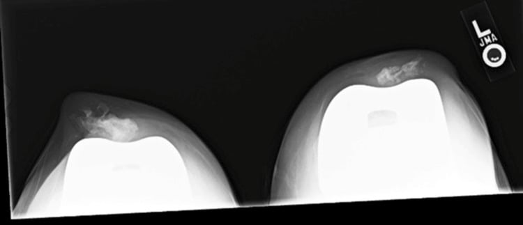
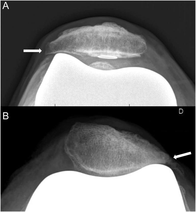
CT rotational profile. CT is indicated for instability or anterior knee pain, and measures the rotational alignment of the femoral and tibial components. Femoral rotation is read from the posterior condylar axis against the transepicondylar axis, tibial rotation from the geometric centre against the tubercle. A confirmed dislocation needs skyline imaging and CT assessment of component rotation before soft-tissue treatment or revision is chosen.
The Berger protocol typically combines femoral and tibial rotation. Internal rotation of the femoral component plus internal rotation of the tibial component gives severe patellar instability, and even minor internal rotation of both can summate to cause significant tracking problems.
Bone scan. For pain with normal radiographs and CT, a bone scan looks for occult loosening. Increased uptake in a "hot" patella may suggest stress fracture or loosening.
Management Algorithm
Principles. Management follows the Goldberg type, and three principles govern it:
- Preserve the extensor mechanism, the first priority
- Preserve bone stock
- Avoid surgery if the mechanism is intact (Type I)
Type I (intact mechanism, stable implant). A cylinder cast or brace in extension for comfort, weight bearing as tolerated, and immediate range of motion if stable to prevent stiffness. Watch for displacement.
Type II (intact mechanism, loose button). Remove the loose button and cement. Recement if the bone is good, greater than 12 mm. If bone stock is poor, it is often better to leave the patella unresurfaced (patelloplasty, a resection arthroplasty) than to recement a new button onto a shell of bone.
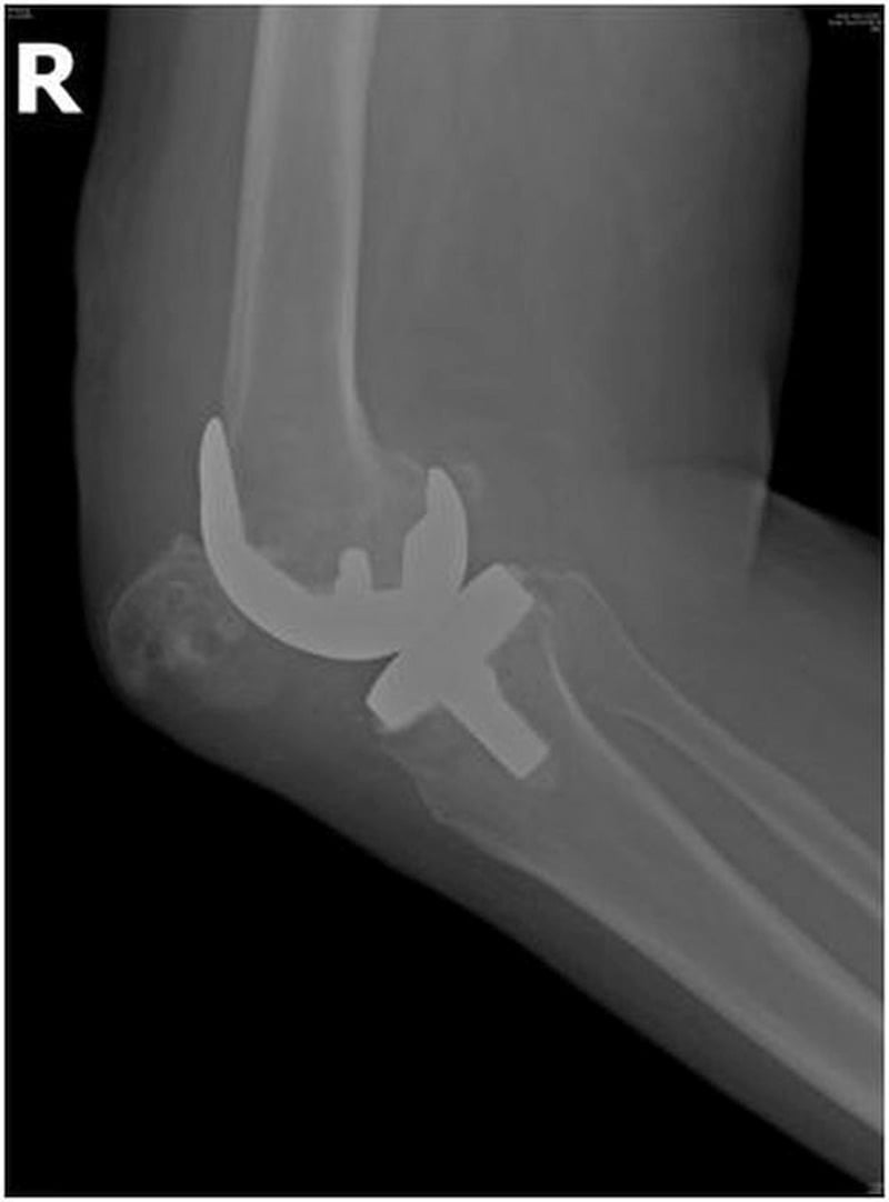
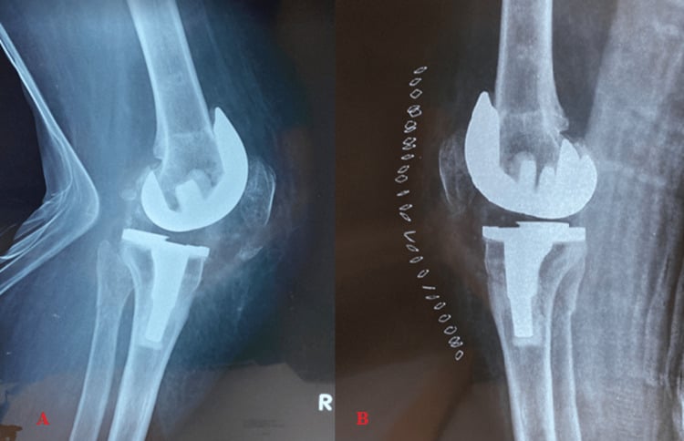
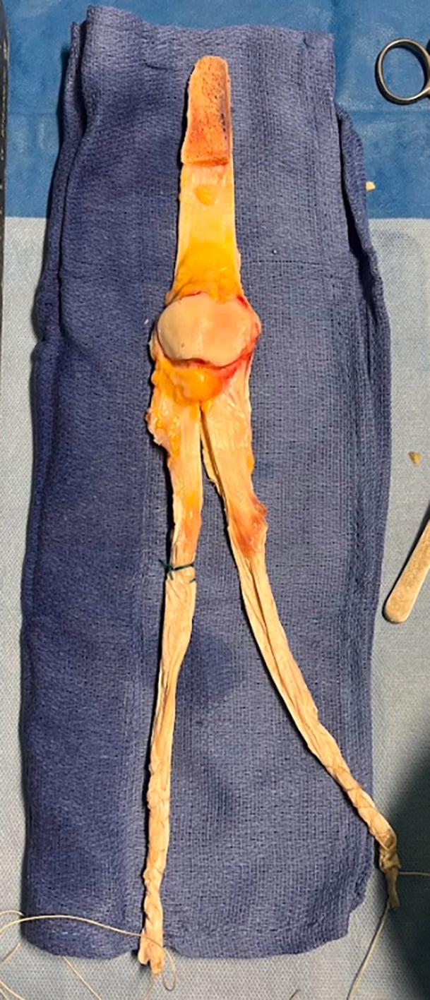
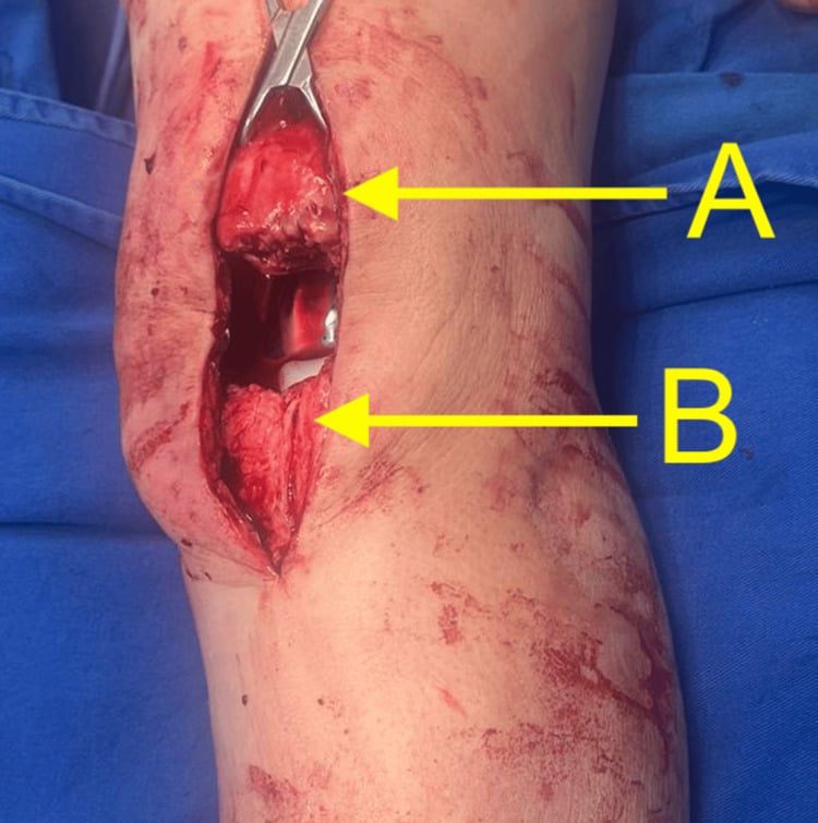
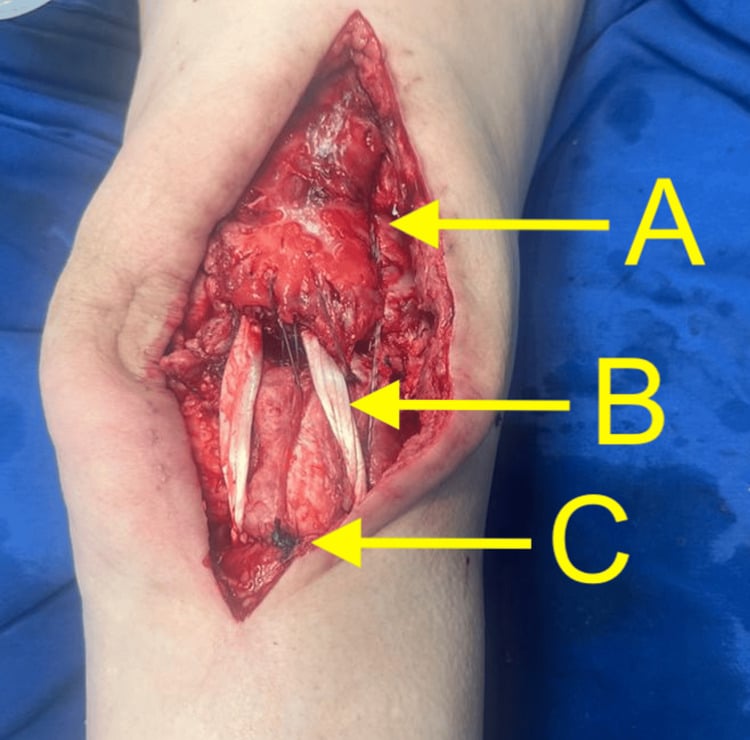
Type III (disrupted mechanism, stable button). Repair the extensor mechanism and fix the fracture (ORIF, tension band). The outcome is poor, with a high failure rate. Consider allograft if the repair is poor. In the case shown below, a displaced distal-pole fracture had interrupted the extensor mechanism despite stable components, and transosseous sutures restored continuity while preserving limited patellar bone stock.
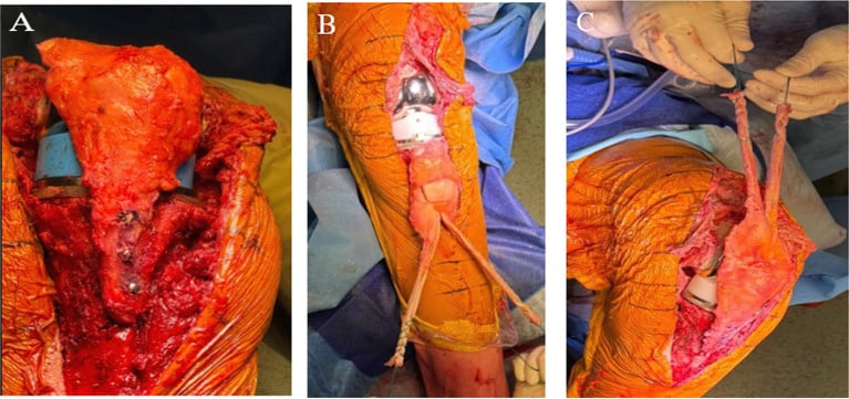
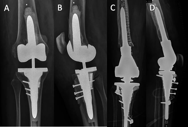
Type IV (disrupted mechanism, loose button). Remove the button and reconstruct the mechanism with allograft or mesh; resection or patellectomy are the alternatives. It typically requires removal of loose bodies and major reconstruction.
Surgical Technique
Revision for patellar instability caused by malrotation runs in five steps:
- Exposure: the standard medial parapatellar approach and an extensive synovectomy, inspecting patellar tracking before dislocating or everting
- Assess rotation: confirm internal rotation of the femoral component against the transepicondylar axis and Whiteside's line
- Component removal: take the femoral component out carefully to preserve bone, with osteotomes or a Gigli saw if cemented
- Re-implantation: set the femoral component in 3 degrees of external rotation relative to the posterior condyles, or parallel to the TEA, and lateralise it with an offset stem or a lateral shift to improve tracking
- Verification: the "no thumbs" test, the patella tracking centrally without thumb pressure throughout the range of motion
Complications
- Risk
- High in Type III/IV
- Management
- Allograft/Mesh Reconstruction
- Prevention
- Avoid aggressive ROM
- Risk
- Lateral Release
- Management
- Observation over Patellectomy
- Prevention
- Preserve Sup Lat Genicular Artery
- Risk
- Common (Poor bone)
- Management
- Accept if painless/stable
- Prevention
- Optimised fixation
- Risk
- Malrotation not addressed
- Management
- Revision TKA
- Prevention
- Correct component rotation
Postoperative Care
Rehabilitation Protocol
- Fracture or repair: hinged knee brace locked in extension
- Weight bearing: as tolerated, with the brace locked in extension
- Revision TKA: often weight bearing as tolerated, with ROM started immediately if stable
- Fracture: passive ROM limited to 0-30 degrees (if the repair is strong)
- Clunk resection: immediate full ROM allowed
- Radiographs at 2 and 6 weeks for alignment and displacement
- Unlock the brace for walking (if SLR control is good)
- Progressive flexion as tolerated
- Quadriceps strengthening starts late (3 months) for fractures
- Full functional activity
- Monitor for late loosening or recurrence of instability
After extensor repair. Strict adherence to range-of-motion limits is required. Serial review must also assess fixation, graft protection and recurrent patella alta.
Outcomes and Prognosis
Fracture. Surgical repair fails in 20-30% of cases. Nonunion is common, at 30-50%, but often painless if Type I, and the best outcome is a Type I fracture treated non-operatively, with 96% success.
Clunk. Arthroscopic debridement has a success rate greater than 95%, and recurrence is rare if enough of the nodule is resected.
Instability. Success depends on identifying the cause. Revising malrotated components succeeds in 80-90%.
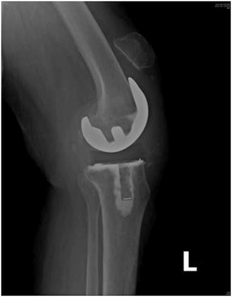
Extensor rupture. The prognosis is poor, and primary repair has a high failure rate. In a TKA, chronic tissue compromise makes reconstruction more reliable than isolated primary repair, and allograft or mesh reconstruction is often required. Arthrodesis may be the end stage.
Guidelines, Registries & Global Practice
Periprosthetic patellar fracture is uncommon (around 0.7% of TKAs; Ortiguera & Berry), but patellofemoral problems collectively remain a leading reason for reoperation. Risk rises with resurfacing, lateral release, over-resection (composite under 12 mm), and component malrotation. Reported rates vary widely with implant design and whether the patella was resurfaced.
- Patellar Resurfacing
- Selective or routine (surgeon-dependent)
- Key Recommendation
- No single mandate; tailor to design, anatomy, anterior knee pain risk
- Patellar Resurfacing
- Either acceptable
- Key Recommendation
- Decision should be informed and design-specific; rule out infection before revision
- Patellar Resurfacing
- N/A (fracture focus)
- Key Recommendation
- Manage periprosthetic patellar fracture by extensor integrity + implant fixation
- Patellar Resurfacing
- Selective
- Key Recommendation
- Optimise component rotation; avoid routine lateral release
- Patellar Resurfacing
- Lower reoperation when resurfaced
- Key Recommendation
- Resurfacing associated with reduced secondary patellar surgery
- Joint registries (NJR UK, AJRR US, AOANJRR Australia, NZJR) consistently show lower rates of secondary patellar surgery when the patella is resurfaced.
- Level I meta-analysis (Chen 2021) concurs: reduced reoperation and noise, no difference in anterior knee pain.
- Rule out PJI first: aspiration plus ESR/CRP before any revision for "pain" or "instability" - a universal standard.
- High-resource: extensor allograft and synthetic mesh available; rotational CT readily obtained.
- Limited-resource: allograft scarce, so autograft/mesh and tertiary referral are emphasised; reliance on skyline radiographs where CT access is limited.
Related pages: TKA Extensor Mechanism Failure is the endpoint the worst of these complications reaches, and the two pages share a governing principle — extensor mechanism integrity decides management; Patella Fractures for the native-knee injury and its fixation, and Patellar Height Abnormalities for the alta and baja that both cause and result from these problems; TKA Alignment Philosophies and TKA Alignment Options carry the rotational landmarks — transepicondylar axis, Whiteside's line, medial third of the tubercle — whose misuse produces the combined internal rotation the Berger card measures; TKA Complications: Instability because patellar maltracking and tibiofemoral instability share malrotation as a cause and are frequently confused; TKA Aseptic Loosening for the patellar component loosening that defines a Goldberg Type III / Ortiguera Type III fracture; Revision TKA for the component revision that malrotation ultimately requires, since no soft-tissue procedure corrects a malrotated component; Knee Arthrofibrosis and TKA Stiffness for the fibrosynovial proliferation at the root of the clunk-crepitus spectrum; Periprosthetic Joint Infection because a painful patellofemoral joint after arthroplasty is infection until excluded; Patellar Instability Pediatric for the native tracking mechanics that the arthroplasty is trying to reproduce; and Polyethylene UHMWPE and XLPE for the bearing material behind patellar component wear.
MCQ Practice Points
Q: Which arterial structure is most at risk during a lateral retinacular release in TKA? A: Superior Lateral Genicular Artery (SLGA) - The SLGA runs in the lateral retinaculum. A wide lateral release can transect this vessel, compromising patellar blood supply and increasing the risk of avascular necrosis and fracture.
Q: A patient with a well-fixed TKA sustains a patellar fracture after a fall. She is able to perform a straight leg raise. The implant appears stable on X-ray. What is the Goldberg classification and appropriate management? A: Type I - Non-operative - This is a Goldberg Type I fracture (Intact extensor mechanism + Stable implant). The active straight leg raise confirms mechanism integrity. Treatment is non-operative (immobilizer/brace) as surgical intervention has a high complication rate.
Q: What is the most likely cause of late-presenting patellar instability after TKA? A: Component Malrotation - While all can cause issues, component malrotation (especially internal rotation of the femoral or tibial component) is a leading cause of mechanical patellar instability. Polyethylene wear can cause late instability but malrotation is the primary mechanical driver.
Q: Why is primary repair of a chronic patellar tendon rupture in TKA contraindicated? A: High Failure Rate - Primary repair of chronic disruptions (especially in revision scenarios) has an excessively high failure rate. Management requires reconstruction using either an extensor mechanism allograft or synthetic mesh (e.g., Marlex), rather than simple suture repair.
Q: How does excessive joint line elevation affect the patella? A: Pseudo-Patella Baja - elevating the joint line (e.g., with thick poly or distal femoral augments) causes "pseudo-patella baja". The patella tendon length is normal, but the patella impinges on the tibial post or poly during flexion, reducing ROM and causing anterior pain.
Exam Viva Scenarios
Practise clinical reasoning and management decisions out loud
“A 68-year-old female, 1 year post-op PS-TKA. Doing well but complains of a painful 'catch' or 'clunk' when she stands up from a chair. No pain at rest. ROM 0-120.”
“A 75-year-old male, 5 years post-TK. Falls onto knee. Pain, swelling. X-ray shows a transverse fracture of the patella. The button appears well-fixed.”
“A patient presents with anterior knee pain and feelings of instability 2 years post-TKA. She feels the kneecap 'jumps'. On exam, she has a positive J-sign and lateral apprehension.”
Key Classifications
- Goldberg I: Intact mech, Stable implant → Non-op
- Goldberg II: Intact mech, Loose implant → Revision/Patelloplasty
- Goldberg III: Disrupted mech, Stable implant → Repair + Fixation
- Goldberg IV: Disrupted mech, Loose implant → Salvage/Reconstruct
Patellar Clunk
- Fibrous nodule at superior pole
- Catches in intercondylar box (PS knees)
- Clunk at 30-45° extension
- Rx: Arthroscopic debridement
Instability Causes
- Internal Rotation of Femoral Comp
- Internal Rotation of Tibial Comp
- Valgus alignment
- Tight lateral retinaculum
- Medialized femoral component
Management Pearls
- Avoid surgery for Type I fractures (high complication rate)
- CT scan critical for instability (assess rotation)
- Lateral release endangers Superior Lateral Genicular Artery
- Berger Protocol: Combined internal rotation predicts instability
Blood Supply
- Genicular anastomosis
- Superior Lateral Genicular Artery (Most important)
- Inferior Lateral Genicular Artery
- Avoid deep lateral dissection
Evidence
- Ortiguera & Berry: nonop for intact mechanism/stable implant; surgery has high complications
- Berger: combined internal rotation predicts patellar complication severity (CT)
- Hozack: defined patellar clunk syndrome (PS designs)
- Chen 2021 / registries: resurfacing lowers reoperation rate
- Scott: lateral release compromises SLGA, risks AVN/stress fracture
Evidence Base
Goldberg Classification of Periprosthetic Patellar Fracture
- The original 1988 paper analysed 36 patellar fractures in 35 condylar TKAs (mean 4.5-year follow-up)
- Classified by fracture location and involvement of the extensor mechanism and implant/cement composite (Types I, II, IIIA, IIIB and lateral fracture-dislocation)
- Fractures sparing the implant/cement and quadriceps (Type I) and nondisplaced inferior-pole fractures (Type IIIB) did well non-operatively; operatively treated disrupted/dislocated types had more failures
- This underpins the widely-taught treatment framework used here: intact mechanism plus stable implant has the best prognosis; disrupted-mechanism or loose-implant fractures the worst
The Patellar Clunk Syndrome
- Original description in three patients after posterior-stabilised TKA
- Painful 'catch' or 'clunk' on knee extension
- Prominent fibrous nodule at junction of proximal patellar pole and quadriceps tendon
- Surgical removal of the nodule resolved symptoms
Periprosthetic Patellar Fracture: Classification & Outcomes
- 85 fractures (77 patients) after 12,464 TKAs - overall prevalence 0.68%
- Significantly more common in men (1.01%) than women (0.40%)
- Type I (intact mechanism + stable implant, n=38): nonoperative treatment with only one late failure
- Operative treatment (disrupted mechanism or loose component) had high complication and reoperation rates
Malrotation Causing Patellofemoral Complications
- 30 patients with patellofemoral complications vs 20 well-functioning controls
- Complication group had excessive combined (femoral + tibial) internal rotation; controls were in external rotation
- Combined internal rotation 1-4 deg: lateral tracking/tilt; 3-8 deg: subluxation; 7-17 deg: dislocation or late prosthesis failure
- Transepicondylar axis and tibial tubercle are reproducible CT landmarks
Lateral Release, Patellar AVN & Stress Fracture
- Patellar stress fracture incidence 0.7% in rheumatoid vs 3.5% in osteoarthritic knees
- Osteonecrosis occurred in at least three cases where lateral release had sacrificed the superior lateral genicular artery
- Vessel should be preserved to protect patellar and lateral skin flap blood supply
- Preserving peripheral cortex and bone stock reduces fracture risk
Patellar Resurfacing vs Non-resurfacing: Meta-analysis
- 32 randomized trials, 6887 knees
- Resurfacing significantly reduced reoperation (overall and at 5+ years) and noise
- Higher Knee Society and function scores with resurfacing
- No significant difference in anterior knee pain, ROM or patient satisfaction