Synovitis-Driven Deformity | Tendon Rupture | Surgical Sequencing
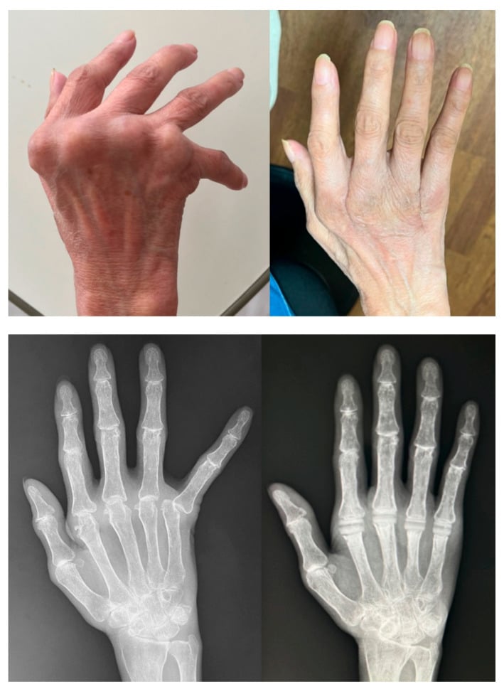
- RA synovitis destroys ligaments, tendons and cartilage producing characteristic zig-zag deformities
- Caput ulnae = dorsal DRUJ subluxation; attrition ruptures extensor tendons (Vaughan-Jackson); Mannerfelt is the flexor-side equivalent, FPL over the scaphoid
- Nalebuff classification guides thumb reconstruction: Type I (MCP boutonniere), Type II (MCP+CMC), Type III (CMC swan-neck), Type IV (MCP ulnar collateral ligament laxity), Type V (MCP volar plate laxity), Type VI (arthritis mutilans)
- Surgical sequence = DIP/PIP first, then MCP, then wrist, then elbow/shoulder (distal-to-proximal)
- Medical optimisation with DMARDs is prerequisite before elective surgical intervention
- “Vaughan-Jackson = EDM rupture from attrition over caput ulnae
- “Mannerfelt-Norman = FPL rupture over scaphoid osteophyte
- “Ulnar drift worsened by radial deviation of wrist (zig-zag collapse)
- “Surgical sequence is distal-to-proximal; wrist before MCP if both needed
Overview and Epidemiology
Rheumatoid arthritis involves the hand and wrist in up to 90% of patients, more than any other region, and the hand deformities are the visible hallmark of the disease. The pattern is bilateral and symmetrical, with MCP and PIP synovitis and progressive deformity, in contrast to the DIP-predominant pattern of osteoarthritis.
Who. RA affects approximately 0.5-1% of the global population, women three times as often as men (3:1). Onset peaks at 30-50 years, though all ages are affected.
What it costs the hand. Grip strength is reduced by up to 70% in advanced disease, and pinch weakens, key pinch especially once the thumb is involved. Buttoning, writing and opening jars are affected early. Deformity then progresses in a predictable pattern, from synovitis to ligament destruction to joint subluxation.
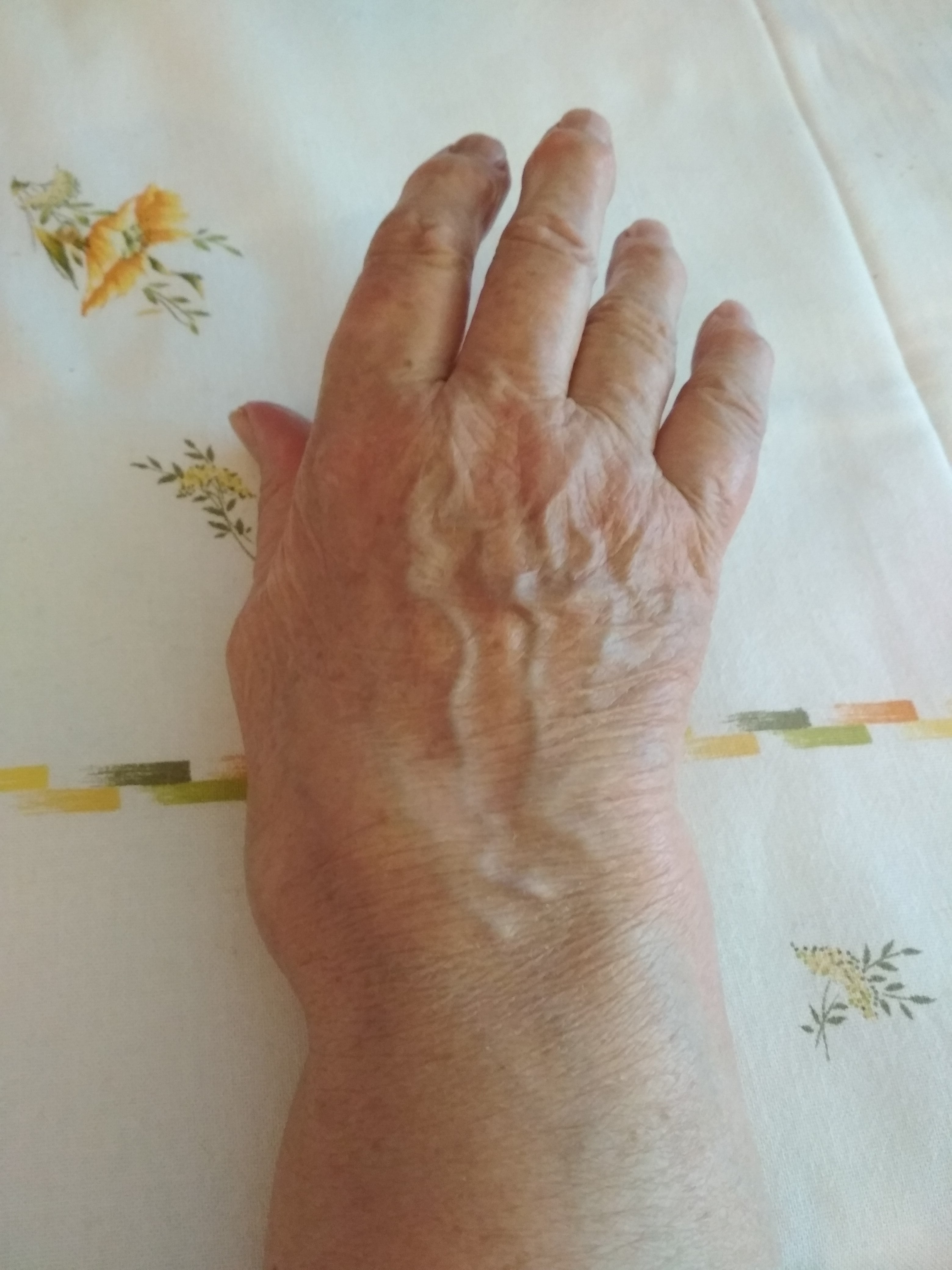
Pathophysiology
Synovitis drives everything. Dorsal synovitis starts at the wrist and MCP joints, and produces hypertrophied pannus that invades and destroys periarticular structures through enzymatic digestion and mechanical pressure. Pannus erodes articular cartilage and bone, and the destruction of stabilising ligaments and tendons leaves an imbalance between flexors and extensors that produces the classic zig-zag deformities. Medical control of synovitis is therefore the foundation of management. In the hand and wrist the process targets:
- Joint capsules and ligaments - instability, subluxation and ulnar drift
- Extensor tendons - attrition rupture over the caput ulnae
- Flexor tendons - FPL especially, in the carpal tunnel over the scaphoid (the Mannerfelt lesion)
- Articular cartilage and bone - marginal erosions, cartilage loss, osteopenia
- Intrinsic muscles - fibrosis and contracture drive swan-neck deformity and contribute to MCP subluxation
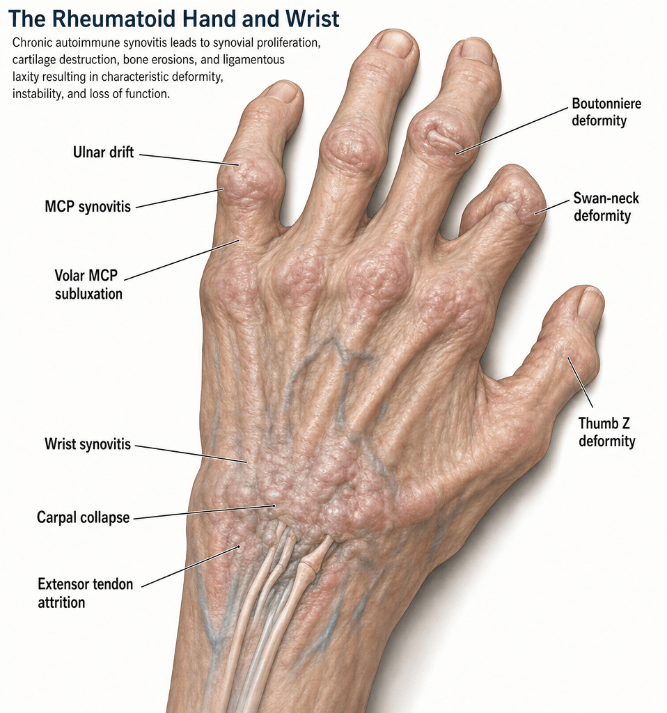
- Primary Structure Damaged
- Dorsal capsule, collateral ligaments, volar plate
- Mechanism
- Synovitis stretches dorsal capsule; flexor tendons pull proximal phalanx volarly
- Resulting Posture
- Proximal phalanx subluxes volarly on metacarpal head
- Primary Structure Damaged
- Radial collateral ligament, radial sagittal band
- Mechanism
- Radial-sided structures attenuate; intrinsic muscles pull ulnarly; extensor tendons slip ulnar to MCP axis
- Resulting Posture
- Fingers deviate ulnarly at MCP joints
- Primary Structure Damaged
- Volar plate, FDS insertion, intrinsic muscles
- Mechanism
- Volar plate laxity + intrinsic tightness + FDS rupture/attenuation allows PIP hyperextension
- Resulting Posture
- PIP hyperextension + DIP flexion
- Primary Structure Damaged
- Central slip (extensor mechanism)
- Mechanism
- Synovitis at PIP attenuates central slip; lateral bands migrate volar
- Resulting Posture
- PIP flexion + DIP hyperextension
- Primary Structure Damaged
- DRUJ capsule, TFCC, ECU tendon sheath
- Mechanism
- DRUJ synovitis leads to dorsal ulnar head subluxation and supination of carpus
- Resulting Posture
- Prominent dorsally subluxed ulnar head
Zig-zag collapse. When the carpus supinates and deviates radially as RA destroys it, the metacarpals point more ulnarly, so radial deviation of the wrist drives ulnar deviation at the MCP joints. Wrist position governs MCP alignment, and correcting the wrist is critical before or during MCP surgery.
Why the ulnar side fails first. The DRUJ and the ulnar-sided wrist structures are the first to be affected in RA:
- The ulnar styloid and head have thin overlying soft tissue
- The ECU tendon sheath is a common site of early synovitis
- The TFCC is vulnerable to pannus invasion
- The extensor tendons lie directly over the subluxing ulnar head, EDM especially, as the most ulnar tendon at the DRUJ
Classification and Types
The rheumatoid thumb. The Nalebuff classification is the most commonly examined system for RA thumb deformity. The thumb is classified by the joint that is the primary site of destruction, which then dictates the surgical plan. There are six types, and Type I, the boutonniere, is by far the most common.
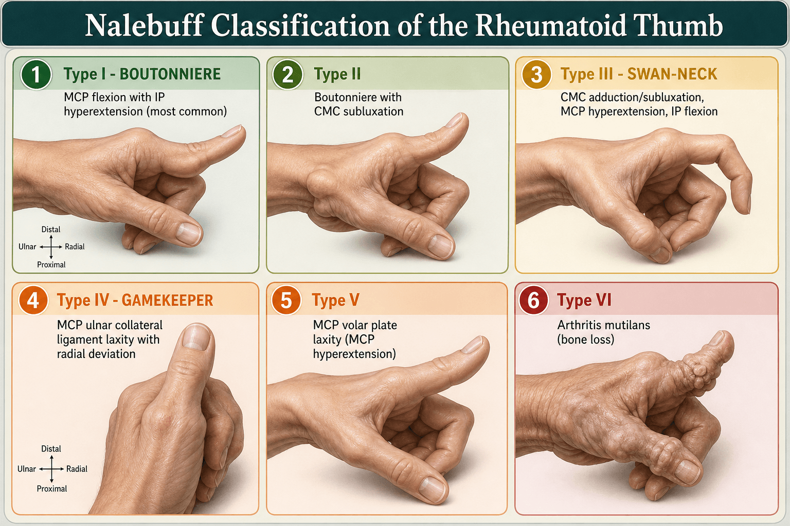
Nalebuff Classification of RA Thumb Deformity
- Primary Site
- MCP joint
- Pathoanatomy
- MCP synovitis stretches dorsal capsule and collateral ligaments; IP hyperextends to compensate
- Deformity
- MCP flexion + IP hyperextension (boutonniere-type)
- Primary Site
- MCP + CMC
- Pathoanatomy
- Combined MCP boutonniere and CMC subluxation
- Deformity
- MCP flexion + CMC dorsal subluxation + IP hyperextension
- Primary Site
- CMC joint
- Pathoanatomy
- CMC synovitis leads to dorsal subluxation; MCP hyperextends and adducts (swan-neck type)
- Deformity
- CMC subluxation + MCP hyperextension + IP flexion
- Primary Site
- MCP ulnar collateral ligament
- Pathoanatomy
- MCP ulnar collateral ligament laxity
- Deformity
- Gamekeeper deformity
- Primary Site
- MCP volar plate
- Pathoanatomy
- Attenuation of the MCP volar plate, without the primary CMC disease that defines Type III
- Deformity
- MCP hyperextension; the IP joint compensates by flexing
- Primary Site
- Skeletal collapse
- Pathoanatomy
- Gross bone loss and resorption (opera-glass hand, main en lorgnette); the end-stage destructive pattern
- Deformity
- Flail, telescoping, grossly shortened thumb ray
Both show MCP hyperextension. In Type III it is secondary to CMC collapse and adduction; in Type V the MCP volar plate is primarily lax and the CMC is normal. The discriminator is whether the CMC joint is the driver, because it changes the operation: CMC arthroplasty for III, MCP-level stabilisation for V.
Clinical Assessment
History. The questions that shape the plan:
- Disease duration and treatment - DMARDs, biologics, steroid history
- Hand function - grip, pinch and ADLs such as buttoning, key turning and writing
- Deformity progression - the rate of change, and which fingers were affected first
- Pain - localised to specific joints (wrist, MCP, PIP)
- Tendon function - sudden loss of extension (rupture), triggering
Examination. Inspect for the zig-zag deformity pattern, MCP ulnar drift, wrist supination and thumb posture, and palpate for boggy synovitis, the caput ulnae and the crepitus of tendon fraying. Test active finger extension tendon by tendon and look at the flexion cascade. Then test passive correctability: can the MCP joints be reduced, and is the PIP deformity flexible or fixed?
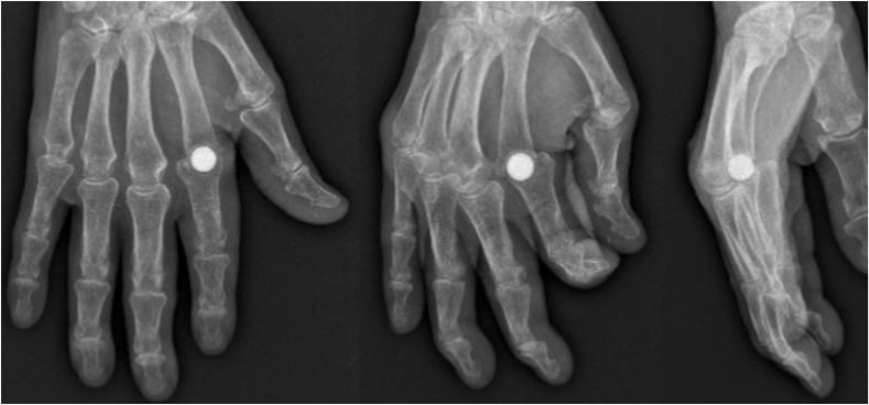
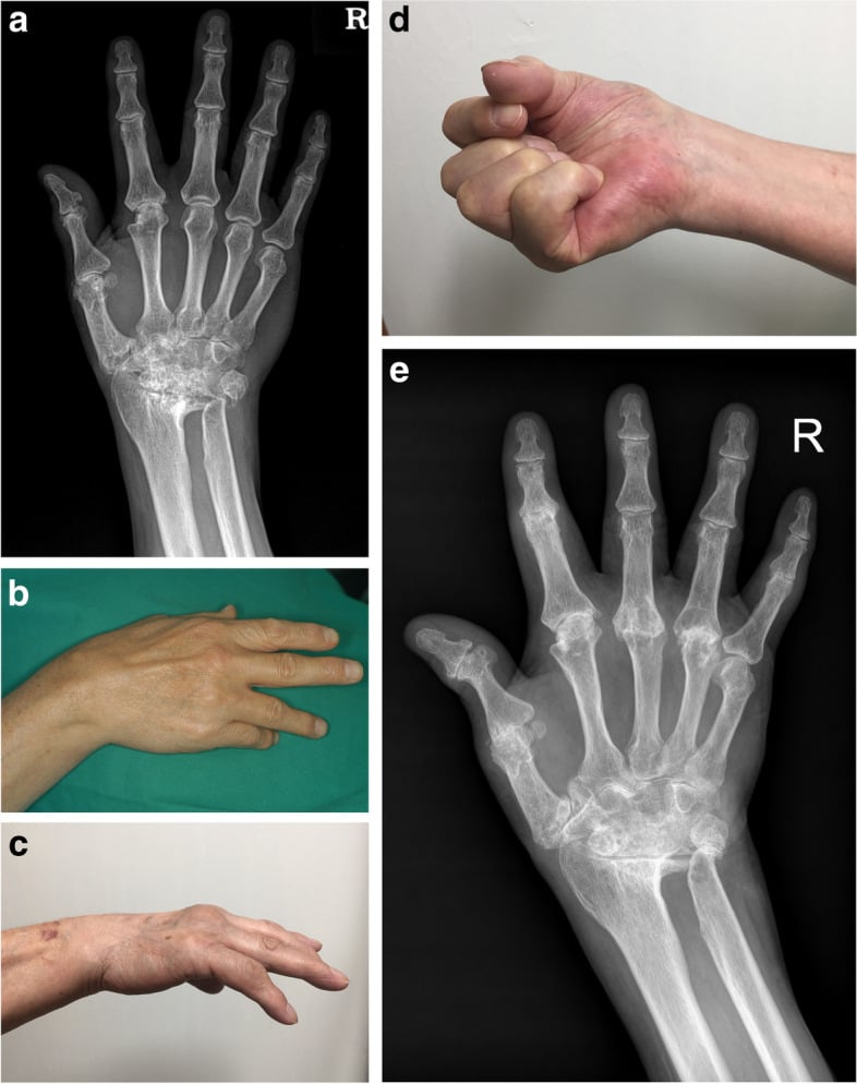
The caput ulnae. A dorsally subluxed ulnar head at the DRUJ. The caput ulnae sign is the prominent ulnar head, visible and palpable dorsally, especially with the forearm pronated. The piano key test demonstrates the subluxation: press the ulnar head and it depresses like a piano key, rebounding when released. The sharp bony prominence it creates attritions the extensor tendons, EDM and the EDC to the small and ring fingers especially.
Testing the tendons. Test each finger's active extension independently. Test EDM by asking the patient to extend the small finger while the others are held flexed: inability is the Vaughan-Jackson rupture, and extension is then lost progressively from ulnar to radial digits. For FPL, test thumb IP flexion against resistance.
Sudden loss of extension is a rupture until proven otherwise. Do not attribute it to a disease flare without ultrasound or MRI confirmation.
- Tendon(s) Involved
- EDM first, then EDC (small, ring, middle sequentially)
- Mechanism
- Attrition over dorsally subluxed ulnar head
- Site of Attrition
- Dorsal DRUJ (caput ulnae)
- Tendon(s) Involved
- Flexor pollicis longus (FPL)
- Mechanism
- Attrition over scaphoid osteophyte or rough bone in carpal tunnel
- Site of Attrition
- Volar wrist / carpal tunnel (scaphoid ridge)
- Tendon(s) Involved
- ECU rupture or subluxation
- Mechanism
- Synovitis of ECU sheath, tendon attenuation
- Site of Attrition
- Dorsal ulnar wrist
The Mannerfelt lesion is easily missed. FPL rupture in RA is often painless and may present as an incidental finding, the patient simply unable to flex the thumb IP joint. Unlike the extensor ruptures it is a flexor-side attrition in the carpal tunnel, so always examine FPL function: loss of thumb IP flexion against resistance is diagnostic. Caught early, carpal tunnel release alone may prevent further ruptures.
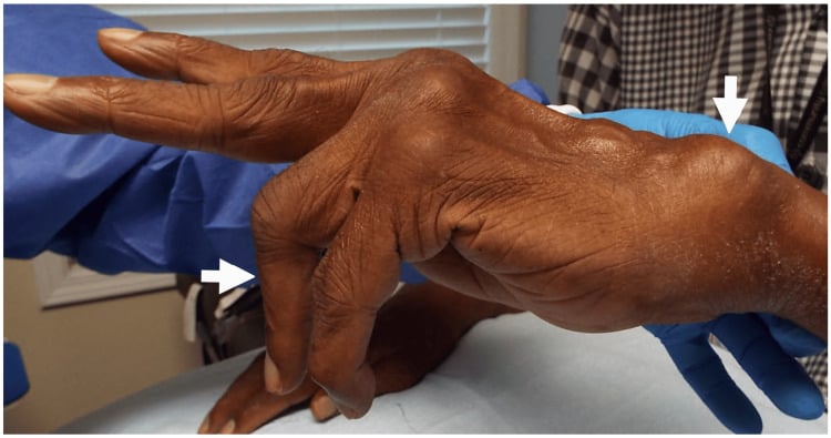
"This rheumatoid patient cannot extend their fingers - why?" is a classic examiner trap, because the answer is not automatically tendon rupture. There are three distinct causes, and they are managed completely differently:
- True extensor tendon rupture (attrition - Vaughan-Jackson). The tendon is discontinuous. Test the tenodesis effect: passive wrist flexion should produce passive finger extension, and if the finger stays flexed the tendon is ruptured. The patient can neither initiate nor hold extension.
- Extensor tendon subluxation off the MCP head (sagittal band attenuation). The tendon is intact but has slipped ulnarly into the intermetacarpal valley, so it can no longer extend the MCP from a flexed position. If you passively extend the finger the patient can hold it (the relocated tendon works) but cannot initiate extension from flexion, and tenodesis is intact. This needs sagittal-band reconstruction or extensor re-centralisation, not a tendon transfer.
- Posterior interosseous nerve (PIN) palsy. Radiocapitellar or elbow synovitis, or a proximal forearm lesion, denervates the digital extensors. Multiple fingers and the thumb lose extension with an intact tenodesis effect and (often) preserved wrist extension in radial deviation (ECRL spared). Examine the elbow for a PIN cause before assuming the problem is at the wrist.
The tenodesis test and the "can hold but cannot initiate" sign separate the three at the bedside: rupture (no hold, no tenodesis), subluxation (can hold, tenodesis intact), PIN palsy (multiple digits, tenodesis intact, look proximally). Operating as if it were a rupture when it is a subluxation or a PIN palsy is a recognised error.
Investigations
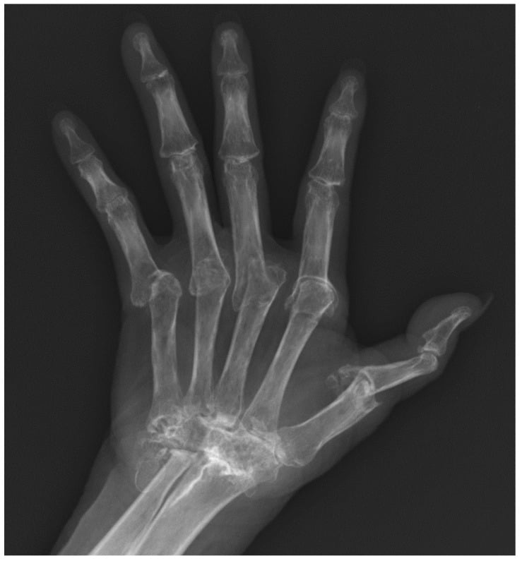
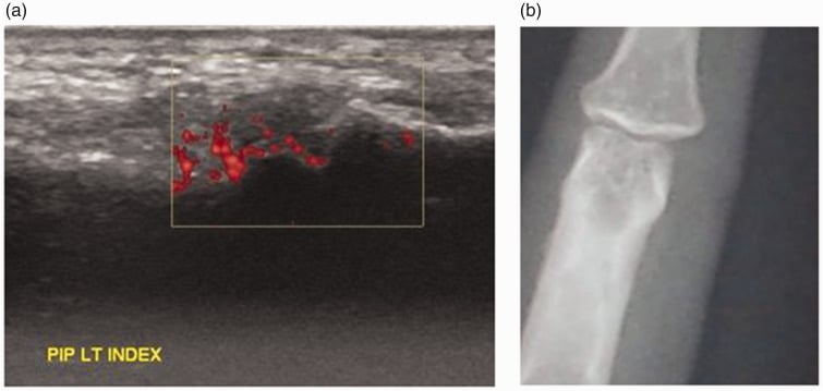
Radiographs first. PA, lateral and oblique views of both hands and wrists, bilateral for comparison and to document the symmetrical pattern typical of RA. Add the Norgard (ball-catcher) view, the most sensitive plain radiograph for early RA erosions in the hands: it profiles the MCP joints and wrist in slight oblique and ulnar-deviated position, revealing erosions that may be missed on a standard PA. The oblique projection exposes erosions and malalignment that can be underestimated on a single frontal view. Look for:
- Periarticular osteopenia
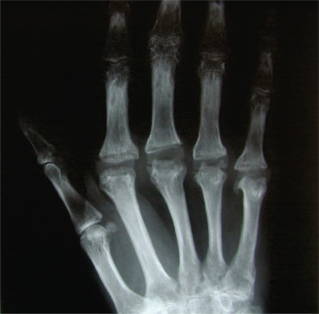
- Joint space narrowing and marginal erosions
- Carpal collapse, ulnar translocation and scapholunate dissociation
- MCP volar subluxation and ulnar drift
Larsen grading is a standardised scoring system for RA radiographic progression.
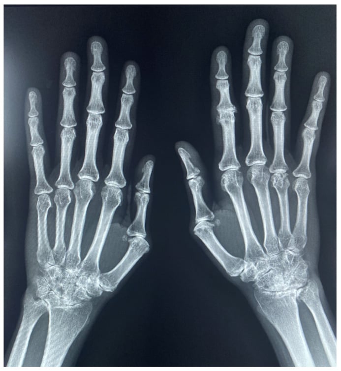
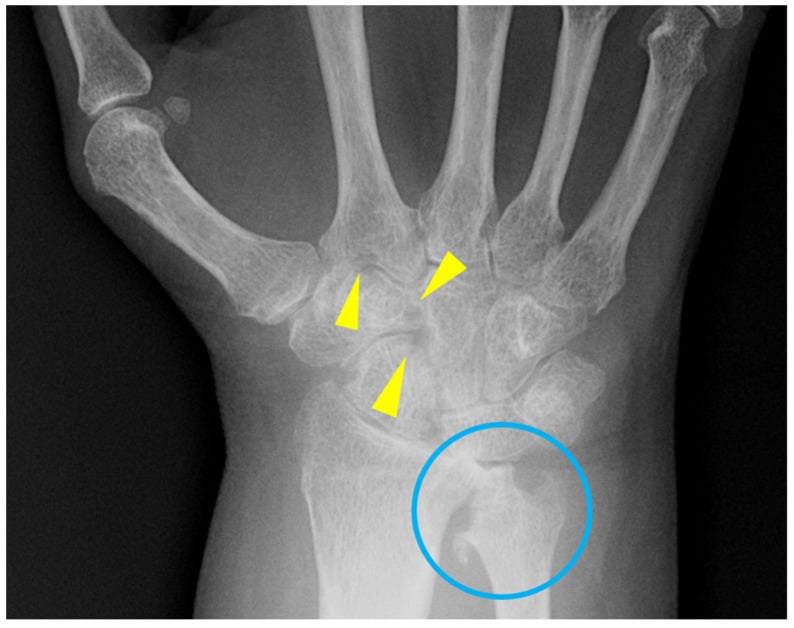
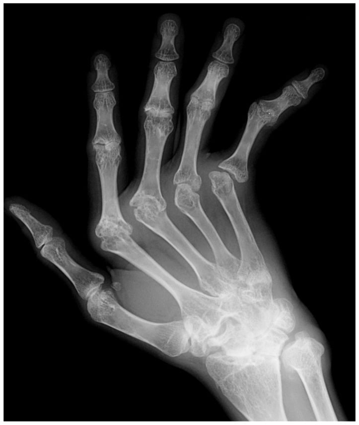
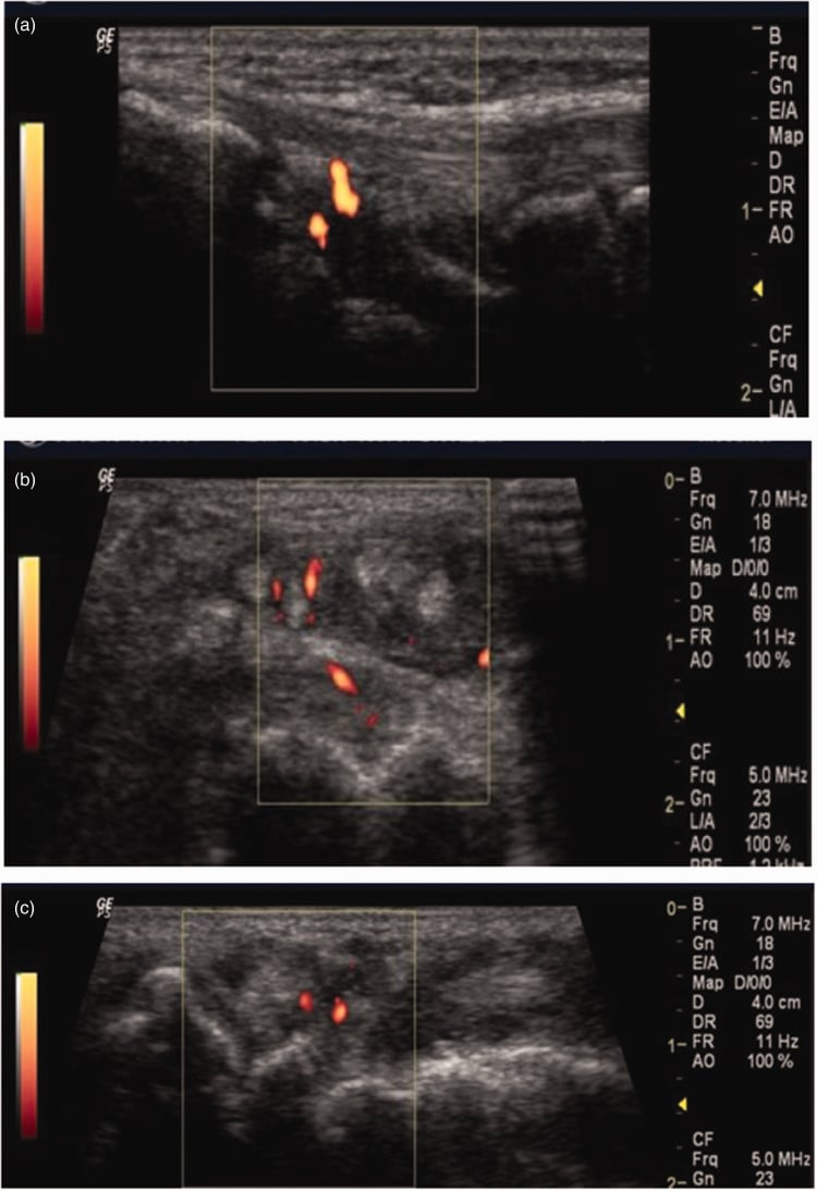
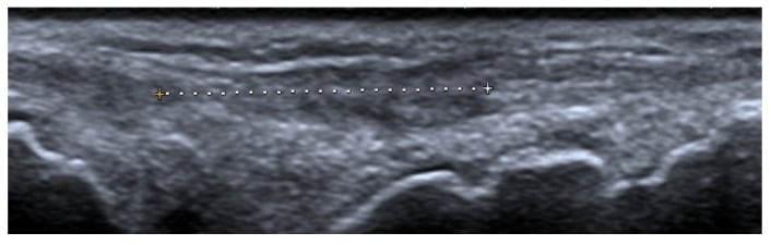
Ultrasound or MRI if a rupture is suspected. Ultrasound gives a dynamic assessment of tendon integrity, synovitis (power Doppler) and tenosynovitis, and detects active disease before advanced radiographic destruction. MRI is the gold standard for early erosions, pannus and bone marrow oedema, and assesses tendon quality. The key findings are tendon discontinuity, surrounding synovitis and the bony spurs causing attrition.
CT for planning (optional). CT assesses bone stock for arthroplasty or arthrodesis and the severity of carpal bone erosion. It helps plan wrist arthrodesis fixation and evaluate the architecture of the distal radius and ulnar head.
Management Algorithm
Medicine before surgery. Medical optimisation with DMARDs is the prerequisite to elective surgery, and the rheumatologist and hand surgeon must work as a team. Surgery on an actively inflamed joint has higher complication rates, so the general principle is to optimise medical management first, then operate on the structural consequences. Exceptions include acute tendon ruptures, which need urgent surgery to prevent further attrition, and severe nerve compression.
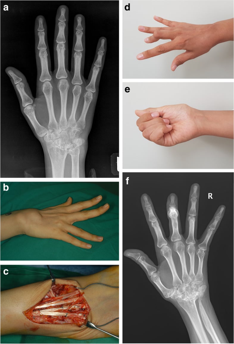
The surgical sequence. The rule is distal to proximal: DIP and PIP deformities first, then the MCP joints, then the wrist, with proximal procedures (elbow, shoulder) performed last. The wrist is the exception. Significant wrist disease drives MCP deformity, so the wrist is stabilised before or at the same time as MCP surgery, and procedures that use the same approach are combined when possible, such as a dorsal wrist operation with the extensor repair.
DMARD Therapy Timeline
- Methotrexate, the first-line conventional synthetic DMARD (csDMARD)
- Adjuncts: hydroxychloroquine, sulfasalazine, leflunomide (combination therapy common)
- Bridging: short-term low-dose prednisone while awaiting DMARD effect (6-12 weeks)
- Biologic DMARDs (bDMARDs): TNF inhibitors (adalimumab, etanercept), IL-6 inhibitors (tocilizumab), B-cell therapy (rituximab)
- JAK inhibitors: tofacitinib, baricitinib, upadacitinib (targeted synthetic DMARDs)
- Treat-to-target: aim for remission or low disease activity
- Methotrexate: generally continue through surgery (evidence supports safety)
- Biologics: withhold 2-5 half-lives before surgery; restart once the wound has healed (usually 2 weeks)
- Prednisone: stress dose coverage if on chronic steroids
- Perioperative DMARD management is a joint decision with rheumatology
Complications
- Incidence / Risk
- Common in poorly controlled disease
- Risk Factors
- Inadequate DMARD therapy, poor soft tissue quality
- Management
- Optimise medical management, revision surgery if functional
- Incidence / Risk
- 63 percent broken plus 22 percent deformed at a mean of 14 years
- Risk Factors
- High demand, poor bone stock, inadequate soft tissue balance
- Management
- Not benign - fracture tracks with recurrent ulnar drift (p less than 0.001) and lower satisfaction; revise for pain or recurrent deformity, not for the radiograph alone
- Incidence / Risk
- Reported in 10-25 percent long-term
- Risk Factors
- Aggressive distal ulnar resection, insufficient ligament repair
- Management
- Sauve-Kapandji preferred if possible; partial repair if identified
- Incidence / Risk
- 5-15 percent
- Risk Factors
- Persistent bony spurs, ongoing synovitis
- Management
- Ensure attrition source addressed at index surgery
- Incidence / Risk
- Higher than non-RA population
- Risk Factors
- Chronic steroids, poor nutrition, vasculitis
- Management
- Pre-operative skin assessment; steroid management; meticulous technique
- Incidence / Risk
- Elevated with DMARDs / biologics
- Risk Factors
- Immunosuppression, steroids, poor glycaemic control
- Management
- Perioperative antibiotic; withhold biologics; coordinate with rheumatology
Prevention. Control synovitis before elective surgery, and coordinate perioperative DMARD management with rheumatology to balance infection risk against flare. Handle the soft tissues meticulously, because RA tissues are fragile and prone to failure, and remove the bony attrition sources and diseased tenosynovium at the index operation.
Outcomes and Prognosis
- Pain Relief
- Over 90 percent good-excellent pain relief
- Functional Outcome
- Stable platform for hand function; improved grip
- Survival / Durability
- Fusion rate over 95 percent; durable long-term
- Pain Relief
- Good pain relief in 80-90 percent
- Functional Outcome
- Improved alignment and appearance; modest grip improvement
- Survival / Durability
- 10-year implant survival approximately 70-80 percent
- Pain Relief
- Reliable restoration of small finger extension
- Functional Outcome
- Depends on quality of donor and recipient tissue
- Survival / Durability
- Good durability if ulnar head addressed
- Pain Relief
- Good relief of DRUJ pain
- Functional Outcome
- Improved forearm rotation, eliminates attrition
- Survival / Durability
- Risk of ulnar translocation long-term
- Pain Relief
- Excellent pain relief
- Functional Outcome
- Reliable pinch restoration, stable thumb column
- Survival / Durability
- High fusion rate, durable
Read the silicone row alongside the 14-year series under MCP Joint before quoting it to a patient.
What predicts the result. The best outcomes come with well-controlled disease on DMARDs, intervention before deformity becomes fixed, good bone stock, compliance with therapy and not smoking. Active synovitis at the time of surgery, steroid dependence, multiple previous surgeries, poor bone quality and vasculitis predict poor ones.
Setting expectations. RA hand surgery is reconstructive, not curative. It improves function and reduces pain but rarely restores normal hand mechanics, and its goals are pain relief, improved function and prevention of further deformity; patients who understand this have better satisfaction.
Guidelines, Registries & Global Practice
- RA prevalence: 0.5-1 percent worldwide; higher in Northern European and North American populations
- Hand involvement: The most common site of RA manifestation across all ethnicities
- Declining surgery rates: Biologic DMARDs have dramatically reduced the need for reconstructive RA hand surgery in well-resourced settings
- Delayed presentation: In resource-limited settings, patients often present with established deformities
- High-resource: Early biologic therapy, hand therapy teams, silicone arthroplasty, total wrist arthroplasty as an option
- Limited-resource: Conventional DMARDs, arthrodesis preferred over arthroplasty (no implant costs, no need for long-term follow-up of implants)
- Universal principle: Tendon attrition must be addressed surgically regardless of resource setting - delay leads to further ruptures
- Surgical training: RA hand reconstruction is increasingly concentrated in specialist centres as caseloads decline
- Medical Management Emphasis
- Treat-to-target with DMARDs; biologics for inadequate response at 6 months
- Surgical Indications
- Surgery for structural damage unresponsive to medical therapy; urgent for tendon rupture
- Implant Preference
- Silicone MCP arthroplasty well-established; wrist arthrodesis preferred over arthroplasty
- Medical Management Emphasis
- Methotrexate first-line; early biologic escalation; JAK inhibitors as option
- Surgical Indications
- Functional impairment and pain despite optimal medical therapy
- Implant Preference
- Silicone MCP arthroplasty; wrist arthroplasty gaining acceptance in select patients
- Medical Management Emphasis
- Treat-to-target within 6 months; combination csDMARDs before biologics
- Surgical Indications
- Multidisciplinary decision; surgery for irreversible structural damage
- Implant Preference
- No specific implant guidance; arthrodesis vs arthroplasty by surgeon preference
- Medical Management Emphasis
- Coordinate perioperative DMARD management with rheumatology
- Surgical Indications
- Tendon rupture is semi-urgent; electives when disease quiescent
- Implant Preference
- Silicone MCP standard; pyrolytic carbon an alternative in some centres
Joint replacement registries (NJR UK, AOANJRR Australia, AJRR US) now track wrist and small joint arthroplasty. Data consistently show that silicone MCP implants have 10-year survival of approximately 70-80 percent, with fracture being the primary mode of failure. Total wrist arthrodesis remains the benchmark against which wrist arthroplasty is measured. The overall volume of RA hand surgery has declined dramatically in countries with early biologic access, making surgical training and maintenance of expertise an ongoing concern.
Coordinate with rheumatology for every RA surgical case:
- Continue: Methotrexate through surgery (evidence supports safety)
- Withhold biologics: 2-5 half-lives pre-operatively; restart 2 weeks post-op if wound healed
- Prednisone: Stress dose coverage for patients on chronic steroids
- JAK inhibitors: Withhold 3-7 days before surgery (short half-life)
- Postpone elective surgery if active infection or poorly controlled disease
- Document: DMARD plan in the operation note and communicate to the anaesthetic team
Controversies & Areas of Uncertainty
The Darrach procedure removes the attrition source but risks ulnar translocation of the carpus over time (10-25 percent). Sauve-Kapandji preserves the ulnar buttress but creates a pseudarthrosis that can be unstable. Choice depends on bone quality, DRUJ congruency, and surgeon preference. No high-quality RCTs compare the two.
Total wrist arthrodesis is reliable and durable but sacrifices all motion. Total wrist arthroplasty preserves some flexion-extension but has higher revision rates and is unsuitable for high-demand patients. In RA (low demand, bilateral disease), arthroplasty is increasingly considered for the non-dominant side while fusing the dominant wrist.
Silicone (Swanson) implants have long track records but fracture over time and may cause particulate synovitis. Pyrolytic carbon implants aim for more anatomical reconstruction but have limited long-term data in RA. Most surgeons still use silicone for RA MCP arthroplasty due to familiarity and published outcomes.
With effective biologic DMARDs, the need for RA hand surgery has declined markedly in well-resourced settings. This creates a training challenge: fewer cases mean fewer surgeons gaining expertise. The paradox is that when surgery is needed, it is often more complex (revision-type scenarios) and requires subspecialist skills.
Exam Viva Scenarios
Practise clinical reasoning and management decisions out loud
“A 58-year-old woman with long-standing seropositive RA presents with sudden inability to extend her small and ring fingers of the right hand over the past 2 weeks. She reports no pain. On examination, she has a prominent dorsally subluxed ulnar head, active extension of index and middle fingers is preserved, but she cannot actively extend the small and ring fingers. Passive extension is full. Wrist radiographs show advanced RA changes with dorsal subluxation of the ulnar head and carpal collapse. What is the diagnosis and management?”
“A 52-year-old woman with seropositive RA presents with bilateral hand deformities. The right hand shows: fixed MCP volar subluxation of all four fingers with 30 degrees ulnar drift, extensor tendon rupture of the small finger, caput ulnae with carpal collapse, and a Type I Nalebuff thumb deformity. Her disease is well-controlled on methotrexate and adalimumab. She requests surgical reconstruction. Describe your surgical plan and the order of procedures.”
Pathomechanics
- Synovial pannus destroys ligaments, tendons, and cartilage producing zig-zag deformities
- Caput ulnae = dorsal DRUJ subluxation; causes attrition ruptures of extensors
- Wrist radial deviation drives MCP ulnar drift (zig-zag collapse concept)
- Ulnar-sided structures fail first (ECU sheath synovitis, DRUJ, EDM attrition)
Key Deformities
- MCP: volar subluxation + ulnar drift from ligament destruction and flexor pull
- Swan-neck: PIP hyperextension + DIP flexion (intrinsic tightness, volar plate laxity, FDS rupture)
- Boutonniere: PIP flexion + DIP hyperextension (central slip attenuation, lateral bands volar)
- Thumb: Nalebuff Type I (MCP boutonniere) most common; Type III (CMC swan-neck) second
Tendon Ruptures
- Vaughan-Jackson: EDM then EDC small, ring, middle - ulnar to radial attrition over caput ulnae
- Mannerfelt-Norman: FPL rupture from attrition over scaphoid osteophyte in carpal tunnel
- Treatment principle: address the bone causing attrition before or during tendon transfer
- EIP is workhorse donor for EDM; FDS ring finger for FPL reconstruction
Surgical Sequence
- Distal-to-proximal rule: DIP/PIP first, then MCP, then wrist, then elbow/shoulder
- Exception: stabilise wrist before or with MCP surgery (wrist drives MCP position)
- Combine procedures using same approach when possible (dorsal wrist + extensor repair)
- Medical optimisation (DMARDs) is prerequisite before elective surgery
Key Procedures
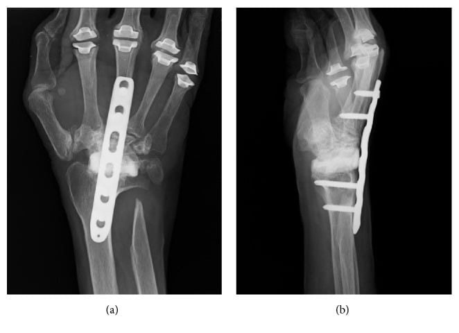
- Total wrist arthrodesis: gold standard for end-stage RA wrist (fusion in 10-20 degrees extension)
- Darrach: distal ulnar excision for caput ulnae; Sauve-Kapandji preserves ulnar buttress
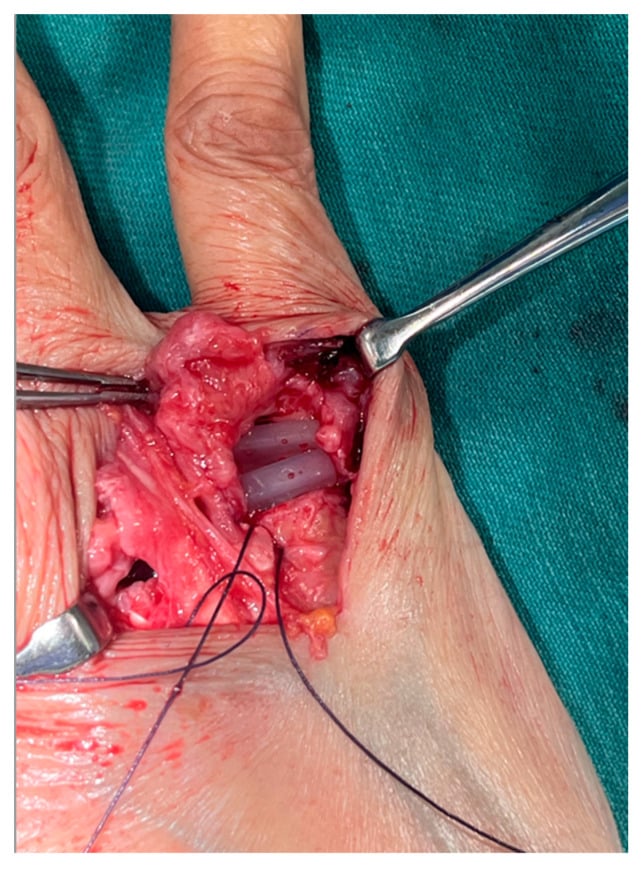
- Silicone MCP arthroplasty: for fixed MCP subluxation; 10-year survival approximately 70-80 percent
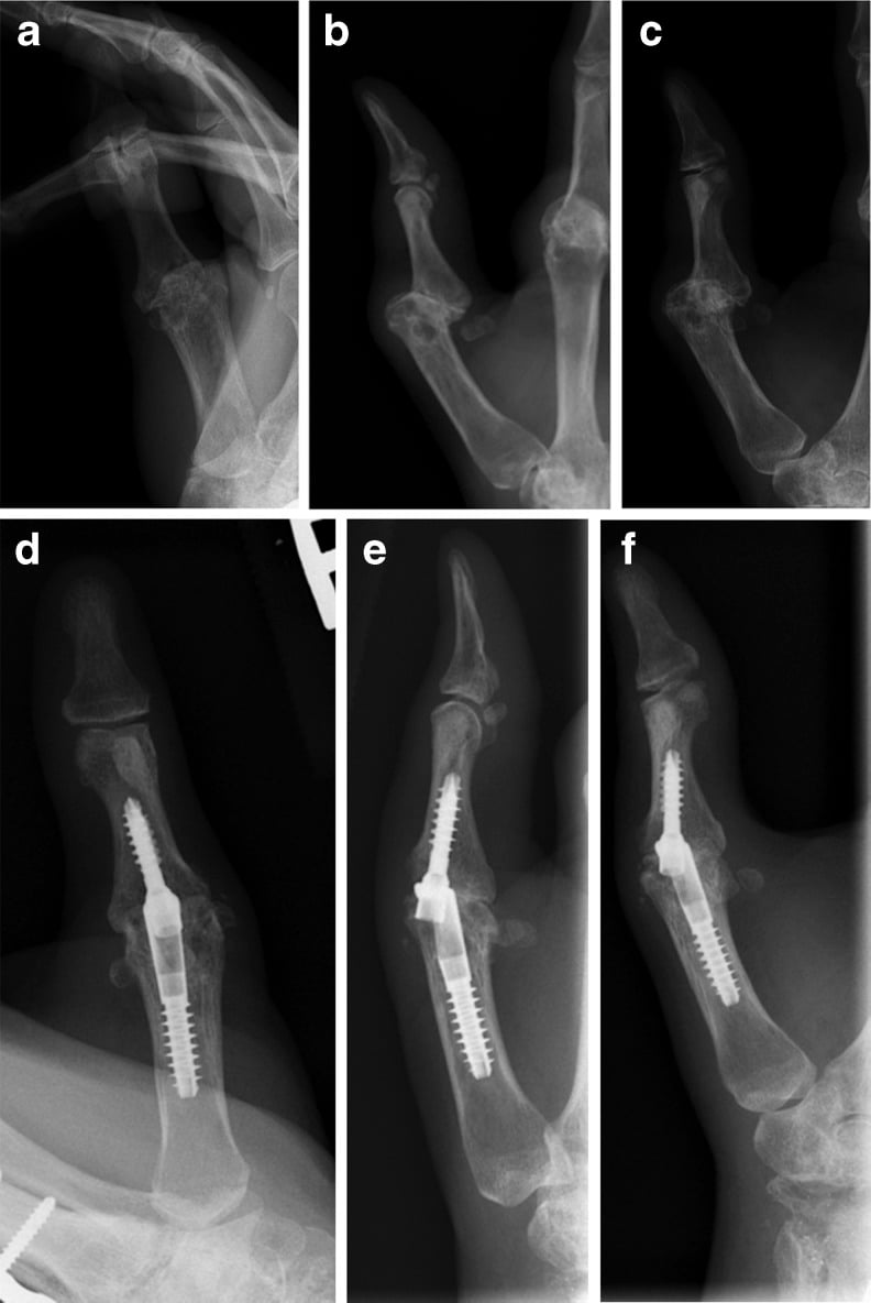
- Thumb MCP arthrodesis: for Nalebuff Type I (most common thumb deformity)
Perioperative Management
- Continue methotrexate through surgery (evidence supports safety)
- Withhold biologics 2-5 half-lives pre-operatively; restart at 2 weeks post-op
- Stress dose steroids for patients on chronic prednisone
- Sudden painless loss of extension = rupture until proven otherwise (do not wait)
Evidence Base and Key Trials
Rupture of extensor tendons by attrition at the inferior radio-ulnar joint: report of two cases
- Original description of sequential extensor tendon rupture by attrition at the distal radio-ulnar joint in rheumatoid disease
- Rupture begins with extensor digiti minimi (EDM) due to attrition over the dorsally subluxed ulnar head (caput ulnae)
- Progresses radially: EDM, then extensor digitorum communis to small, ring, and middle fingers in sequence
- Identified dorsal subluxation of the ulnar head as the mechanical cause of attrition
Scaphoid exostosis causing rupture of the flexor pollicis longus
- Describes attrition rupture of flexor pollicis longus over a scaphoid exostosis (osteophyte) within the carpal tunnel in rheumatoid disease
- Mechanism parallels the Mannerfelt-Norman lesion: rough bone on the volar scaphoid ridge abrades the FPL during flexion
- Often associated with carpal tunnel syndrome, requiring concurrent release and spur excision
- FPL reconstruction by tendon transfer or graft can restore thumb IP flexion