Extensor Hood Disruption | MCP Instability | Radial vs Ulnar Subluxation
- Sagittal bands stabilise the EDC at MCP level - rupture causes subluxation into the intermetacarpal valley
- Long finger most commonly affected (Rayan-Murray series: long, then small, then index, then ring)
- Ulnar EDC subluxation is most common, and it is the RADIAL sagittal band that fails (Young & Rayan biomechanics)
- Rayan Type I-II often respond to extension splinting alone for 4-6 weeks
- Chronic cases (over 6 weeks) usually require surgical reconstruction
- “Painful snapping at MCP with active extension = sagittal band injury until proven otherwise
- “Elson test: normal PIP extension with weak MCP extension differentiates it from central slip rupture
- “Surgical repair within 3 weeks has better outcomes than delayed reconstruction
- “Juncturae tendinum can mask single finger EDC injury - examine each finger independently
Overview and Epidemiology
Sagittal band injuries are uncommon but functionally significant, because they disrupt the balance of the extensor mechanism at the MCP joint. The sagittal bands are the dorsal stabilisers of the extensor tendon there, keeping it out of the intermetacarpal valleys as the finger flexes and extends. Without them the extensor digitorum communis (EDC) loses its mechanical advantage, and MCP extension is weak even though the tendon is in continuity.
Which finger. The long finger (the third ray: "long" and "middle" finger are the same digit) is the single most commonly injured, which is attributed to its greater excursion and central position. In the Rayan-Murray series the order was long, then small, then index, then ring. Biomechanically, instability after sagittal band division is most pronounced in the long finger and least in the small finger.
Who and how. Typically young to middle-aged active adults injured at sport or at work, with boxers, martial artists and manual workers over-represented because of the forced-flexion mechanism.
- Forced MCP flexion, as in punching (the "boxer knuckle") or ball sports, or forced extension
- A direct blow
- Laceration
- Spontaneous attenuation in rheumatoid arthritis
What it costs. Loss of effective MCP extension weakens grasp and release, and the tendon snaps painfully with gripping and active extension. Time to treatment is what drives the prognosis.
Anatomy and Pathomechanics
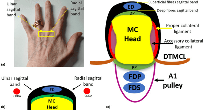
The band. The sagittal band is not a simple ligament. It is a confluence of transverse fibres from the volar plate, longitudinal fibres from the interosseous fascia and contributions from the juncturae tendinum, a dynamic stabilising system that centralises the EDC through the full arc of MCP motion. The radial and ulnar bands arise from the volar plate of the MCP joint and the interosseous fascia, wrap around either side of the EDC and merge with the extensor hood dorsally, anchoring the tendon to the metacarpal.
What it does. The band has four jobs:
- Prevents EDC subluxation into the intermetacarpal valley
- Keeps the EDC centred dynamically through the full range of motion
- Transfers intrinsic muscle forces to the extensor mechanism
- Acts as a direct MCP extensor, independent of the EDC
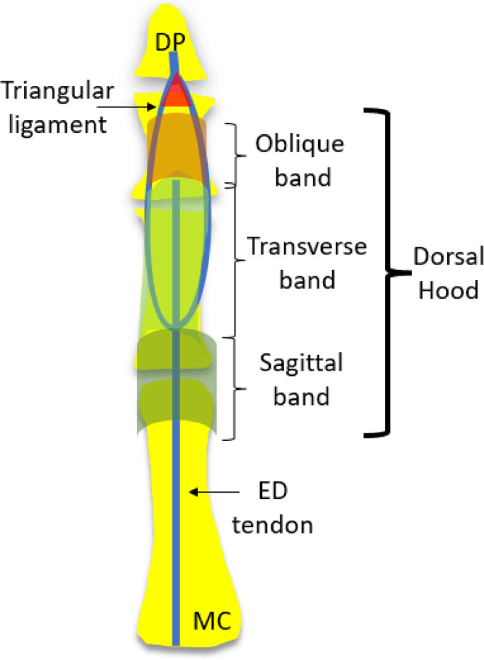
The rest of the hood. The extensor hood covers the dorsum of the MCP joint and is organised from proximal to distal into sagittal, transverse and oblique bands. The EDC continues distally as the central slip and terminal extension. The lateral bands carry the intrinsic contributions of the lumbricals and interossei, and the triangular ligament connects them distally.
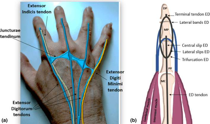
Juncturae and neighbouring tendons. The juncturae tendinum, which interconnect adjacent EDC tendons, can partially compensate for a sagittal band disruption in some cases, and they can mask a single-finger injury.
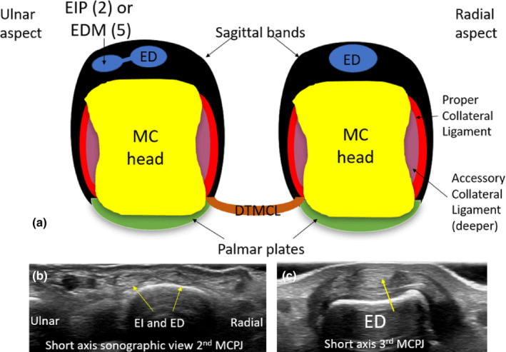
Why the tendon goes ulnar. Ulnar subluxation of the EDC is the usual pattern, in about 90% of cases, and it follows failure of the radial band. In Young and Rayan's cadaveric study, dividing the ulnar band alone produced no instability, partial division of the proximal radial fibres caused subluxation, and complete radial division caused dislocation; the proximal radial fibres are the critical restraint. The natural ulnar resting drift of the EDC and ulnarly directed forces during pinch and grip add to it.
- Normal Mechanism
- EDC centralised dorsally
- After RADIAL Band Rupture (common)
- EDC tends to displace ULNARLY
- After ULNAR Band Rupture (rare)
- EDC tends to displace radially (minimal in cadaver models)
- Normal Mechanism
- EDC maintained dorsal by sagittal bands
- After RADIAL Band Rupture (common)
- EDC subluxates ULNARLY into the intermetacarpal valley (typical pattern)
- After ULNAR Band Rupture (rare)
- EDC subluxates radially over the metacarpal head (uncommon)
- Normal Mechanism
- Smooth EDC excursion, strong extension
- After RADIAL Band Rupture (common)
- Painful snap as tendon reduces from the ulnar valley, weak extension
- After ULNAR Band Rupture (rare)
- Painful snap as tendon reduces radially
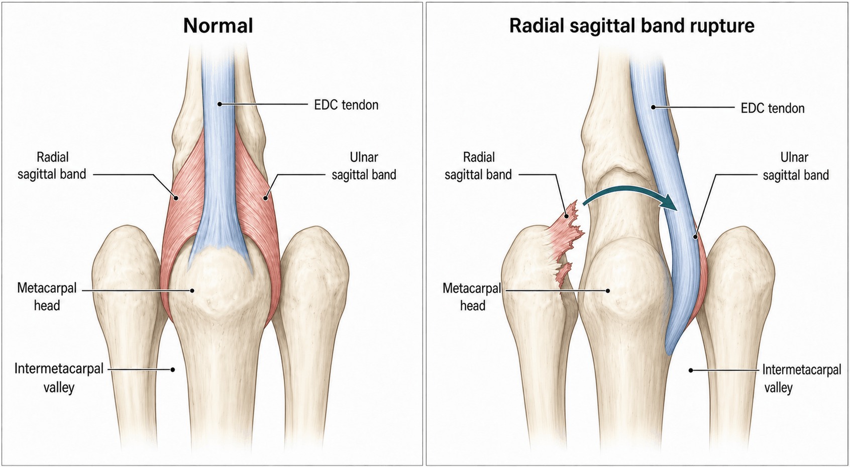
Ulnar subluxation means the RADIAL band has failed. Many texts still teach the inverted explanation, that a "thicker radial band" protects against ulnar slip. Examiners may probe this: quote the biomechanics (Young & Rayan), not the folklore.
Classification Systems
The Rayan-Murray classification (1994), the most widely used, grades the injury by the severity and reducibility of EDC subluxation, and it guides treatment.
- Pathology
- Partial tear or attenuated sagittal band
- Clinical Findings
- Pain, mild swelling, NO visible subluxation, active extension maintained
- Treatment
- Splint MCP extension 4-6 weeks, NSAIDs
- Prognosis
- 85-90% excellent outcomes
- Pathology
- Complete tear, tendon subluxates but reducible
- Clinical Findings
- Visible/palpable subluxation with MCP flexion that reduces with extension, painful snap, weak active extension
- Treatment
- Splint (low-demand) vs early surgery (athletes, manual workers)
- Prognosis
- 70-80% good with splinting, 90% with surgery
- Pathology
- Complete tear with frank tendon dislocation into the intermetacarpal valley
- Clinical Findings
- Tendon displaced, weak or absent active MCP extension, painful snap
- Treatment
- Acute closed: trial relative-motion splint (can succeed). Irreducible/chronic/lacerated: surgical reduction and repair
- Prognosis
- Good with early treatment; worse if chronic (over 6 weeks)
The key distinction is reducibility. Type II subluxates with MCP flexion and reduces with extension, a dynamic instability. A Type III tendon locked in the valley is a static deformity that cannot extend the MCP actively and needs surgery, although an acute closed dislocation can still succeed in a relative-motion splint (Catalano).
Clinical Assessment
History. The patient reports painful snapping at the MCP joint, weakness extending the finger and swelling over the joint. The answers that shape management:
- Time since injury: acute (days), subacute (weeks) or chronic (months)
- Hand dominance, which affects treatment urgency
- Occupation: manual labour or desk work
- Red flags: rheumatoid arthritis (pathological rupture) or more than one finger involved
Examination. The classic triad is pain over the MCP joint with active extension, a palpable or visible tendon subluxation (the snap), and weak active MCP extension. Look for swelling over the joint and the posture of the finger at rest, and feel for tenderness over the radial or ulnar band and a snap with motion. PIP and DIP extension are normal, and the neurovascular examination is intact, as neurovascular deficit is not a feature of an isolated sagittal band injury. Because the juncturae can mask a single-finger injury, examine each finger independently.
Special tests. Three manoeuvres:
- Dynamic subluxation test (pathognomonic). The patient makes a fist, flexing all the MCP joints, then actively extends the fingers while the examiner's finger rests over the dorsal MCP joint. Visible or palpable EDC subluxation with flexion, usually ulnar, followed by a painful snap as it reduces on extension, confirms sagittal band disruption with dynamic instability.
- Resisted MCP extension. Stabilise the proximal phalanx in slight flexion and ask the patient to extend the MCP against resistance. Extension is weak and painful compared with the normal adjacent fingers, because the eccentric EDC has lost its mechanical advantage.
- Elson test. Flex the PIP joint over the edge of the table and ask the patient to extend it against resistance. In a sagittal band injury the PIP extends normally, the central slip being intact, while MCP extension is weak; a central slip rupture shows weak PIP extension with rigid DIP extension. The test separates MCP-level pathology from a PIP-level injury.
- Level / Site
- MCP joint (extensor hood)
- Active Extension Pattern
- Weak but present MCP extension; normal PIP/DIP
- Key Discriminator
- Dynamic EDC subluxation with painful snap on clenched fist
- Confirmatory Test
- Dynamic clenched-fist ultrasound
- Level / Site
- PIP joint
- Active Extension Pattern
- Weak PIP extension; MCP normal
- Key Discriminator
- Positive Elson test, evolving boutonniere
- Confirmatory Test
- Elson test; lateral PIP radiograph
- Level / Site
- Over the metacarpal/MCP
- Active Extension Pattern
- Complete loss of MCP extension
- Key Discriminator
- Open wound, no dynamic snap, fixed deficit
- Confirmatory Test
- Wound exploration
- Level / Site
- MCP joint, often multiple digits
- Active Extension Pattern
- Progressive loss of MCP extension
- Key Discriminator
- Atraumatic, multi-digit, systemic disease, synovitis, ulnar drift
- Confirmatory Test
- Tenodesis test, inflammatory markers, US
- Level / Site
- MCP joint
- Active Extension Pattern
- Mechanical block or stiffness with limited ROM, not weakness
- Key Discriminator
- Catching/locking without tendon snap
- Confirmatory Test
- Radiographs
Investigations
Sagittal band injury is a clinical diagnosis. Imaging is mainly there to exclude other pathology, such as fracture or arthritis, rather than to confirm the rupture, and the dynamic subluxation test is more sensitive and specific than any imaging modality.
Radiographs come first: PA, lateral and oblique views of the affected digit to exclude bony pathology and assess the MCP joint. They are usually normal, this being a soft-tissue injury, though chronic arthritis may show as MCP joint space narrowing. A dynamic lateral with active MCP flexion may show the tendon subluxation, but is rarely needed.
Ultrasound is the next step if the diagnosis is uncertain. It assesses EDC subluxation dynamically and in real time and shows the integrity of the band: with a dorsal longitudinal view over the MCP joint while the patient flexes and extends, a positive study shows the EDC moving into the intermetacarpal valley with flexion and discontinuity of the band fibres. A partial radial-band tear appears thickened and hypoechoic. It is operator-dependent and not widely used, though its use is increasing.
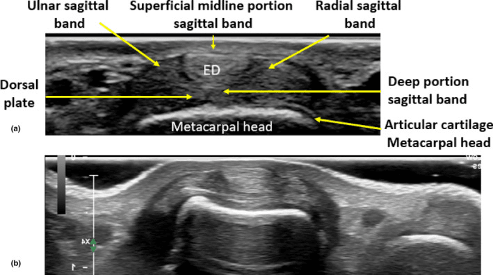
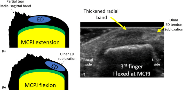
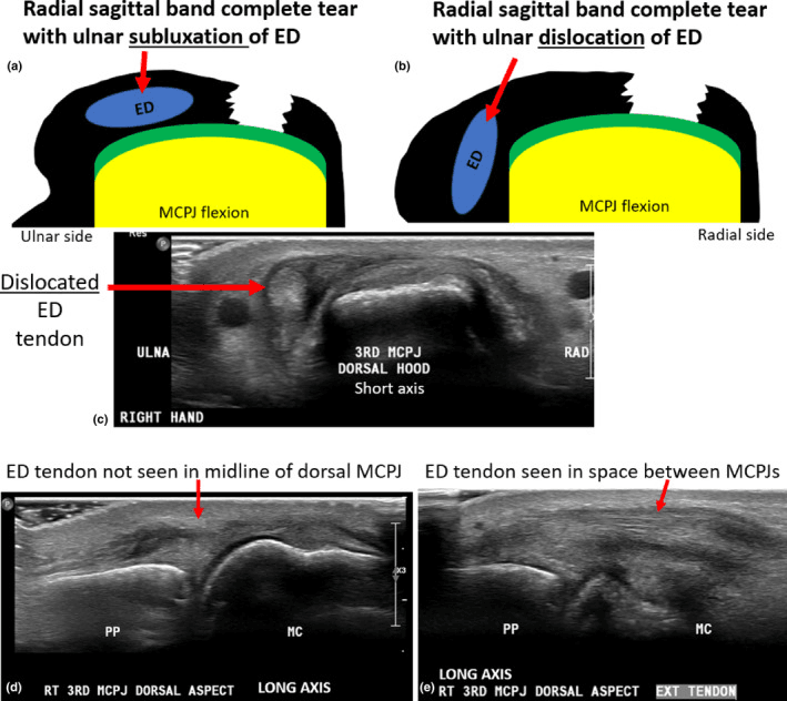
MRI is for chronic cases, atypical presentations and pre-operative planning. It shows disruption of the band fibres, fluid in the MCP joint and the position of the EDC, and it can assess tissue quality, acute tear or chronic scarring, for surgical planning. It is expensive and rarely necessary when the clinical diagnosis is clear.
Management Algorithm
The Rayan type, the time since injury and the demands of the patient decide between a splint and an operation. Splinting has the best results in early presentations, and manual labourers and athletes fail it more often.
Who splints. The candidates for a splint are:
- Rayan Type I: partial tear, no subluxation
- Rayan Type II, with reducible subluxation, in a low-demand patient
- Acute presentation, within 2-3 weeks of injury
- Non-manual occupation, older age or medical comorbidities
Who does not. Splinting is contraindicated in:
- Rayan Type III with irreducible subluxation
- Chronic injury, over 6 weeks
- High-demand patients, athletes and manual labourers who need reliable grip strength
- Recurrent subluxation after an adequate splinting trial
Rigid extension splinting. A custom thermoplastic dorsal blocking splint or a commercial aluminium foam splint holds the MCP in full extension (0 degrees) with the PIP and DIP joints free to move.
- Weeks 0-4: worn continuously, 24 hours a day, off only for hygiene; no gripping, lifting or forceful hand use; weekly review of compliance and skin, adjusting the splint
- Weeks 4-6: off during the day for light activities if there is no pain or subluxation, on at night; gentle active range of motion and blocked MCP extension; no forceful gripping or weight-bearing on the hand
- Weeks 6-8: discontinued if the MCP is stable and pain-free; progressive grip strengthening with putty and therapy balls; watch for any recurrence of snapping or subluxation; gradual return to activity by symptom tolerance
- Week 8 onward: full activity, including manual labour, once grip strength reaches 80% with no pain or subluxation; protective taping for contact sports for a further 4 weeks; if symptoms recur, consider surgical reconstruction
Relative-motion (yoke) splinting. A yoke splint links three or four fingers but holds the injured finger's MCP in relative hyperextension, roughly 15 degrees more extended than its neighbours; Catalano's sagittal band bridge held it in 25-35 degrees of hyperextension relative to the adjacent MCPs. The EDC tendons act as a linked group, so positioning the injured MCP more extended reduces the relative excursion and tension that the injured band and EDC must bear during active flexion. The tendon is effectively unloaded at the injury while the other fingers do the work.
The protection is dynamic. The patient can make a light active fist and move immediately rather than being locked out for 4-6 weeks, and early controlled motion prevents the extensor adhesions, MCP extension contracture and stiffness that follow prolonged rigid splinting, while the tendon is held centralised over the metacarpal head so the band heals reduced.
What it treats. Traditionally reserved for Type I-II, relative-motion splinting has healed even acute closed Type III (dislocated) injuries when started early in a compliant patient (Catalano), which makes it a genuine first-line alternative to surgery rather than an adjunct. It depends on early presentation and good compliance, is worn for a similar 4-6 week protected period, and is unsuitable for irreducible, lacerated, chronic or rheumatoid injuries, which still need operative repair or reconstruction.
Surgical Technique
Position. Supine with a hand table: the shoulder abducted 80-90 degrees, the elbow extended on the arm board, the forearm pronated so the dorsum of the hand is up, and the wrist neutral to slightly extended.
Tourniquet. A forearm tourniquet is preferred for better visualisation, at 200-225 mmHg, after exsanguination by 2 minutes of elevation or an Esmarch bandage. The limit is 90 minutes; most cases are completed in 30-45 minutes.
Prep and drape. Prepare from the fingertips to mid-forearm with betadine or chlorhexidine. A transparent hand drape lets you watch finger motion during the operation, and all the digits are exposed so the juncturae and EDC continuity can be assessed.
Equipment. A hand surgery set with fine scissors and forceps, loupes at 2.5-3.5x (helpful but not mandatory), palmaris longus harvested if a reconstruction is needed, and thermoplastic for the postoperative extension splint.
Consent. Recurrence, stiffness, infection and incomplete return of strength, at the rates in the Complications section, with recurrence higher in chronic cases and MCP joint stiffness in 5-10% if the repair is overly tight. About 10% need revision by juncturae transfer or reconstruction.
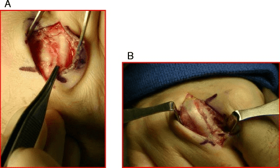
Incision. A 3-4 cm longitudinal incision over the dorsum of the affected MCP joint, centred over the EDC and running from the distal metacarpal to the proximal phalanx. Raise minimal skin flaps to preserve their vascularity.
The dorsal digital neurovascular bundles lie radial and ulnar to the midline, and the radial and ulnar dorsal digital nerves cross the surgical field deep to the skin in loose areolar tissue. Careful dissection with blunt spreading minimises the risk. Nerve injury causes a painful neuroma and dorsal finger numbness.
Find the lesion. Identify the EDC and whether it is centralised or subluxated. Locate the tear, usually in the radial band, and judge the tissue: a fresh acute tear, or chronic, scarred and attenuated. Assess the adjacent juncturae in case a transfer is needed for augmentation.
Reduce the tendon. In chronic cases adhesions may need lysis to mobilise the EDC out of the intermetacarpal valley. Draw it dorsally with gentle traction to centralise it over the joint, then range the MCP passively to check that it glides smoothly. An irreducible tendon may need release of contracted tissue, which is rare.
Complications
- Incidence
- 10-15%
- Risk Factors
- Chronic injury, inadequate repair tension, early mobilisation, heavy manual labour
- Management
- Revision repair with juncturae augmentation or tendon graft reconstruction
- Incidence
- 10%
- Risk Factors
- Over-tight repair, prolonged immobilisation over 6 weeks
- Management
- Hand therapy with passive flexion stretching, rarely requires surgical release
- Incidence
- 5-10%
- Risk Factors
- Excessive scar formation, adhesions
- Management
- Aggressive hand therapy, tenolysis if persistent at 6 months
- Incidence
- 2-5%
- Risk Factors
- Open injury, diabetes, immunosuppression
- Management
- Antibiotics, irrigation and debridement if deep, delayed repair
- Incidence
- 3-5%
- Risk Factors
- Iatrogenic during dissection
- Management
- Neuroma excision and burial if symptomatic painful neuroma
- Incidence
- 15-20%
- Risk Factors
- Chronic injury, muscle atrophy, patient factors
- Management
- Prolonged hand therapy (up to 6 months), accept functional outcome
Preventing recurrence. Recurrent EDC subluxation is the most common complication. The prevention strategies are:
- Correct tension at repair, with the EDC centralised and the MCP extended
- Juncturae augmentation in subacute and chronic cases
- Strict compliance with the 4-6 week immobilisation
- A gradual return to heavy activity over 12 weeks
Postoperative Care and Rehabilitation
The protocol. Therapy starts at 2 weeks and progresses under supervision:
- Weeks 0-4, immobilisation: MCP extension splint worn continuously, removed only by the therapist for wound care, with sutures out and the wound checked at 10-14 days; PIP and DIP motion encouraged to prevent stiffness in those joints; no gripping or weight-bearing on the hand
- Weeks 4-6, protected mobilisation: splint off for supervised therapy sessions only and on between them and at night; gentle active MCP flexion and extension within a pain-free range, with blocked exercises isolating the MCP by stabilising the proximal phalanx; goal 60-70 degrees of MCP flexion without pain or subluxation
- Weeks 6-8, active range: daytime splint discontinued, night splint continued; full active range, composite fist and intrinsic stretching; light resistance with putty and soft therapy balls; any recurrence of snapping or subluxation means a return to splinting
- Weeks 8-12, strengthening: splint discontinued if the range is full and there is no subluxation; progressive, graded grip strengthening and activity-specific work simulation; goal 80% of contralateral grip strength
- Week 12 onward: return to manual labour once the strength criteria are met; gradual return to sport, with protective taping for contact sports for a further 4 weeks; expect 85-90% of baseline function
Compliance with the 4-6 week immobilisation is CRITICAL. Mobilising before 4 weeks significantly increases the risk of recurrent subluxation.
The wrist. These protocols control only the MCP, yet Young and Rayan concluded that "wrist flexion contributes to extensor tendon instability after sagittal band disruption and may exacerbate its injury". The EDC crosses the wrist, so wrist flexion tightens the extensor tendons (the tenodesis effect) and increases their tension, and their ulnarly directed displacing force, at the MCP. A splint or activity that lets the wrist flex therefore loads the healing radial band and pulls the tendon toward re-subluxation.
During the protected phase the wrist should be held in slight extension, or at least neutral, rather than left free to flex, and the patient should avoid gripping with a flexed wrist. For higher-risk repairs some surgeons extend the splint to include the wrist in slight extension, and hand therapists cue a neutral-to-extended wrist during early active-motion (relative-motion) programmes. Inadequate control of the displacing forces is one driver of recurrence, and keeping the wrist out of flexion removes one of the main forces that re-tensions the healing band.
Outcomes and Prognosis
- Patient Group
- Low-demand, office workers
- Expected Outcomes
- 80-85% good, minimal recurrence
- Predictors of Poor Outcome
- Manual labour, poor compliance, presentation over 3 weeks
- Patient Group
- Athletes, manual workers
- Expected Outcomes
- 90% return to sport/work at pre-injury level
- Predictors of Poor Outcome
- Delayed surgery over 6 weeks, inadequate tension at repair
- Patient Group
- All patients
- Expected Outcomes
- 60-70% good, higher stiffness and recurrence
- Predictors of Poor Outcome
- Over 6 months delay, multiple previous failed repairs, rheumatoid arthritis
Best outcomes: acute presentation (within 3 weeks), Rayan Type I-II, surgical repair with adequate tension, compliance with immobilisation, and an experienced hand surgeon and therapist. Worst outcomes: chronic injury (over 6 weeks), failed prior repair, rheumatoid arthritis, and the manual labourer who returns to heavy work too early.
Guidelines, Registries & Global Practice
Sagittal band injury is a clinical (not registry-tracked) diagnosis, so there is no implant or arthroplasty registry data and no formal Level-1 society guideline. Practice is governed by classic case series and consensus expert opinion, which is broadly consistent worldwide.
- Distribution: Worldwide; no geographic predilection. Over-represented in combat-sport athletes (boxing, MMA, "boxer knuckle") and manual workers
- Long finger (third ray) is the most frequently injured digit across published series
- Rheumatoid presentation is declining globally as early DMARD/biologic therapy and tenosynovectomy reduce tendon-centred deformity
- Limited-resource settings: Reliance on clinical diagnosis and relative-motion/extension splinting is an advantage - no advanced imaging required
- Shared principles (AAOS, BSSH/BOA, federation of European hand societies, ASSH teaching): trial of splinting first for closed acute injuries presenting within ~3 weeks; surgery for irreducible, chronic, lacerated, or failed-conservative cases
- Relative-motion (yoke / sagittal band bridge) splinting increasingly favoured over rigid MCP immobilisation, allowing early controlled motion
- Rheumatoid disease: multidisciplinary rheumatology co-management and disease control before elective reconstruction
- Prognosis counselling: acute repair outcomes good; chronic reconstruction and RA cases more guarded
Because there is no registry or graded guideline, examiners expect you to anchor decisions on the Rayan-Murray classification (1994), the 3-week window for non-operative success, and the biomechanical principle that the radial band fails to allow ulnar subluxation. Relative-motion splinting is the modern, resource-light first-line approach worldwide.
mechanism (sport/assault/occupational/spontaneous in RA), Rayan-Murray type, reducibility, time since injury, and a clear record of the operative vs non-operative discussion with realistic outcome expectations and consent for recurrence, stiffness and incomplete return of strength.
- Missing the diagnosis as a "simple contusion" so the patient presents late with fixed subluxation
- Failing to differentiate from central slip injury (always check PIP/DIP extension)
- Over-tight repair causing an MCP extension contracture
- Inadequate splint education leading to non-operative failure
Related pages: Extensor Tendon Injuries is the parent topic - sagittal band disruption is the zone V lesion that produces subluxation without discontinuity, which is why the tendon is intact but the finger will not extend; Metacarpal Fractures for the injury that shares the punching mechanism and must be excluded on radiographs before the swelling is attributed to the hood; Human Bites and Fight Bite for the same clenched-fist mechanism with an open wound over the MCP joint, where the diagnosis to make first is septic arthritis rather than a band tear; Rheumatoid Hand and Wrist for the attritional form, where the band attenuates from synovitis and the operation is a transfer rather than a repair; MCP Joint Arthritis for the degenerative mimic of a painful swollen knuckle; and Extensor Mechanism Ruptures for the same principle of a competent tendon rendered useless by loss of its restraint.
Controversies and Areas of Uncertainty
The evidence base is almost entirely small retrospective series, case reports and cadaveric work, with no randomised trials, so these genuine areas of debate are high-yield for discussion vivas.
Rigid or relative-motion splinting. Traditional teaching used continuous full MCP-extension immobilisation for 4-6 weeks. Catalano's sagittal band bridge and the relative-motion splints that followed permit early active motion instead. The optimal splint design and duration remain unsettled.
Can an acute Type III be treated closed? Classic doctrine made tendon dislocation an operative indication, but Catalano showed that many acute closed Type III injuries succeed with relative-motion splinting if treated early. Whether to operate primarily or trial a splint is a genuine decision point.
Which reconstruction. Direct repair, junctura tendinum transfer, distally based EDC tendon flaps and free tendon grafts all have advocates, and there are no comparative data. The anchor point, the deep transverse metacarpal ligament or the adjacent volar plate, is debated, with biomechanical modelling suggesting that volar-plate anchoring lowers repair tension in flexion.
MCQ Practice Points
Q: A patient has visible EDC tendon subluxation with MCP flexion that reduces with extension, and weak but present active MCP extension. What is the Rayan classification and recommended treatment? A: Rayan Type II (complete tear with reducible subluxation). Treatment depends on patient factors: splinting for low-demand patients (70-80% success), surgical repair for high-demand patients/athletes/manual laborers (90% success).
Q: Why is ulnar subluxation of the EDC more common than radial subluxation in sagittal band injuries? A: The high-yield answer is the cadaveric biomechanics of Young & Rayan: it is the RADIAL sagittal band whose disruption permits the tendon to slide ulnarly. Dividing the ulnar band alone produced no instability; partial proximal radial-band division caused subluxation and complete radial division caused dislocation. The natural ulnar resting drift of the EDC and ulnarly directed grip/pinch forces reinforce this. Hence ulnar subluxation is the typical pattern.
Q: What is the optimal timing for treatment of sagittal band injuries and why? A: Treatment (operative or non-operative) is best started within 3 weeks of injury, when tissue quality is good and direct repair or splinting is most likely to succeed (Rayan-Murray). Beyond 6 weeks, scarring and attenuation make direct repair difficult, often requiring reconstruction with inferior outcomes.
Exam Viva Scenarios
Practise clinical reasoning and management decisions out loud
“A 35-year-old manual laborer presents 5 days after punching a wall in anger. He has pain over the dorsum of his right long finger MCP joint and complains of a snapping sensation when he makes a fist. On examination, you observe the EDC tendon subluxating ulnarly with MCP flexion and snapping back with extension. He has weak but present active MCP extension. What is your assessment and management?”
“A 42-year-old female was treated with MCP extension splinting for 6 weeks for a Rayan Type II sagittal band injury of her index finger. She was initially compliant but now, 2 weeks after discontinuing the splint, she has recurrent painful snapping and visible EDC subluxation with gripping. She is frustrated and wants definitive treatment. How would you manage this case?”
“A 55-year-old patient with rheumatoid arthritis presents with progressive ulnar subluxation of the EDC tendons of her long and ring fingers over the past 6 months. She has difficulty extending her MCPs and reports that the tendons slip off the side of the knuckles when she tries to grip. Examination shows fixed ulnar subluxation of EDC with inability to actively extend the MCPs. How would you manage this?”
Key Anatomy
- Sagittal bands = transverse fibers from volar plate wrapping around EDC to stabilize at MCP level
- RADIAL band disruption causes ULNAR EDC instability (the common pattern); proximal radial fibers are the key restraint
- Long finger (third ray) most commonly affected; greatest instability after division (least in small finger)
- Function: Centralize EDC, prevent subluxation into intermetacarpal valley, transmit intrinsic forces
Rayan Classification
- Type I = Partial tear, NO subluxation, active extension maintained → Splint 4-6 weeks
- Type II = Complete tear, REDUCIBLE subluxation → Splint (low-demand) vs Surgery (high-demand)
- Type III = Complete tear, IRREDUCIBLE subluxation → Surgery required
- Dynamic subluxation test: EDC displaces ulnarly with MCP flexion, snaps back with extension
Treatment Algorithm
- Type I: Splint MCP extension 4-6 weeks (85-90% success)
- Type II: Surgery for athletes/manual workers, splinting for office workers
- Type III: Surgical reduction and repair (cannot extend MCP actively)
- Chronic (greater than 6 weeks): Reconstruction with tendon graft (worse outcomes 60-70%)
- Optimal surgical timing: 1-3 weeks (90% good outcomes)
Surgical Pearls
- Direct repair: Horizontal mattress sutures with 3-0 non-absorbable, tension with MCP extended
- Juncturae transfer: Augment weak tissue by transferring adjacent juncturae
- Tendon graft: Palmaris longus reconstruction for chronic/RA cases
- Critical: Appropriate tensioning (too loose = re-subluxation, too tight = extension contracture)
- Test repair: Passive MCP flexion to 90 degrees should not cause subluxation
Rehabilitation
- Immobilization: MCP extension splint 4-6 weeks CONTINUOUSLY (PIP/DIP free)
- Early mobilization before 4 weeks increases recurrence risk 20-30%
- Weeks 4-6: Protected supervised ROM
- Weeks 6-12: Progressive strengthening
- Return to manual work: 10-12 weeks
Outcomes and Complications
- Acute repair: 85-90% good outcomes
- Chronic reconstruction: 60-70% good outcomes
- Recurrence: 10-15% (higher in chronic cases, inadequate repair tension)
- MCP stiffness: 10% (over-tight repair)
- Predictors of poor outcome: Chronic injury greater than 6 weeks, RA, failed previous repair, manual labor with early return to work
Evidence Base and Key Studies
Classification and Treatment of Closed Sagittal Band Injuries
- Retrospective series of 28 nonrheumatoid patients with sagittal band injuries
- Defined the three clinical types still used today: Type I (no extensor tendon instability), Type II (tendon subluxation), Type III (tendon dislocation)
- Digit frequency in this cohort: long, then small, then index, then ring
- 8 of 9 small-finger injuries involved the RADIAL sagittal band, 4 with small-finger abduction deformity
- Satisfactory results with non-operative (splint) treatment when started within 3 weeks of injury; 10 patients required surgery (extensor centralization or tendon transfer)
The Sagittal Band: Anatomic and Biomechanical Study
- Cadaveric study of 48 digits defining sagittal band anatomy and the mechanism of injury
- Extensor instability after sagittal band division was most common in the long finger and least common in the small finger
- Ulnar extensor instability results from RADIAL sagittal band disruption; serial division of the ulnar band alone produced no instability
- Partial proximal (not distal) radial band division caused subluxation; complete radial division caused dislocation
- Greatest forces on the sagittal band occurred at full MCP extension (and full flexion); wrist flexion worsened instability
Closed Treatment of Nonrheumatoid Extensor Tendon Dislocations (Sagittal Band Bridge Splint)
- Retrospective review of 10 patients (11 digits) with ACUTE complete EDC dislocation (Rayan-Murray Type III)
- Treated with a relative-motion splint holding the injured MCP in 25-35 degrees of hyperextension relative to adjacent MCPs (the sagittal band bridge)
- Immediate active PIP/DIP motion; mean follow-up 14 months
- All patients regained full flexion-extension range; 8 of 10 pain-free; 3 treatment failures (moderate residual subluxation, 1 needed reconstruction)
- Shows that even acute Type III injuries can often be managed non-operatively with relative-motion splinting
MCP Joint Extensor Tendon Subluxation: A Reconstructive Stabilization Technique
- Describes a junctura-tendinum-based reconstruction that lengthens an EDC slip to recreate a sagittal-band sling and centralize the extensor tendon
- Designed to reproduce native anatomy with minimal local soft-tissue disruption
- Aims for a strong repair without the MCP stiffness associated with tighter techniques
- Reported as simple and effective in the illustrated case